. 2020 Jan-Dec:29:963689720970456.
doi: 10.1177/0963689720970456.

Intravenously Injected Pluripotent Stem Cell-derived Cells Form Fetomaternal Vasculature and Prevent Miscarriage in Mouse

Affiliations

Intravenously Injected Pluripotent Stem Cell-derived Cells Form Fetomaternal Vasculature and Prevent Miscarriage in Mouse

Atsushi Daimon et al. Cell Transplant. 2020 Jan-Dec.

Abstract

Miscarriage is the most common complication of pregnancy, and about 1% of pregnant women suffer a recurrence. Using a widely used mouse miscarriage model, we previously showed that intravenous injection of bone marrow (BM)-derived endothelial progenitor cells (EPCs) may prevent miscarriage. However, preparing enough BM-derived EPCs to treat a patient might be problematic. Here, we demonstrated the generation of mouse pluripotent stem cells (PSCs), propagation of sufficient PSC-derived cells with endothelial potential (PSC-EPs), and intravenous injection of the PSC-EPs into the mouse miscarriage model. We found that the injection prevented miscarriage. Three-dimensional reconstruction images of the decidua after tissue cleaning revealed robust fetomaternal neovascularization induced by the PSC-EP injection. Additionally, the injected PSC-EPs directly formed spiral arteries. These findings suggest that intravenous injection of PSC-EPs could become a promising remedy for recurrent miscarriage.

Keywords: cellular therapy; embryonic stem cells; endothelial progenitor cells; induced pluripotent stem cells; stem cell therapy.


Conflict of interest statement

Declaration of Conflicting Interests: The author(s) declared no potential conflicts of interest with respect to the research, authorship, and/or publication of this article.

Figures

Figure 1.
Figure 1.
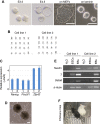
Establishment and characterization of CBA/J-derived ESC lines. (A) Images for preimplantation embryos and colonies of cells established from the embryos. Size bar = 100 μm. (B) Karyotype of two cell lines by G banding. (C) Expression of pluripotency marker genes. Reverse transcription and quantitative polymerase chain reaction was performed using RNA extracted from the cell lines showing a normal karyotype in (B). Probes are indicated. Ct values obtained with the indicated probes were normalized to those with Gapdh (ΔCt) and then normalized with ΔCts from authentic ESCs E14 (ΔΔCts). The negative values of the ΔΔCts are shown in the graph. Mean ± SD. n = 3. (D, E) The ability of the cell lines to differentiate in vitro by EB formation. Image of EBs (D). Real-time polymerase chain reaction was performed with RNA from attached EB-derived cells and revealed that ectoderm (Nestin), mesoderm (T), and endoderm (Gata6) maker genes were induced, compared with those in ESCs. H2O samples were obtained from PCRs with no RNA and served as negative controls. Size bar = 100 μm. (F) Photo of newborn chimeras after blastocyst injection of the CBA/J-derived ESCs. The brown coat color of the mouse indicates the high contribution of the ESCs to mouse development. EB: embryoid body; ESC: embryonic stem cell.
Figure 2.
Figure 2.
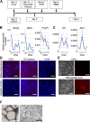
Differentiation of ESCs into the endothelial lineage. (A) Endothelial differentiation protocol. (B, C) Changes in the expression of pluripotent markers (Nanog, Zfp42, and Pou5f1) (B) and vascular endothelial markers (Tal1 and Egr4) (C) during the differentiation into endothelial lineages. Reverse transcription and quantitative polymerase chain reaction was performed using RNA extracted from cells at the indicated time points during differentiation. Probes for qPCR are indicated. Ct values obtained with the indicated probes were normalized by those with Gapdh (ΔCt) to calculate the relative expression values, and the values are plotted. Mean ± SEM. n = 3. # indicates the time at which one sample was undetectable. (D) Immunofluorescence staining (red) of differentiated cells using a specific antibody against CD31 (PECAM1), CD45, or VE-cadherin. DNA (blue) was stained with 4′,6-diamidino-2-phenylindole. The size bar = 50 μm. (E) Fluorescent microscopic images for DiI-Ac-LDL uptake (red). ESCs (top images) or day 4 differentiated cells (bottom) were treated with DiI-Ac-LDL for 4 h. After washing, the bright-field (left) or fluorescent (right) images were taken. Size bar = 50 μm. The fluorescence images were taken with the same condition. (F) Bright-field microscopic images of the differentiated cells forming a tube-like structure on a Matrigel-coated plate. Size bar = 20 µm (left) and 300 µm (right). Two images were taken 24 h after the day 4 differentiated cells were seeded on the plate. Two different microscopes were used for taking the images to clarify the tube-like structures formed by the differentiated cells. DiI-Ac-LDL: 1,10-dioctadecy1-3,3,30,30-tetramethlyindocarbocyanineperchlorate-acetylated–low-density lipoprotein; ESC: embryonic stem cell.
Figure 3.
Figure 3.
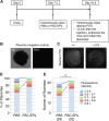
A decrease in number of placentae that exhibit hypoperfusion by intravenous injection of PSC-EPs. (A) The experimental design. (B, C) Images of placentae without (B) or with (C) the injection of fluorescence-conjugated dextran. The fluorescent image in (B) serves as a negative control for (C). (C) shows the examples of the fluorescent signals in placentae; the left shows the fluorescence signals whose intensity is close to the mean intensity among the placentae analyzed, and the right shows a decrease in the fluorescence intensity by <0.6 of the mean. (D, E) Classification of the placentae by fluorescence signals from the injected dextran. The fluorescence intensities used for the classification and numbers of the placentae analyzed are shown in (E). n = 29 (PBS) and 32 (PSC-EPs) placentae. Chi-squared test for the placentae that exhibited reduced fluorescence intensities by <0.8 of the mean in each group. *P < 0.05. PBS: phosphate-buffered saline; PSC-EPs: PSC-derived cells with endothelial potential.
Figure 4.
Figure 4.
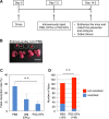
Reduction of fetal resorption by intravenous injection of PSC-EPs. (A) The experimental design. (B) Images of embryos, including placentae, collected from a DBA/2-mated CBA/J female mouse at day 14.5 of pregnancy. Circles in the image indicate resorptions. Size bar = 13 mm. (C) Fetal resorption rates with the indicated treatments. Rates were calculated by dividing the number of resorbed embryos with a total number of embryos in a pregnant mouse, and then the mean rate in each condition was calculated. Mean ± SD. n = 17 pregnant mice (PBS), 5 (BM EPCs), and 22 (PSC-EPs). Wilcoxon rank-sum test. **P < 0.01. (D) Number of embryos that were resorbed or not resorbed. n = 124 (PBS) and 149 (PSC-EPs) embryos. Chi-squared test for the resorbed embryos in each group. **P < 0.01. BM: bone marrow; EPC: endothelial progenitor cell; PBS: phosphate-buffered saline; PSC-EPs: PSC-derived cells with endothelial potential.
Figure 5.
Figure 5.
Robust establishment of spiral arteries by intravenous injection of PSC-EPs. (A, C) The reconstruction of the tiling Z-stack fluorescent images of the placenta collected from a PBS or PSC-EP-injected female mouse. In (A), the placenta was stained with an antibody against CD31 (red) before the imaging. In (C), PBS or PSC-EPs from GFP-labeled PSCs were intravenously injected into a DBA/2-mated CBA/J female mouse. The placenta was collected from the mouse, and GFP signals (green) in the reconstructed placenta images were captured. The top images show the entire placenta captured from an embryo. The bottom images are enlarged ones from the top. Size bar = 750 (top) and 300 µm (bottom) in (A), 300 (top) and 200 µm (bottom) in (C). (B) Number, diameter, or density of spiral arteries in placenta images. Mean (short blue bar) ± SD (long blue bars). n = 3 for each group (number and density). For diameter, n = 57 (PBS) vs. n = 96 (PSC-EPs). t-test. *P < 0.05; **P < 0.01. GFP: green fluorescent protein; PBS: phosphate-buffered saline; PSC-EPs: PSC-derived cells with endothelial potential.
Figure 6.
Figure 6.
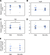
PSC-EP injection does not substantially affect the angiogenic factor balance in DBA/2-mated CBA/J females. (A-D) mRNA expression of the indicated genes in the placenta. RNA was extracted from a placenta of a PBS- or a PSC-EP-injected female mouse. Reverse transcription and quantitative polymerase chain reaction was performed using the extracted RNA. The probes used are indicated. The Ct values obtained with the indicated probes were normalized by those with Gapdh (ΔCt) and then normalized with ΔCts from the PBS-injected samples (ΔΔCts). The negative values of the ΔΔCts are shown in the graph. Mean (short blue bar) ± SD (long blue bars). In (A, B, and D), n = 26 for each group. In (C), n = 23 (PBS) vs. n = 26 (PSC-EPs). t-test. *P < 0.05. (E) Expression of sFlt1 protein in the blood. Blood was collected from a pregnant female mouse, and sFlt1 levels in the blood were quantified by enzyme-linked immunosorbent assay. Mean (short blue bar) ± SD (long blue bars). n = 8 (PBS) vs. n = 10 (PSC-EPs). PBS: phosphate-buffered saline; PSC-EPs: PSC-derived cells with endothelial potential.

Similar articles

Cited by

References

    1. Practice Committee of the American Society for Reproductive M. Evaluation and treatment of recurrent pregnancy loss: a committee opinion. Fertil Steril. 2012;98(5):1103–1111. - PubMed
    1. Shahine L, Lathi R. Recurrent pregnancy loss: evaluation and treatment. Obstet Gynecol Clin North Am. 2015;42(1):117–134. - PubMed
    1. Empson M, Lassere M, Craig J, Scott J. Prevention of recurrent miscarriage for women with antiphospholipid antibody or lupus anticoagulant. Cochrane Database Syst Rev. 2005;2005(2):CD002859. - PMC - PubMed
    1. Bobe P, Chaouat G, Stanislawski M, Kiger N. Immunogenetic studies of spontaneous abortion in mice. II. Antiabortive effects are independent of systemic regulatory mechanisms. Cell Immunol. 1986;98(2):477–485. - PubMed
    1. Clark DA, Chaouat G, Arck PC, Mittruecker HW, Levy GA. Cytokine-dependent abortion in CBA x DBA/2 mice is mediated by the procoagulant fgl2 prothrombinase [correction of prothombinase]. J Immunol. 1998;160(2):545–549. - PubMed

Publication types

LinkOut - more resources