Review
. 2021 Apr;46(4):284-300.
doi: 10.1016/j.tibs.2020.10.004. Epub 2020 Dec 18.

Big-Data Glycomics: Tools to Connect Glycan Biosynthesis to Extracellular Communication

Affiliations
Review

Big-Data Glycomics: Tools to Connect Glycan Biosynthesis to Extracellular Communication

Benjamin P Kellman et al. Trends Biochem Sci. 2021 Apr.

Abstract

Characteristically, cells must sense and respond to environmental cues. Despite the importance of cell-cell communication, our understanding remains limited and often lacks glycans. Glycans decorate proteins and cell membranes at the cell-environment interface, and modulate intercellular communication, from development to pathogenesis. Providing further challenges, glycan biosynthesis and cellular behavior are co-regulating systems. Here, we discuss how glycosylation contributes to extracellular responses and signaling. We further organize approaches for disentangling the roles of glycans in multicellular interactions using newly available datasets and tools, including glycan biosynthesis models, omics datasets, and systems-level analyses. Thus, emerging tools in big data analytics and systems biology are facilitating novel insights on glycans and their relationship with multicellular behavior.

Keywords: bioinformatics; cell–cell interaction; extracellular matrix; glycobiology; glycomics; systems biology.


Figures

Figure 1 (Key figure) -
Figure 1 (Key figure) -. The feedback between glycan dependent receptor sensing and glycan biosynthesis regulate cellular communication and environmental response.
Here we discuss (A) the system-level impacts of glycosylation, (B) tools to study glycan biosynthesis given various data types, and (C) databases and analytical strategies to explore the dynamic co-regulatory systems of glycan biosynthesis and intercellular communication. Diverse bioinformatics approaches and resources are now making it easier to consider glycosylation in diverse research fields.
Figure 2 -
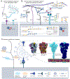
Figure 2 -. The Glycocalyx extends into the extracellular matrix.
(A) Common depictions of glycans and glycoconjugates in the extracellular matrix often censor structural and functional diversity. Panel B mirrors panel A with the structural detail unmasked glycoproteins describe proteins with branched N-or O-glycans, where carbon one of the first monosaccharide is covalently linked to the asparagine amine(N-), or serine/threonine hydroxyl(O-). Proteoglycans describe large linear glycosaminoglycans (GAGs) covalently bound to either a secretory granule protein, a protein with a transmembrane (e.g. syndecans) or glycosylphosphatidylinositol (GPI) anchor (e.g. glypicans), a pericellular protein or a Hyaluronan binding extracellular protein; GAGs like heparan and chondroitin sulfate present functional groupings of sulfation patterns. Hyaluronan-bound chondroitin sulfate proteoglycans are shown connecting two collagen fibers. Other common O-type glycans include glycolipids--mono or oligosaccharides often bound to glycerol or sphingosine backbones, and unconjugated lactation-secreted oligosaccharides. Each class is synthesized a little differently, for example, the acceptor for N-glycans, many O-glycans and GAGs is an amino acid while the conjugate for a glycolipid is a ceramide. Despite these distinctions, each of these classes follow general principles of glycan biosynthesis. Inside the cell, an O-GlcNAc modified a serine residue phosphorylation site. (C) A current example of glycans involved in SARS-CoV-2 attachment Heparan Sulfate facilitates target-cell attachment, while both the Spike protein and ACE2 are glycosylated; details cryo-EM struggles to resolve. Partially glycosylated (dark-blue) closed cryo-EM structure [19], 3D model of the open structure showing glycan range of motion (purple, green, orange, yellow) [14], and a simulation of the complete structure with and without glycans (dark-blue) [15] were adapted with permission.
Figure 3 -
Figure 3 -. Glycans & glycoconjugates mediate multiple types of interactions modulating cell state.
(A) Transitions between cell states (tan and brown) can be modulated by differential glycosylation (e.g. cell-cycle or epithelial-mesenchymal transition [75,76,153,154]). (B) Transition between two cell states with low or high MAN2A1 (mannosidase necessary to escape the hybrid glycan) expression could result in differential abundance of hybrid and complex glycans respectively. Differential glycosylation could modulate cell state in an oscillatory fashion (C). For example, differential expression of alpha-mannosidase II (MAN2A1; i & vi) would change both mannosylation and complexity of N-linked glycans [155,156]. As a result, each cell state produces a different dominant glycan: a hybrid biantennary structure (ii) and a sialylated biantennary structure (vii). The production of different glycans could result in the differential attachment of fibronectin to the integrin [24,157] thereby facilitating (iii) or disrupting (viii) ligand recruitment [16,158-160]. Differential glycosylation of a receptor can also directly impact receptor-ligand binding by changing receptor conformation (iv & ix) [23,25,161]. Differential receptor activation can induce the activation (v) or inhibition (x) of pathways and transcription factors (red circles are activated signalling cascade elements, blue circles are inactive elements) ultimately inducing differential expression of MAN2A1 (i & vi). In this theoretical system, transcription of MAN2A1 (vi) will move the cell to the complex-glycan state (vi-x) and this state ultimately leads to the inactivation of the signal transduction (x) and the subsequent inhibition of MAN2A expression (i) moving the cell back to the hybrid state (i-v). Thus, through basic principles of cell function and glycosylation, we have constructed a theoretical glycan-modulated oscillating cell-state system.
Figure 4 -
Figure 4 -. Characteristics of glycan biosynthesis.
Generally, glycans are covalently bound to a glycoconjugate and built by iterative addition, and occasional removal, of monosaccharides by highly-specific glycosyltransferases (GT), as the glycoconjugate passes through the endoplasmic reticulum (ER) and Golgi; most glycan products reactants for later reactions. GTs are retained in different endomembrane compartments thereby increasing biosynthetic diversity and control. N-glycosylation occurs in approximately 3 stages: addition and pruning of a large oligomannose structure in the ER, further pruning and GIcNAc-capping in the cis/medial Golgi, and GlcNAc-capped branch maturation. In specific models, (i) nonlinear kinetics can capture complex reaction behavior and incorporate variation over time (ii) In the absence of temporal data, simpler reaction behaviors can be adequately described with linear kinetics. (iii) Glycogenes like glycosyltransferases, nucleotide-sugar biosynthesis, and transport proteins must be present in the genome, epigenetically accessible, expressed and translated to perform their functions. Both linear and nonlinear models can be improved by including information on the availability of glycogenes. Other models can be created using only information on the availability of glycogenes. (iv) Glycan structure can also be used to supplement comprehensive modeling because the glycan structure is a complete description of every biosynthesis reaction a glycan undergoes. (v) New evidence suggests that steric interactions between glycoproteins and glycosyltransferases add additional constraints to glycan biosynthesis; sterics can decide which glycosyltransferases can access a growing glycan. (vi) Finally, metabolism can determine the availability of monosaccharide precursors thereby limiting the diversity of additions that may occur.
Figure 5 -
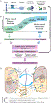
Figure 5 -. The progression of model complexity and predictions appropriate, given various common data types.
(A) Various datatypes of increasing complexity and rarity necessary to train different models. (B) The relationship between model specificity and model complexity. As the complexity increases and the magnitude and rarity of input data increases, so does the specificity of the model. Though lower complexity models are not as specific, they can be beneficially generalizable. (C) Finally, once the glycoprofile predictions are complete, they can be compared to lectin and substructure databases to predict what receptor-ligand interactions the differential glycosylation may impact. Panel D describes two theoretical cells (Figure 3C) co-regulating through differential glycosylation. If a reasonable differential expression signature can be inferred (due to differential interference or promotion of a receptor-ligand interaction), we can generate a “gene-availability” based prediction of updated glycosylation, thus exploring the sustainability of a glycosylation pattern. Labels A, B, and C correspond to methods illustrated in panels in A, B, and C respectively: (A) The omics assessment of the intracellular environment, (B) modeling of the glycan biosynthetic implications of the intracellular environment, (C) differential glycosylation of peptides and receptors and (A-C) the propagation of that information to the intercellular interface.

Similar articles

Cited by

References

    1. Davies HW, Trotter MD. Synthesis and turnover of membrane glycoconjugates in monolayer culture of pig and human epidermal cells. Br J Dermatol. 1981; 104: 649–658. - PubMed
    1. Mathieson T, Franken H, Kosinski J, Kurzawa N, Zinn N, Sweetman G, et al. Systematic analysis of protein turnover in primary cells. Nat Commun. 2018;9: 689. - PMC - PubMed
    1. Martinez-Palomo A, Braislovsky C, Bernhard W. Ultrastructural modifications of the cell surface and intercellular contacts of some transformed cell strains. Cancer Res. 1969;29: 925–937. - PubMed
    1. Aebersold R, Agar JN, Amster IJ, Baker MS, Bertozzi CR, Boja ES, et al. How many human proteoforms are there? Nat Chem Biol. 2018; 14: 206–214. - PMC - PubMed
    1. Lombard V, Golaconda Ramulu H, Drula E, Coutinho PM, Henrissat B. The carbohydrate-active enzymes database (CAZy) in 2013. Nucleic Acids Res. 2014;42: D490–5. - PMC - PubMed

Publication types

Substances