. 2021 Jan;24(1):47-60.
doi: 10.1038/s41593-020-00757-6. Epub 2020 Dec 21.

Microglia facilitate repair of demyelinated lesions via post-squalene sterol synthesis

Affiliations

Microglia facilitate repair of demyelinated lesions via post-squalene sterol synthesis

Stefan A Berghoff et al. Nat Neurosci. 2021 Jan.

Abstract

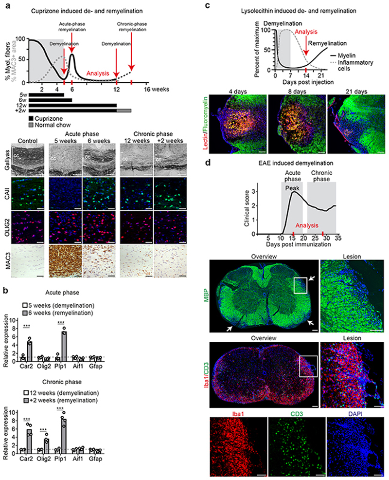
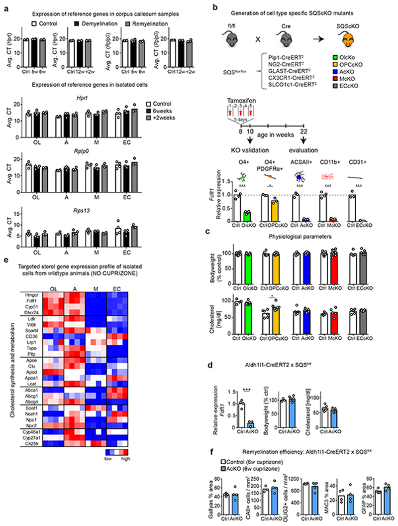
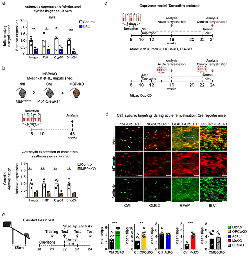
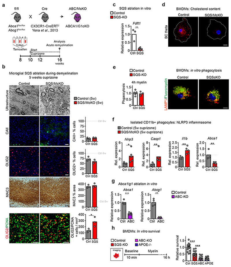
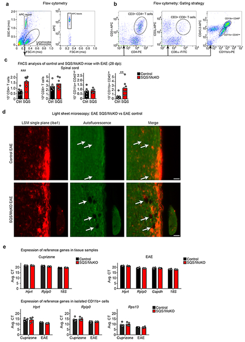
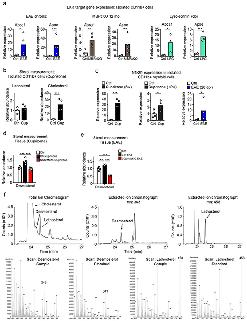
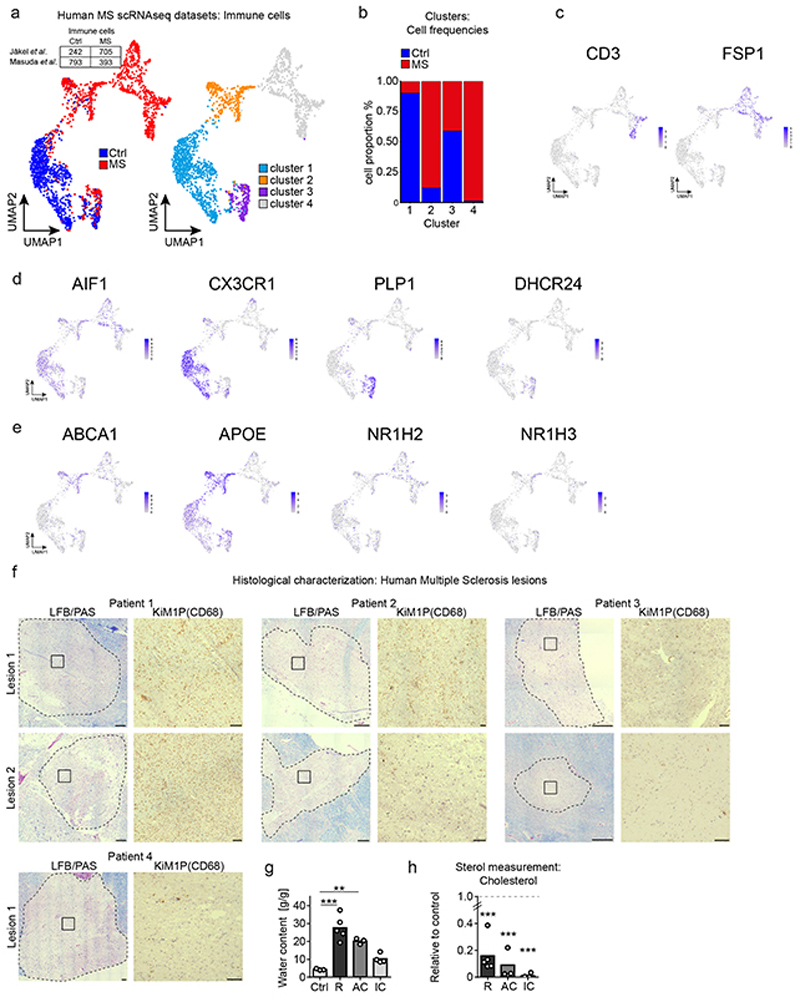
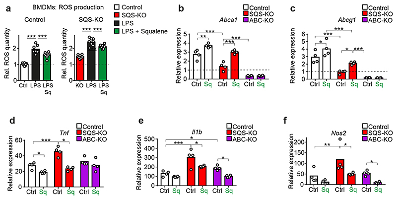
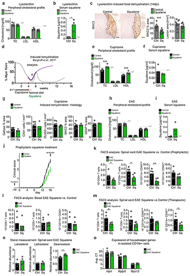
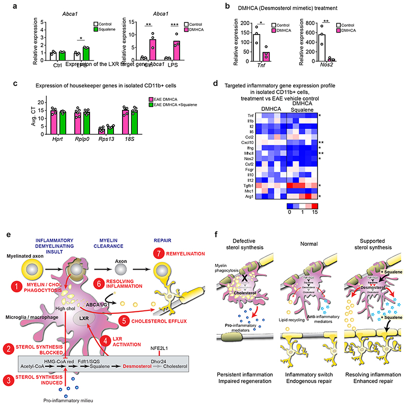
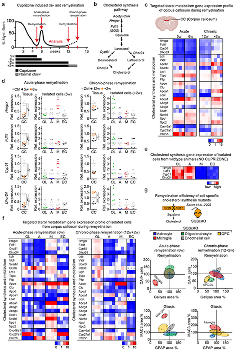
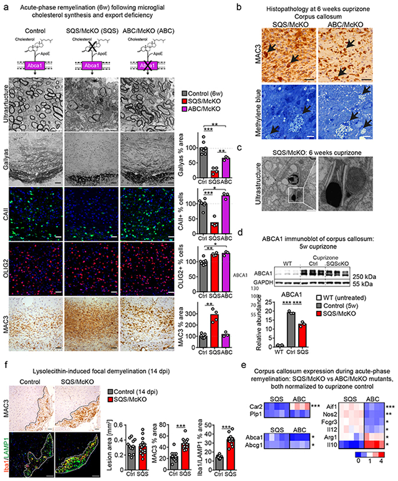
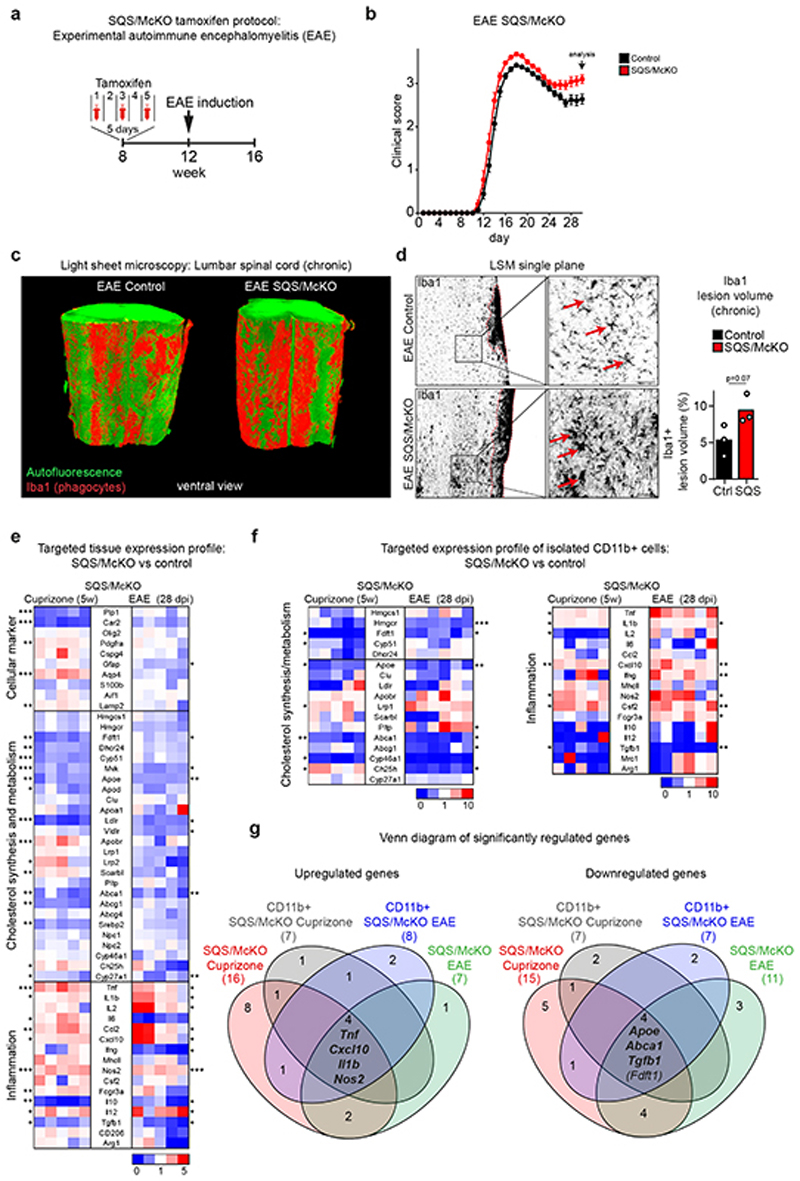
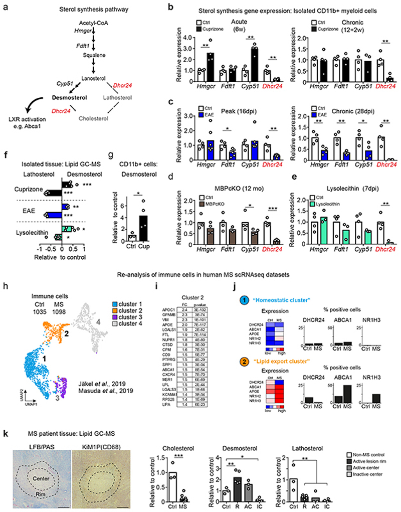
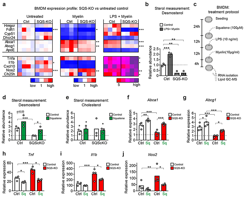
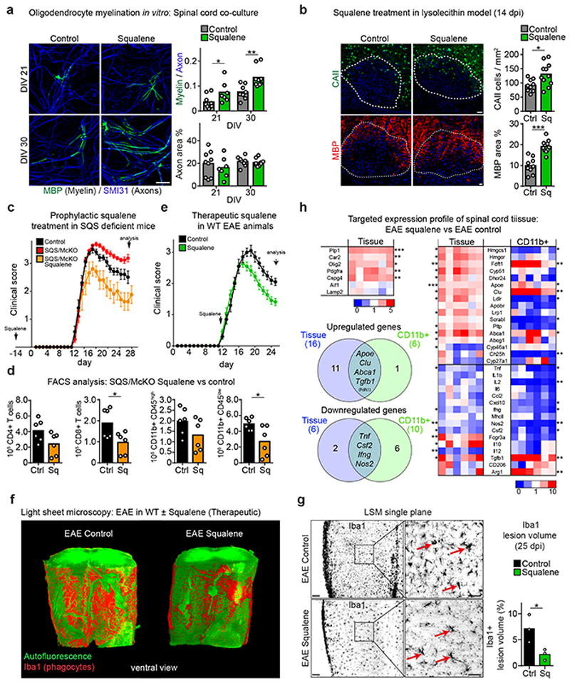
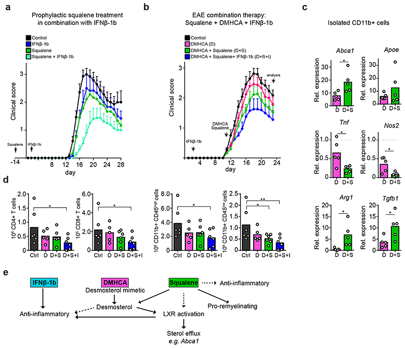
The repair of inflamed, demyelinated lesions as in multiple sclerosis (MS) necessitates the clearance of cholesterol-rich myelin debris by microglia/macrophages and the switch from a pro-inflammatory to an anti-inflammatory lesion environment. Subsequently, oligodendrocytes increase cholesterol levels as a prerequisite for synthesizing new myelin membranes. We hypothesized that lesion resolution is regulated by the fate of cholesterol from damaged myelin and oligodendroglial sterol synthesis. By integrating gene expression profiling, genetics and comprehensive phenotyping, we found that, paradoxically, sterol synthesis in myelin-phagocytosing microglia/macrophages determines the repair of acutely demyelinated lesions. Rather than producing cholesterol, microglia/macrophages synthesized desmosterol, the immediate cholesterol precursor. Desmosterol activated liver X receptor (LXR) signaling to resolve inflammation, creating a permissive environment for oligodendrocyte differentiation. Moreover, LXR target gene products facilitated the efflux of lipid and cholesterol from lipid-laden microglia/macrophages to support remyelination by oligodendrocytes. Consequently, pharmacological stimulation of sterol synthesis boosted the repair of demyelinated lesions, suggesting novel therapeutic strategies for myelin repair in MS.


Conflict of interest statement

Competing interests

SAB and GS are listed as inventors on pending patent claims (PCT###) filed by MPG covering the application of squalene in demyelinating disorders.

Figures

Extended Data Fig. 1
Extended Data Fig. 1. Mouse models of demyelination and remyelination
(a) The cuprizone model of demyelination and remyelination exhibits some similarities to pattern III MS lesions. Although the exact molecular cause of the cuprizone induced pathology remains elusive, copper chelation induced dysfunction of mitochondrial enzymes could selectively lead to apoptosis in oligodendrocytes (Praet et al., 2014). Cuprizone (bis-cyclohexanone oxaldihydrazone) is administered to adult mice as a 0.2% dietary supplement, which leads to a highly reproducible oligodendropathy followed by demyelination of distinct brain regions, predominantly the corpus callosum (CC). The graphical representation of myelination and microgliosis is based on histochemical stainings as shown in lower panels (further details to this model, and Matsushima and Morell, 2001). Feeding mice with cuprizone induces progressive loss of mature oligodendrocytes (CAII) that is already detectable two to three days following cuprizone administration and accompanied by gliosis and blood-brain barrier impairment (Berghoff et al., 2017, Stumpf et al., 2019). Loss of oligodendrocytes induces demyelination (Gallyas) in the corpus callosum starting after 2-3 weeks of cuprizone treatment. Loss of mature oligodendrocytes to less than 10% of control numbers and maximum demyelination of the corpus callosum is achieved after 5 weeks of continuous cuprizone supplementation (5w, acute demyelination). During the 5 weeks, OPCs proliferate (OLIG2) and remain largely undifferentiated. Whether OPCs do not differentiate during this disease period or differentiate, attempt to remyelinate and subsequently die is unclear. Demyelination is accompanied by progressive microgliosis (MAC3, dashed gray line). Following the peak at 5w, microgliosis quickly resolves within the following week despite ongoing cuprizone administration, leading to moderate microgliosis at 6 weeks cuprizone treatment (about 30% of MAC3 levels at 5w). This coincides with a brief remission phase characterized by oligodendrocyte maturation. The corpus callosum remyelinates reaching levels of about 50% myelin content of untreated controls (6w, acute-phase remyelination). In case of ongoing cuprizone treatment, remyelinated myelin sheaths subsequently degenerate. Microglial numbers progressively decrease despite ongoing degeneration leading to residual numbers of microglia at 12 weeks cuprizone. At this stage, the corpus callosum is chronically demyelinated (12w, chronic demyelination). CAII numbers of ~30% of untreated controls might also reflect a second spontaneous but minor remyelination phase. When cuprizone is withdrawn at this point and mice are fed normal chow, remyelination commences and reaches ~20% myelin content of untreated controls after 2 weeks recovery (12+2 weeks, chronic-phase remyelination) (Scale 50 μm). (b) Quantitative RT-PCR analysis determining the expression of oligodendrocyte and myelin related genes (Car2, Plp1, Olig2), and marker genes for microglia (Aif1) and astrocytes (Gfap) in corpus callosum samples during acute and chronic remyelination. Bars represent the means with individual data points normalized to the demyelination time point 5w cuprizone (n=4 animals, set to 1, dotted line). Acute-phase (6w, n=4 animals) and chronic-phase (12+2w, n=4 animals) remyelination is reflected in increased transcripts of genes related to oligodendrocyte maturation and myelination (Car2, Plp1) in corpus callosum samples. The resolving microgliosis between 5w and 6w of cuprizone feeding is reflected in an about 40% drop of Aif1 transcripts. Asterisks mark significant changes, ***p<0.001 (Student’s t-test, two-sided). (c) The lysolecithin model of demyelination and remyelination in the spinal cord (Bjelobaba et al., 2018). Focal spinal cord demyelinating lesions are induced by stereotactic injection of 1 μl lysolecithin (1%) into the left and right ventro-lateral funiculus between Th3 and Th4. Demyelination starts within hours and proceeds until about 5-7 days post injection, which is paralleled by microgliosis and OPC proliferation. Then phagocyte activity progressively resolves resulting in low levels of activated microglia at 3 weeks post induction. After clearance of myelin debris, oligodendrocytes differentiate and remyelination starts after about 10 days post induction. Like other toxic models, pathology in the lysolecithin is not mediated by inflammatory immune cell infiltration, although some invasion of T- and B-lymphocytes occur and the blood-brain barrier is disrupted by the experimental procedure (Yazdi et al., 2015) (Scale 100 μm). In contrast to the cuprizone model, repeated demyelination does not result in limited remyelination and reduced number of OPCs (Penderis et al., 2003). (d) MOG-EAE (experimental autoimmune encephalomyelitis) causes a monophasic inflammatory demyelinating disease and models predominantly inflammatory aspects of MS pathology (Constantinescu et al., 2011; Ransohoff, 2012). Mice are immunized with the MOG35-55 peptide (amino acid 35-55 of myelin oligodendrocyte protein) that induces expansion of autoreactive T lymphocytes during a preclinical phase. Already during this disease period, the blood-brain barrier is compromised (Paul et al., 2013) and some CNS immune cell entry and cytokine production occur. After 10-13 days post immunization clinical disabilities become evident characterized by ascending paralysis. Histopathology is more pronounced in spinal cord tissue than in brain. Lesions are characterized by marked infiltration of encephalitogenic effector T cells (CD3), as well as recruitment and activation of microglia/macrophages cells (Iba1) and demyelination (MBP). Tissue damage during this acute disease phase of EAE reaches a peak after several days followed by a chronic phase with milder but persistent disability. Remyelination events that are considered to be minor. Scale 50 μm. Berghoff, S.A., Düking, T., Spieth, L., Winchenbach, J., Stumpf, S.K., Gerndt, N., Kusch, K., Ruhwedel, T., Möbius, W., and Saher, G., Acta neuropathologica communications, 2017, 5, 94. Bjelobaba, I., Begovic-Kupresanin, V., Pekovic, S., and Lavrnja, I., J Neurosci Res, 2018, 96, 1021-1042. Constantinescu, C.S., Farooqi, N., O'Brien, K., and Gran, B., Br J Pharmacol, 2011, 164, 1079-1106. Matsushima, G.K., and Morell, P., Brain pathology, 2001, 11, 107-116. Paul, D., Cowan, A.E., Ge, S., and Pachter, J.S., Microvascular research, 2013, 86, 1-10. Penderis, J., Shields, S.A., and Franklin, R.J., Brain : a journal of neurology, 2003, 126, 1382-1391. Praet, J., Guglielmetti, C., Berneman, Z., Van der Linden, A., and Ponsaerts, P., Neurosci Biobehav Rev, 2014, 47, 485-505. Ransohoff, R.M., Nature neuroscience, 2012, 15, 1074-1077. Stumpf, S.K., Berghoff, S.A., Trevisiol, A., Spieth, L., Düking, T., Schneider, L.V., Schlaphoff, L., Dreha-Kulaczewski, S., Bley, A., Burfeind, D., Kusch, K., Mitkovski, M., Ruhwedel, T., Guder, P., Rohse, H., Denecke, J., Gärtner, J., Möbius, W., Nave, K.A., and Saher, G., Acta Neuropathol, 2019, 138, 147-161. Yazdi, A., Baharvand, H., and Javan, M., Neuroscience, 2015, 311, 34-44.
Extended Data Fig. 2
Extended Data Fig. 2. Generation and validation of cell specific cholesterol synthesis mutants
(a) Quantitative RT-PCR analysis determining the expression of housekeeping reference genes in corpus callosum or isolated cells from mice (n=4 animals) treated with cuprizone (A astrocytes, EC endothelial cells, M microglia/macrophages, OL oligodendrocytes). (b) Conditional mutants were generated by crossing SQSflox/flox mice to Cre driver lines. Fdft1 was inactivated by tamoxifen injections, and mice were analyzed at time points as indicated (middle panel). Fdft1 (SQS) expression in acutely isolated cells targeted by the conditional mutation shown as mean of fold values (OLcKO n=4, OPCcKO n=3, AcKO n=3, McKO n=3, ECcKO n=3) normalized to cells from control mice (OLcKO-Ctrl n=4, OPCcKO-Ctrl n=3, AcKO-Ctrl n=3, McKO-Ctrl n=4, ECcKO-Ctrl n=3; set to 1). Asterisks mark significant changes (Student’s t-test, two-sided). (c) Body weight (OLcKO n=15, OPCcKO n=7, AcKO n=5, McKO n=10, ECcKO n=5; OLcKO-Ctrl n=11, OPCcKO-Ctrl n=6, AcKO-Ctrl n=5, McKO-Ctrl n=9, ECcKO-Ctrl n=6) and total serum cholesterol (OLcKO n=7, OPCcKO n=7, AcKO n=5, McKO n=5, ECcKO n=8; OLcKO-Ctrl n=4, OPCcKO-Ctrl n=6, AcKO-Ctrl n=5, McKO-Ctrl n=5, ECcKO-Ctrl n=5) of analyzed cell-specific cholesterol synthesis mutants in (b) at the age of 22 weeks. Bars represent mean of individual animals. Asterisks mark significant changes (Student’s t-test, two-sided). (d) Evaluation of astrocyte-specific cholesterol synthesis mutants (Aldh1l1-CreERT2 driver line, Winchenbach et al., 2016), determining Fdft1 expression in isolated astrocytes, body weight and total serum cholesterol in mutants (isolated astrocytes n=3 animals; body weight n=9 animals, cholesterol n=7 animals and Cre controls (isolated astrocytes n=4 animals; body weight n=3 animals, cholesterol n=8 animals). Bars represent the means with individual data points normalized to controls. Asterisks mark significant changes (Student’s t-test, two-sided). (e) Targeted sterol metabolism gene expression profile of isolated cells from the corpus callosum of n=4 untreated WT animals. The relative expression heat map shows low transcript levels (blue) and high transcript levels (red) for each gene normalized to the mean of all samples. (f) Quantification of histological stainings for myelination (Gallyas), mature oligodendrocytes (CAII), oligodendrocyte linage cells (Olig2), microgliosis (MAC3) and astrogliosis (GFAP) in Aldh1l1-CreERT2 astrocyte cholesterol synthesis mutants (n=4 animals) as in (d) at 6 weeks cuprizone. Bars represent the means with individual data points normalized to controls (n=4 animals) set to 100). ***p < 0.001, **p < 0.01, *p < 0.05 Winchenbach, J., et al. Inducible targeting of CNS astrocytes in Aldh1l1-CreERT2 BAC transgenic mice. F1000Research 5, 2934 (2016).
Extended Data Fig. 3
Extended Data Fig. 3. Remyelination efficiency in conditional mutants
(a) Expression of cholesterol synthesis genes in isolated astrocytes from EAE lesions. Bars represent the means with individual data points (n=4 animals) normalized to isolated cells from controls (n=4 animals, set to 1). Asterisks mark significant changes (Student’s t-test, two-sided). (b) (Upper panel) Generation of MBP conditional mutants (MBPcKO) and (Lower panel) expression of cholesterol synthesis genes in isolated astrocyte 12 month post tamoxifen administration. Bars represent the means with individual data points (n=4 animals) normalized to isolated cells from control (n=4 animals, set to 1). Asterisks mark significant changes (Student’s t-test, two-sided). (c) Tamoxifen protocol to induced recombination during cuprizone paradigm. Tamoxifen was given either before cuprizone application to target SQS in astrocytes, microglia, OPCs and endothelial cells or before remyelination to target oligodendrocytes. (d) TDTO reporter expression during remyelination to evaluate cell targeting in cuprizone fed mice by co-labeling with CAII for oligodendrocytes, OLIG2 for oligodendrocyte linage cells, GFAP for astrocytes and Iba1 for microglia (Scale 50 μm). (e) Elevated beam testing of cholesterol synthesis mutants during chronic remyelination (OLcKO n=8, OPCcKO n=7, AcKO n=5, McKO n=5, ECcKO n=7; OLcKO-Ctrl n=8, OPCcKO-Ctrl n=7, AcKO-Ctrl n=5, McKO-Ctrl n=5, ECcKO-Ctrl n=8). Bars represent the means with individual data points of three testing sessions. Asterisks mark significant changes (Student’s t-test, two-sided). ***p < 0.001, **p < 0.01.
Extended Data Fig. 4
Extended Data Fig. 4. Ablation of microglial cholesterol synthesis does not affect demyelination in the cuprizone model
(a) Generation and cuprizone treatment paradigm of phagocyte-specific Abca1/Abcg1 cholesterol export mutants (ABC/McKO). (b) Representative images from control (n=3 animals) and SQS/McKO mice (n=3 animals) after 5w cuprizone (acute demyelination), showing myelination (ultrastructure, scale 1 μm), oligodendrocytes (CAII), oligodendrocyte lineage cells (OLIG2) and microgliosis (MAC3) (Scale 50 μm) with corresponding quantification on the right. Asterisks mark significant changes (Student’s t-test, two-sided). (c) Fdft1 expression in SQS-KO BMDMs cultures that were recombined in vitro (n=4 cultures). Data normalized to control cells (n=3 cultures, set to 1). Asterisks mark significant changes (Student’s t-test, two-sided). (d) Cholesterol labeling in SQS-KO BMDMs (Ctrl n=3, SQS-KO n=4 cultures) cultured in lipid-depleted media by live staining with BCtheta (Scale 10μm). (e) In vitro Fluoromyelin phagocytosis in control and SQS-KO BMDM cultures after a 4h challenge with myelin (Scale 10μm). Internalized myelin (fluoromyelin) was quantified by co-labeling for Lamp1-positive endo/lysosomes (Ctrl n= 7 cultures, SQS-KO n=4 cultures; 600-1050 cells). (f) Mean gene expression related to NLRP3 inflammasome and cholesterol efflux (Abca1) in isolated phagocytes from SQS/McKO mice (n=5 animals) normalized to cuprizone control (n=3 animals). Asterisks mark significant changes (Student’s t-test, two-sided). (g) Abca1 and Abcg1 expression in ABC-KO BMDMs cultures that were recombined in vitro (n=4 cultures). Data normalized to control cells (n=4 cultures, set to 1). Asterisks mark significant changes (Student’s t-test, two-sided). (h) In vitro survival of BMDM following myelin (10 μg/ml) treatment (n=17 cultures from control mice; n=16 from SQS mutants and ABC mutants, n=12 from APO knockout mice; each with 251-351 cells). About 50% of SQS mutant phagocytes died within 16h of incubation with myelin, with morphology attributable to pyroptosis (Video S1). Of note, in two lipid export mutants, i.e. ABCA1/ABCG1 and ApoE-deficient phagocytes, pyroptotic cell death was even more pronounced, whereas control phagocyte numbers did not change. Asterisks mark significant changes (one-way ANOVA with Tukey’s post test). ***p < 0.001, **p < 0.01, *p < 0.05
Extended Data Fig. 5
Extended Data Fig. 5. EAE pathology in SQS/McKO animals
(a) Representative pictures showing gating strategy to analyze lymphocytes and monocytes by flow cytometry. (b) Flow cytometry plots depicting CD4+ T cells (left), CD8+ T cells (middle) and myeloid subpopulations (right) consisting of macrophages (CD11b+/CD45high) and microglia (CD11b+/CD45low). (c) Flow cytometric quantification of inflammatory cell abundance at chronic EAE of SQS/McKO (n=8 animals) and control (n=8 animals) spinal cord samples. Asterisks mark significant changes (Student’s t-test, two-sided). (d) Light sheet microcopy of SQS/McKO (n=3 animals) and control (n=3 animals) spinal cord samples stained for Iba1+ phagocytes (red) and autofluorescence imaging. Arrows point to circular areas with low optical density suggestive of focal edema that was only occasionally observed in EAE controls (Scale 100μm). (e) Quantitative RT-PCR analysis determining the expression housekeeping reference genes in tissue and isolated CD11b+ cells from cuprizone treated SQS/McKO (n=5 animals), cuprizone control mice (n=4 animals), EAE (n=6 animals) and EAE control mice (n=5 animals). ***p < 0.001, **p < 0.01, *p < 0.05
Extended Data Fig. 6
Extended Data Fig. 6. LXR target gene upregulation and desmosterol accumulation in demyelinating mouse models
(a) Quantitative RT-PCR analysis determining expression of Abca1 and Apoe in isolated CD11b+ cells from chronic EAE (left panel, n=4 animals), MBPcKO (middle panel, n=4 animals) and lysolecithin (right panel, n=3 animals) lesions. Bars represent the means with individual data points normalized to isolated cells from control (EAE n= 4 animals, MBPcKO-Ctrl n=4 animals, Lysolecithin Ctrl n=4 animals, set to 1) (Student’s t-test, two-sided). (b) Relative abundance of lanosterol and cholesterol normalized to cell number in isolated CD11b+ myeloid cells during acute cuprizone-mediated demyelination (5w cuprizone), measured by lipid mass spectroscopy (Ctrl n=3 animals, cuprizone n=4 animals) (Student’s t-test, two-sided). (c) Relative expression of Nrf1/Nfe2l1 in isolated microglia isolated from control mice and diseased mice (n=4 mice per group) after feeding cuprizone for 6 weeks (left), 12+2 weeks (middle) and 28d after induction of EAE (right) (Student’s t-test, two-sided). (d) Relative abundance of desmosterol in corpus callosum during acute demyelination (5w cuprizone) of SQS/McKO animals (n=5 animals) and cuprizone controls (n=5 animals) measured by lipid mass spectrometry. Data normalized to non-disease controls (n=3 animals, set to 1) (one-way ANOVA with Tukey’s post test). (e) Relative abundance of desmosterol in spinal cord tissue during chronic EAE (28 dpi) of SQS/McKO animals (n=4 animals) and EAE controls (n=3 animals) normalized to untreated controls (n=4 animals, set to 1) measured by lipid mass spectroscopy (one-way ANOVA with Tukey’s post test). (f) Total ion chromatogram, selected ion mass chromatograms and spectra from representative human tissue sample and commercial standards analyzed by GC-MS. Time axes of chromatograms are broken to show only the elution of sterols. Lower panel shows background subtracted (adjacent to peak) mass spectra of N-Methyl-N (trimethylsilyl) trifluoroacetamide (MSTFA)-derivated metabolites. Asterisks mark significant changes, ***p < 0.001, **p < 0.01, *p < 0.05
Extended Data Fig. 7
Extended Data Fig. 7. Identity of immune cell cluster in human MS scRNA-seq datasets and characterization of analyzed human MS lesions
(a) Distribution of immune cells from control and MS patients (left) and k-means clustering (right) of cells (resolution 0.1) on UMAP plot of merged external scRNA-seq datasets. (b) Frequency distribution of cells from control and MS patients in each cluster. (c) Expression of selected marker genes in T cells (CD3) and non-microglia/macrophages (FSP1, Fibroblast-Specific Protein-1) in cluster 4 of sc-RNAseq datasets. (d) Expression of selected marker genes in myeloid cells (AIF1, CX3CR1), oligodendrocytes (PLP1) and DHCR24 expressing cells in analyzed immune cells of sc-RNAseq datasets. (e) Expression of genes related to cholesterol export in analyzed immune cells of sc-RNAseq datasets. (f) Histological characterization of isolated lesions from individual patients. Dotted line indicates lesion rim defined by myelin staining (LFB/PAS).Center activity (square) was determined by presents of phagocytic cells (KiM1P) (Scale 500μm). (g) Tissue water (g/g dry tissue) of control white (n=3 patients) matter and MS lesion material (n=4 patients). R: Active lesion rim (n=5 lesions); AC: Active lesion center (n=3 lesions); IC: Inactive lesion center (n=4 lesions). Asterisks mark significant changes (one-way ANOVA with Holm-Sidak post test). **p < 0.01,*p < 0.05. (h) Relative abundance of cholesterol normalized to input tissue weight and standard (controls set to 1) of control white (n=3 patients) matter and MS lesion material (n=4 patients). R: Active lesion rim (n=5 lesions); AC: Active lesion center (n=3 lesions); IC: Inactive lesion center (n=4 lesions). Asterisks mark significant changes (one-way ANOVA with Holm-Sidak post test). ***p < 0.001, **p < 0.01, *p < 0.05
Extended Data Fig. 8
Extended Data Fig. 8. Inflammatory response in wild type and SQS-KO BMDMs challenged with LPS
(a) Mean ROS levels in control (left) and SQS-KO BMDMs (right) stimulated with or without LPS and treated with or without squalene (100μM) (n=8 cultures per condition). Data normalized to untreated control (set to 1). Asterisks mark significant changes (two-way ANOVA with Holm-Sidak post test). (b-f) Relative expression of cholesterol efflux (b, c) and inflammatory (d-f) genes in control and SQS-KO BMDMs treated with myelin+LPS with or without squalene (as depicted in Fig. 5c), normalized to untreated WT cultures(n=4 cultures) (two-way ANOVA with Holm-Sidak post test). Asterisks mark significant changes, ***p < 0.001, **p < 0.01, *p < 0.05
Extended Data Fig. 9
Extended Data Fig. 9. Squalene affects serum squalene levels in lysolecithin and EAE models of MS but not in cuprizone-treated mice
(a) Peripheral serum cholesterol profile of control (n=5 animals) and squalene-treated WT (n=6 animals) animals 14 days post injection with lysolecithin. Measurement shows total cholesterol (TC), low-density lipoprotein cholesterol (LDL) and high-density lipoprotein cholesterol (HDL). Dotted lines indicate values of non-diseased WT mice (Student’s t-test, two-sided). (b) Serum squalene concentration measured by HPLC in control and squalene-treated lysolecithin animals treated as in (a) (n=3 animals each group) (Student’s t-test, two-sided). (c) Histopathological quantification of the lesion size and MAC3+ microglia/macrophages in the lesions from control (n=9 lesions from 5 animals) and squalene treated animals (n=11 lesions from 6 animals) at 14 dpi lysolecithin (Scale 100μm) (Student’s t-test, two-sided). (d) Scheme depicting induced remyelination paradigm in the cuprizone model. Mice were fed cuprizone for 4 weeks after which mice received chow supplemented with vehicle or squalene for one week (dotted line) followed by analysis. The gray line depicts the degree of myelination in case of continued cuprizone feeding. (e) Peripheral serum cholesterol profile determining total cholesterol (TC), low-density lipoprotein cholesterol (LDL) and high-density lipoprotein cholesterol (HDL) in control (n=5 animals) and cuprizone (n=4 animals) mice treated as in (d). Dotted lines indicate values of non-disease WT mice (Student’s t-test, two-sided). (f) Serum squalene concentration measured by HPLC in control and squalene-treated lysolecithin animals treated as in (d) (n=3 animals each group). Dotted line indicates values of non-disease controls. (g) Histological quantification of myelination (Gallyas), mature oligodendrocytes (CAII), oligodendrocyte linage cells (OLIG2), microgliosis (MAC3) and astrogliosis (GFAP) at induced remyelination with squalene supplementation (n=3 animals) compared to control (n=7 animals) in mice treated as in (d). (h) Peripheral serum cholesterol profile of control (n=10 animals) and squalene-treated (n=11 animals) EAE animals during peak of disease (16 dpi). Measurement shows total cholesterol (TC), low-density lipoprotein cholesterol (LDL) and high-density lipoprotein cholesterol (HDL). Dotted lines indicate values of non-disease WT mice. (i) Serum squalene concentration of control (n=2 animals) and squalene-treated (n=3 animals) EAE animals during peak of disease (16 dpi) measured by HPLC. (j) Clinical score ± SEM of control (n=6) and prophylactically squalene-treated (n=6) EAE animals analyzed at peak of disease (16 dpi). (k) Number of CD4+ and CD8+ T cells, CD11b/CD45high macrophages and CD11b/CD45low microglia per gram spinal cord determined by flow cytometry at peak of EAE (16 dpi) in control (n=6 animals) and squalene-treated animals (n=6 animals) (Student’s t-test, two-sided). (l) Quantification of blood inflammatory cells by flow cytometry from animals as in (j) during peak of EAE disease (n=6 animals each group). (m) Analysis of inflammatory cells in spinal cord tissue by flow cytometry from control (n=6 animals) and therapeutically squalene-treated (n=5 animals) EAE animals (Fig. 6e) (Student’s t-test, two-sided). (n) Relative abundance of sterol intermediates in spinal cord of squalene-treated EAE animals (n=4 animals) normalized to untreated EAE controls (n=4 animals, set to 1) (Student’s t-test, two-sided). (o) Quantitative RT-PCR analysis determining the expression housekeeping reference genes in isolated CD11b+ cells from spinal cord from EAE control (n=6 animals) and EAE mice treated with squalene (n=6 animals). Bar graphs represent means with individual data points. Asterisks mark significant changes, ***p < 0.001, **p < 0.01, *p < 0.05
Extended Data Fig. 10
Extended Data Fig. 10. Working model
(a) Abca1 expression following squalene (100 μM, left) and DMHCA (10 μM, right) treatment in LPS activated BMDMs (n=3 cultures) (Student’s t-test, two-sided). (b) Expression of inflammatory genes (Tnf, Nos2) in LPS (10ng/ml) activated WT BMDMs (n=3 cultures) 24h after DMHCA (10μM) treatment. Data normalized to untreated control (n=3 cultures, set to 1) (two-way ANOVA with Holm-Sidak post test). (c) Quantitative RT-PCR analysis determining the expression housekeeping reference genes in spinal cord from EAE mice treated with DMHCA (n=6 animals) and squalene (n=6 animals). (d) Fold expression profiles of inflammatory genes in CD11b+ cells isolated from spinal cord of EAE animals treated with DMHCA (n=6 animals) and squalene (n=6 animals) (normalized to vehicle-treated EAE mice, n=6 animals). Asterisks indicate significant differences between DMHCA and DMHCA/squalene co-treatment (Student’s t-test, two-sided). (e) Resulting working model of the relationship between sterol metabolism in microglia/macrophages in an inflammatory demyelinating lesion and myelin repair. (1) Phagocytosis of lipid-rich myelin increases the cellular cholesterol content in microglia/macrophages, which (2) inhibits sterol synthesis by negative feedback regulation. (3) In demyelinating lesions, pro-inflammatory mediators counteract this inhibition and induce sterol synthesis. NRF1/NFE2L1 likely senses intracellular cholesterol excess and represses Dhcr24, leading to the synthesis of the sterol intermediate desmosterol. (4) Increasing desmosterol levels activate LXR-signaling. (5) This leads to LXR-mediated export of cholesterol via ABC transporters (cholesterol recycling) and (6) dampening of the pro-inflammatory phenotype of microglia/macrophages. (7) Resolved inflammation and increased disposition of cholesterol facilitates remyelination by oligodendrocytes. (f) In case of normal repair of demyelinated lesions, microglia/macrophages are capable of lipid/cholesterol recycling and switching to an anti-inflammatory signature that depends on desmosterol synthesis. We speculate that this conserved mechanism mediates “silent” regeneration in asymptomatic individuals and in the preclinical phase of MS. When endogenous sterol synthesis is impaired as in our SQS conditional mutants or presumably also during aging or advanced disease, these repair strategies fail leading to cholesterol overloading of microglia/macrophages, persistent inflammation and impaired regeneration. Dietary squalene supplementation supports sterol synthesis. This increases desmosterol levels in microglia/macrophages that facilitates to resolve the local inflammation and to recycle lipid/cholesterol. In addition, squalene promotes cholesterol synthesis by oligodendrocytes for remyelination that collectively enhances repair. Asterisks mark significant changes, ***p < 0.001, **p < 0.01, *p < 0.05.
Figure 1
Figure 1. Cholesterol metabolism during de- and remyelination
(a) Time course of demyelination/remyelination in the cuprizone model with treatment paradigms. See also Extended Data Fig. 1a-b. (b) Cholesterol synthesis pathway with major enzymes and sterol intermediates. (c) Gene expression profile in the corpus callosum following cuprizone treatment (see selected genes in (d), Supplementary Table 1 shows numeric data). (d) Expression of sterol synthesis genes in corpus callosum and isolated cells from cuprizone-treated animals in comparison to untreated controls (16 weeks). Data points represent individual mice. Only significant changes are indicated (one-way ANOVA with Tukey’s post test; A astrocytes, CC corpus callosum, EC endothelial cells, M microglia/macrophages, OL oligodendrocytes). (e) Gene expression profile of sterol synthesis genes in cells isolated from the corpus callosum of untreated controls. (f) Gene expression profile in isolated cells from cuprizone-fed animals (selected genes in (d)). (g) Remyelination efficiency and gliosis in conditional cholesterol synthesis mutants (Acute: AcKO n=4, OLcKO n=5, OPCcKO n=4, McKO n=4, ECcKO n=5; Chronic: AcKO n=4, OLcKO n=4, OPCcKO n=7, McKO n=5, ECcKO n=5) relative to controls (Acute: AcKO-Ctrl n=4, OLcKO-Ctrl n=6, OPCcKO-Ctrl n=6, McKO-Ctrl n=7, ECcKO-Ctrl n=3; Chronic: AcKO-Ctrl n=5, OLcKO-Ctrl n=4, OPCcKO-Ctrl n=4, McKO-Ctrl n=5, ECcKO-Ctrl n=8, set to 100%). Quantified histochemical stainings for myelin (Gallyas), oligodendrocytes (CAII), microgliosis (MAC3) and astrogliosis (GFAP) are shown as 95% confidence ellipses with data points of individual animals (n). Heat maps show fold expression normalized to controls. Each square represents data obtained from one mouse (n=4). ***p<0.001, **p<0.01, *p<0.05
Figure 2
Figure 2. Impaired remyelination in sterol metabolism mutants
(a) Histopathology of microglial cholesterol synthesis (SQS/McKO, n=4 animals) and cholesterol export (ABC/McKO, n=3 animals) mutants, showing myelin loss (electron microscopic images, Gallyas), mature oligodendrocytes (CAII), oligodendrocyte linage cells (OLIG2) and microgliosis (MAC3) (Scale 50 μm) with quantification normalized to cuprizone controls (n=7 animals). Asterisks mark significant changes (one-way ANOVA with Tukey’s post test). (b) Foamy phagocytes following demyelination, visualized by MAC3 (Scale 50 μm) and methylene blue (Scale 10 μm) staining. Representative image of stainings from SQS/McKO (n=4 animals) and ABC/McKO (n=3) animals. (c) Cholesterol crystals in SQScKO cuprizone mice (n=4 animals, Scale 1 μm). (d) Cropped ABCA1 immunoblot following demyelination (5w cuprizone) of corpus callosum samples from untreated WT, cuprizone control and cuprizone SQS/McKO (n=3 animals). Reprobing for GAPDH confirms comparable protein loading. Asterisks mark significant changes (one-way ANOVA with Tukey’s post test). (e) Gene expression related to oligodendrocytes (Car2, Olig2), sterol efflux (Abca1, Abcg1) and inflammation during acute remyelination in corpus callosum from SQS/McKO (n=5) and ABC/McKO (n=4) mice normalized to cuprizone control (n=4 mice). Asterisks mark significant changes between SQS/McKO and ABC/McKO mutants (Student’s t-test, two-sided). (f) Microgliosis (MAC3, Iba1/LAMP1) in the DAPI-positive lesion area (dotted line) at 14 dpi after lysolecithin-mediated demyelination with quantification (Scale 100 μm). Asterisks mark significant changes between control (n= 12 lesions from 6 animals) and SQS/McKO (n=14 lesions from 7 animals) mutants (Student’s t-test, two-sided). ***p<0.001, **p<0.01, *p<0.05
Figure 3
Figure 3. Non-resolving inflammation in demyelinated microglia-specific cholesterol mutants
(a) Recombination strategy in EAE. Inducing EAE 4 weeks after tamoxifen injections allowed renewal of peripheral monocytes such that only microglia but not monocyte derived macrophages lacked sterol synthesis. (b) Mean clinical score ± SEM of control (n=34 animals) and SQS/McKO (n=27 animals) mice. (c) Representative light sheet microcopy of lumbar spinal cord samples (~2.25 mm length) from SQS/McKO and controls 28d after EAE induction, stained for Iba1+ phagocytes (n=3 animals). (d) Single plane from (c) with detail (Scale 50 μm) and quantification of percent Iba1+ lesion volume of control and SQS/McKO mutants (n=3 animals, Student’s t-test, two-sided). (e) Fold expression levels in corpus callosum of cuprizone (5w) treated SQS/McKO mice (n=5 animals) normalized to control mice (n= 7 animals, set to 1) and lumbar spinal cord of EAE (28 dpi) treated SQS/McKO mice (n=6 animals) normalized to control mice (n=6 animals, set to 1). Each square represents data obtained from one mouse. Asterisks mark significant changes (Student’s t-test, two-sided). Numerical data with statistics is shown in Supplementary Table 6-7. (f) Fold gene expression in isolated CD11b+ cells from SQS/McKO mice normalized (Cuprizone n=5 animals, EAE n=6 animals) to controls (Cuprizone n=4 animals, EAE n=6 animals, set to 1) treated as in (e). Asterisks mark significant changes (Student’s t-test, two-sided). (g) Venn diagram of differentially expressed genes in (e) and (f). ***p<0.001, **p<0.01, *p<0.05
Figure 4
Figure 4. Desmosterol synthesis is a common mechanism in demyelinating disease
(a) Scheme depicting the sterol synthesis pathway with the link to LXR activation via the agonist function of desmosterol. (b-e) Expression of cholesterol synthesis genes in isolated CD11b+ cells from individual mice (n) during cuprizone (b; n=4), EAE (c; control n=4, EAE n=6) MBPcKO (d; n=4) and lysolecithin (e; n=3) treatment (Student’s t-test, two-sided). (f) Mean relative abundance of desmosterol and lathosterol in tissue from cuprizone (5w, n=5 animals), EAE (n=3) and lysolecithin-treated mice (n=5) measured by GC-MS (one-way ANOVA, Tukey’s post test). (g) Mean relative abundance of desmosterol in isolated CD11b+ cells from 5w cuprizone-treated mice (n=4 animals) compared to untreated controls (n=3), measured by GC-MS (Student’s t-test, two-sided). (h) UMAP and unsupervised clustering of immune cell clusters from human MS scRNAseq datasets (GSE118257, GSE124335). (i) Top 20 upregulated genes of cluster 2 compared to cluster 1 showing fold change (FC) and p-value (Wilcoxon Rank Sum test, two sided). (j) Mean expression (related to the mean of cluster 1-3) and expression frequency (% positive nuclei/cells). (k) MS lesions (scale 500 μm) stained for myelin (LFB/PAS) and phagocytes (CD68/KiM1P) to illustrate lesion center and rim (dotted lines). Mean relative abundance of lipids (as indicated) in tissue samples from 4 MS patients, normalized to 3 control individuals. Ctrl: control white matter (n=3 samples); R: Active lesion rim (n=5 lesions); AC: Active lesion center (n=3 lesions); IC: Inactive lesion center (n=4 lesions) (one-way ANOVA, Holm-Sidak post test). Significant changes are marked by asterisks (***p<0.001, **p<0.01, *p<0.05).
Figure 5
Figure 5. Squalene administration restores LXR-signaling in SQS mutant phagocytes in vitro
(a) Relative expression of genes related to cholesterol synthesis, cholesterol export and inflammation in control and SQS-KO BMDMs treated with myelin (10 ng/ml) or LPS (10 ng/ml) + myelin for 16h normalized to untreated control cultures (n=4 cultures set to 1; one-way ANOVA with Tukey’s post test). (b) Desmosterol abundance in control and SQS-KO BMDMs (n=3 cultures) treated as in (a). (c) Scheme of squalene treatment of BMDM cultures. 24h after seeding, cells were treated with squalene or left untreated, followed by administration of LPS and myelin. (d-e) Abundance of desmosterol (d,) and cholesterol (e) in LPS + myelin-treated BMDM cultures with or without squalene administration (as in c), normalized to untreated WT controls (n=3 cultures). Asterisks mark significant changes (Student’s t-test, two-sided). (f-j) Relative expression of cholesterol efflux (f, g) and inflammatory (h-j) genes in control and SQS-KO BMDMs treated with myelin+LPS with or without squalene (as in c), normalized to untreated WT cultures (n=4 cultures, two-way ANOVA with Holm-Sidak post test). Bars represent the mean of individual cultures. ***p<0.001, **p<0.01, *p<0.05.
Figure 6
Figure 6. Exogenous squalene rescues non-resolving inflammation in SQS/McKO mice in vivo and promotes remyelination
(a) Mean myelination in spinal cord cultures at 21-30 days in vitro (DIV) with (DIV21, n=7 cultures; DIV30, n=9) or without squalene (DIV21, n=9; DIV30, n=8) by quantification of MBP+ myelin segments relative to SMI31+ axons (two-way ANOVA, Holm-Sidak post test). Scale, 50 μm. (b) Mean CAII+ oligodendrocytes and MBP+ myelin in lesions from control (n=9 lesions from 5 animals) and squalene-treated animals (n=11 lesions from 6 animals, two-sided Student’s t-test) at 14dpi lysolecithin. Scale, 50 μm. (c) Mean clinical score ± SEM of EAE, comparing controls (n=6), SQS/McKO (n=11) and SQS/McKO treated with prophylactic squalene. (d) Mean density inflammatory cells as in (c) from controls (n=6) and squalene-treated SQS/McKO mice (n=6) by flow cytometry (Student’s t-test, two-sided). (e) Mean clinical score ± SEM of WT EAE animals therapeutically treated with (n=11) or without squalene (n=10). (f) Representative light sheet microcopy of spinal cord samples (~2.25 mm length) from WT EAE mice treated with or without squalene (n=3 animals), stained for Iba1+ phagocytes (red) and autofluorescence imaging. (g) Single plane from (f) with detail and quantification of % Iba1+ spinal cord volume (n=3 animals, Student’s t-test, two-sided). Scale, 50 μm. (h) Fold expression profiles in tissue and CD11b+ cells from EAE animals treated with or without prophylactic squalene (normalized to vehicle-treated EAE mice, n=6), showing cellular markers (upper left) and genes related to sterol metabolism and inflammation (right). Venn diagrams indicate significantly differentially expressed genes (Student’s t-test, two-sided). ***p<0.001, **p<0.01, *p<0.05.
Figure 7
Figure 7. Combination therapy of squalene and interferone ameliorates EAE
(a) Mean clinical scores ± SEM of WT control (n=7), IFNβ-1b-treated (n=6), prophylactically squalene-treated (n=9) and combination-treated (squalene + IFNβ-1b, n=8) EAE animals. (b) Mean clinical scores ± SEM of EAE animals treated therapeutically with DMHCA (n=12), DMHCA + Squalene (n=11), DMHCA + Squalene in combination with IFNβ-1b (n=10) or left untreated animals. (c) Relative expression of LXR target genes and inflammatory genes in isolated CD11b+ cells from treatment groups in (b, n=5 animals), normalized to EAE control (n=6 animals). Asterisks mark significant changes (Student’s t-test, two-sided). (d) Flow cytometric quantification of inflammatory cells at chronic EAE of animals in (b, n=5 animals, one-way ANOVA with Holm-Sidak). (e) Scheme depicting suggested treatment effects of IFNβ-1b, DMHCA and squalene on inflammation, LXR-signaling and remyelination. Data represent means (data points) of individual animals. **p<0.01, *p<0.05.

Similar articles

Cited by

References

    1. Dietschy JM. Central nervous system: cholesterol turnover, brain development and neurodegeneration. Biological chemistry. 2009;390:287–293. - PMC - PubMed
    1. Saher G, et al. High cholesterol level is essential for myelin membrane growth. Nature neuroscience. 2005;8:468–475. - PubMed
    1. Courtney R, Landreth GE. LXR Regulation of Brain Cholesterol: From Development to Disease. Trends in endocrinology and metabolism: TEM. 2016;27:404–414. - PMC - PubMed
    1. Reich DS, Lucchinetti CF, Calabresi PA. Multiple Sclerosis. The New England journal of medicine. 2018;378:169–180. - PMC - PubMed
    1. Plemel JR, Liu WQ, Yong VW. Remyelination therapies: a new direction and challenge in multiple sclerosis. Nature reviews Drug discovery. 2017;16:617–634. - PubMed

Publication types

MeSH terms

LinkOut - more resources