. 2021 May;85(5):2723-2734.
doi: 10.1002/mrm.28622. Epub 2020 Dec 8.

Ensuring both velocity and spatial responses robust to B0/B1+ field inhomogeneities for velocity-selective arterial spin labeling through dynamic phase-cycling

Affiliations

Ensuring both velocity and spatial responses robust to B0/B1+ field inhomogeneities for velocity-selective arterial spin labeling through dynamic phase-cycling

Dapeng Liu et al. Magn Reson Med. 2021 May.

Abstract

Purpose: To evaluate both velocity and spatial responses of velocity-selective arterial spin labeling (VS-ASL), using velocity-insensitive and velocity-compensated waveforms for control modules, as well as a novel dynamic phase-cycling approach, at different B0 / B1+ field inhomogeneities.

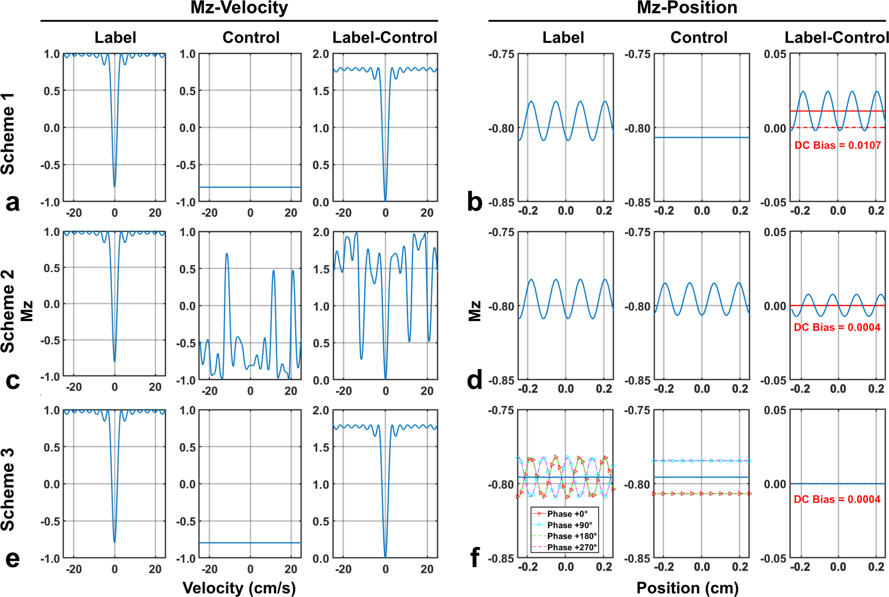
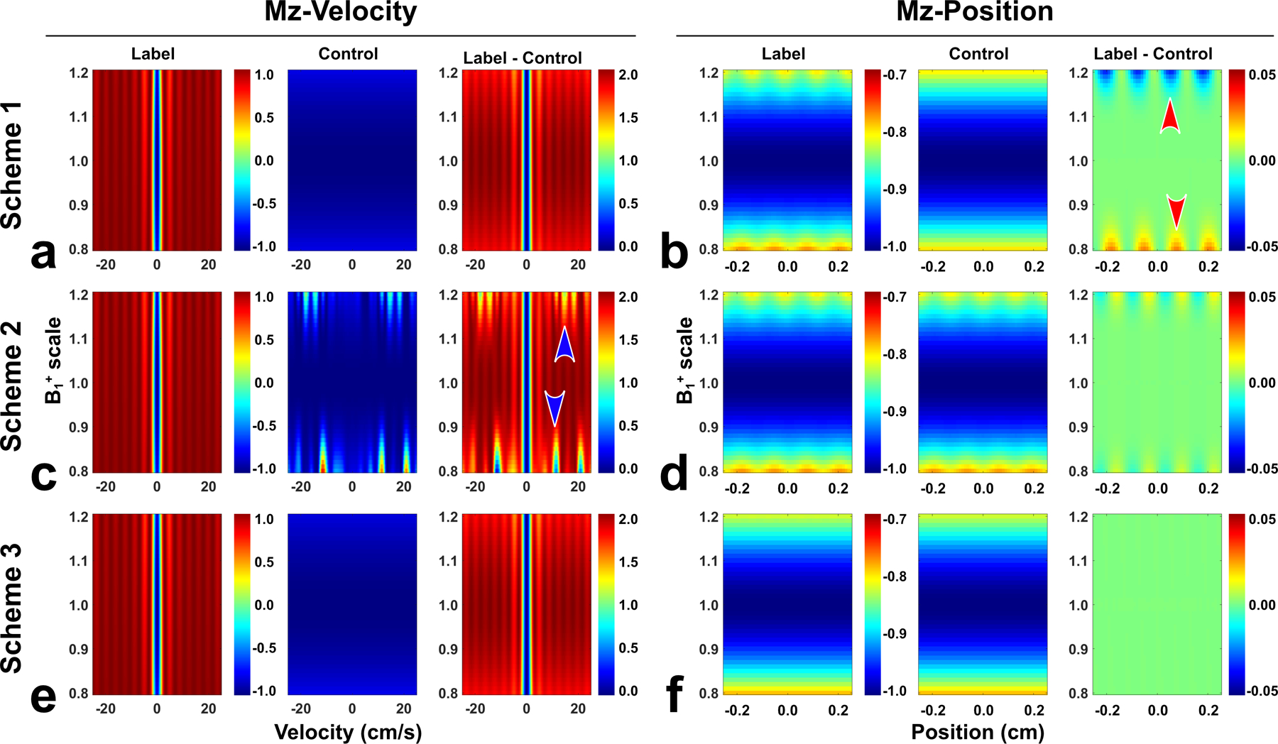
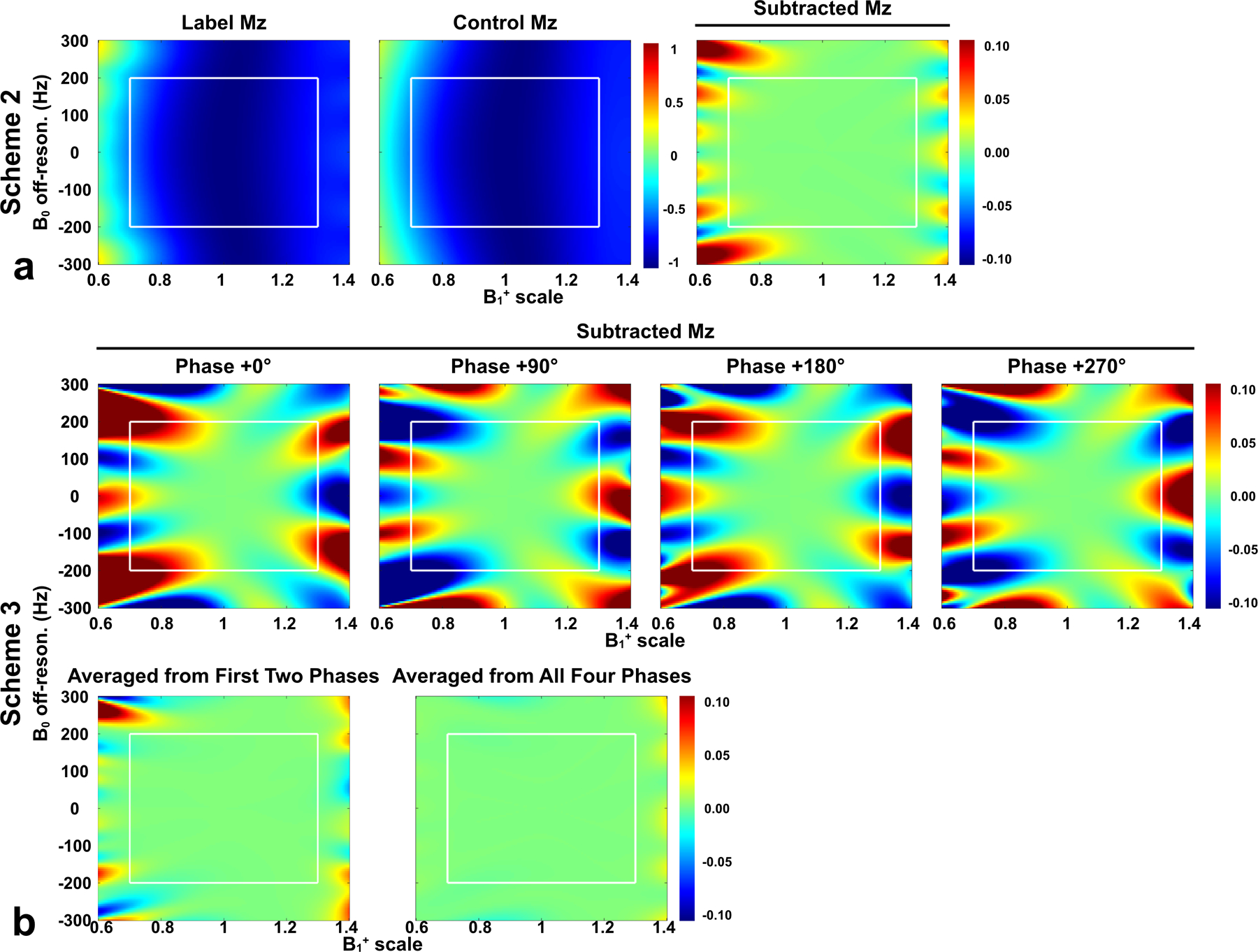
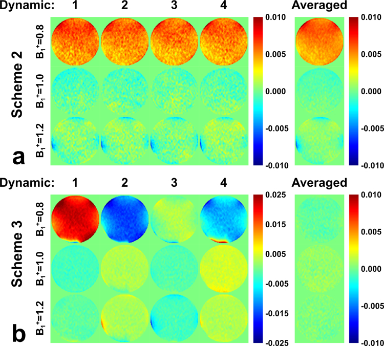
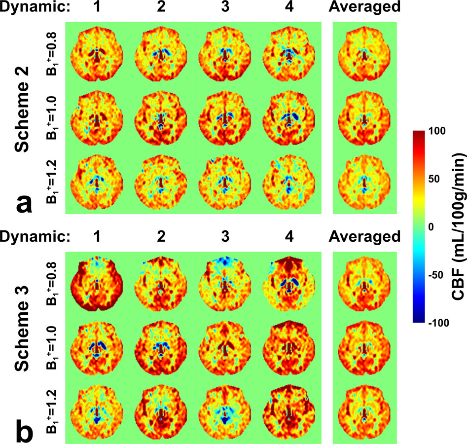
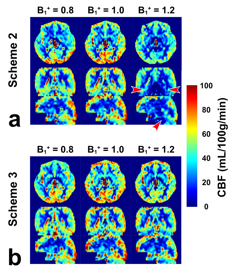
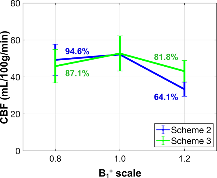
Methods: In the presence of imperfect refocusing, the mechanism of phase-cycling the refocusing pulses through four dynamics was first theoretically analyzed with the conventional velocity-selective saturation (VSS) pulse train. Numerical simulations were then deployed to compare the performance of the Fourier-transform based velocity-selective inversion (FT-VSI) with these three different schemes in terms of both velocity and spatial responses under various B0 / B1+ conditions. Phantom and human brain scans were performed to evaluate the three methods at B1+ scales of 0.8, 1.0, and 1.2.

Results: The simulations of FT-VSI showed that, under nonuniform B0 / B1+ conditions, the scheme with velocity-insensitive control was susceptible to DC bias of the static spins as systematic error, while the scheme with velocity-compensated control had deteriorated velocity-selective labeling profiles and, thus, reduced labeling efficiency. Through numerical simulation, phantom scans, and brain perfusion measurements, the dynamic phase-cycling method demonstrated considerable improvements over these issues.

Conclusion: The proposed dynamic phase-cycling approach was demonstrated for the velocity-selective label and control modules with both velocity and spatial responses robust to a wide range of B0 and B1+ field inhomogeneities.

Keywords: B1+ field inhomogeneity; B0 field inhomogeneity; arterial spin labeling; cerebral blood flow; velocity-selective inversion.


Figures

Figure 1:
Figure 1:
Simulated Mz-velocity profiles (position = 0 cm) and Mz-position profiles (velocity = 0 cm/s) of FT-VSI label and control pulse trains at the B1+ scale of 0.8 and their subtractions with (a,b) scheme 1 using velocity-insensitive control; (c,d) scheme 2 using velocity-compensated control; and (e,f) scheme 3 using dynamic phase-cycling. For both label and control in scheme 3 (f), Mz-position profiles from all four phases were shown by four colored dashed lines. In Mz-position profiles (b,d,f), solid red lines indicate the DC bias averaged from signals at all positions. In contrast to spatial response of scheme 1 (b) and velocity response of scheme 2 (c), scheme 3 showed well-preserved velocity-selective profile (e) as well as minimal spatial stripe artifact and DC bias (f).
Figure 2:
Figure 2:
Simulated Mz-velocity profiles and Mz-position profiles of FT-VSI label and control pulse trains at a B1+ scale range of 0.8 – 1.2 and their subtractions with (a,b) scheme 1 using velocity-insensitive control; (c,d) scheme 2 using velocity-compensated control; and (e,f) scheme 3 using dynamic phase-cycling. Compared to schemes 1 and 2, scheme 3 yielded both velocity and spatial responses with much less susceptibility to B1+ variations.
Figure 3:
Figure 3:
DC bias of Mz-position profiles following simulation of FT-VSI pulse trains with a range of B0 (±300 Hz) and B1+ (±0.4). Label, control, and subtracted Mz were shown for (a) scheme 2 using velocity-compensated control, while only subtracted Mz was shown for (b) scheme 3 using dynamic phase-cycling. In scheme 3, four individual dynamic phases (second row) and averages of first two phases and all four phases (third row) were shown. Note that scheme 3 with the phase of 0° was just scheme 1. White rectangles indicated the typical B0/B1+ range of human brain at 3T (B0: ±200 Hz; B1+ scale: ±0.3). Compared to the other two schemes, scheme 3 considerably mitigated the DC bias as false perfusion signal.
Figure 4:
Figure 4:
Evaluations of DC bias on the phantom with FT-VSI prepared ASL (post labeling delay = 0 ms) at B1+ scale of 0.8, 1.0, and 1.2 with (a) scheme 2 using velocity-compensated control and (b) scheme 3 using dynamic phase-cycling. Normalized subtracted signal from each dynamic label/control pairs are shown along with their averages. For scheme 3, although each dynamic generated strong false signal, they were mostly canceled after averaging.
Figure 5:
Figure 5:
Evaluations of DC bias using FT-VSI prepared brain ASL (post labeling delay = 1500 ms, one axial slice) at the B1+ scale of 0.8, 1.0, and 1.2 with (a) scheme 2 using velocity-compensated control and (b) scheme 3 using dynamic phase-cycling. Calculated CBF maps from each dynamic label/control pairs are shown along with their averages. Compared to scheme 2, scheme 3 generated some variable signal through dynamics in some brain regions, such as in frontal cortex, but after averaging, delivered CBF maps with more consistency across different B1+ scales.
Figure 6:
Figure 6:
CBF maps acquired from the FT-VSI prepared brain ASL along axial, sagittal and coronal views at the B1+ scale of 0.8, 1.0, and 1.2 with (a) scheme 2 using velocity-compensated control and (b) scheme 3 using dynamic phase-cycling. Scheme 3 markedly reduced the pseudo perfusion deficit at the base of the brain produced by scheme 2 with the B1+ scale of 1.2 (a, red arrowhead).
Figure 7:
Figure 7:
Averaged gray matter CBF of all five subjects, quantified from the FT-VSI prepared brain ASL at B1+ scale of 0.8, 1.0, and 1.2, with scheme 2 using velocity-compensated control and scheme 3 using dynamic phase-cycling. The respective signal levels at B1+ scales of 0.8 and 1.2 relative to the ones from B1+ = 1.0 are labeled in %.

Similar articles

Cited by

References

    1. Wong EC, Cronin M, Wu WC, Inglis B, Frank LR, Liu TT. Velocity-selective arterial spin labeling. Magn. Reson. Med 2006;55:1334–1341. - PubMed
    1. Duhamel G, de Bazelaire C, Alsop DC. Evaluation of systematic quantification errors in velocity-selective arterial spin labeling of the brain. Magn. Reson. Med 2003;50:145–153. - PubMed
    1. Wu WC, Wong EC. Intravascular effect in velocity-selective arterial spin labeling: The choice of inflow time and cutoff velocity. Neuroimage 2006;32:122–128. - PubMed
    1. Qiu D, Straka M, Zun Z, Bammer R, Moseley ME, Zaharchuk G. CBF measurements using multidelay pseudocontinuous and velocity-selective arterial spin labeling in patients with long arterial transit delays: Comparison with xenon CT CBF. J. Magn. Reson. Imaging 2012;36:110–119. - PMC - PubMed
    1. Bolar DS, Gagoski B, Orbach DB, et al. Comparison of CBF measured with combined velocity-selective arterial spin-labeling and pulsed arterial spin-labeling to blood flow patterns assessed by conventional angiography in pediatric Moyamoya. Am. J. Neuroradiol 2019;40:1842–1849. - PMC - PubMed

Publication types