. 2021 Jan 29;41(1):BSR20202514.
doi: 10.1042/BSR20202514.

Optimization of the isolation procedure and culturing conditions for hepatic stellate cells obtained from mouse

Affiliations

Optimization of the isolation procedure and culturing conditions for hepatic stellate cells obtained from mouse

Thanh Minh Dang et al. Biosci Rep. .

Abstract

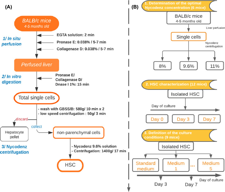
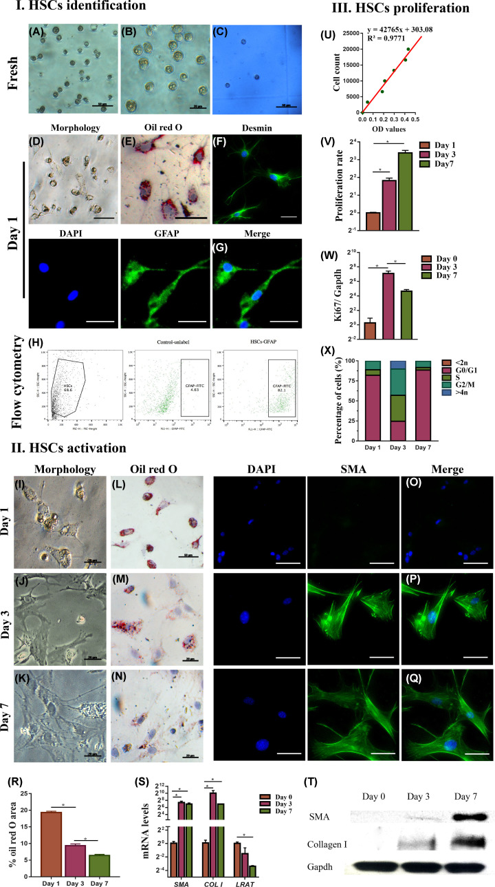
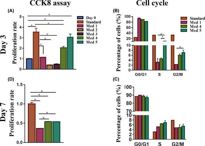
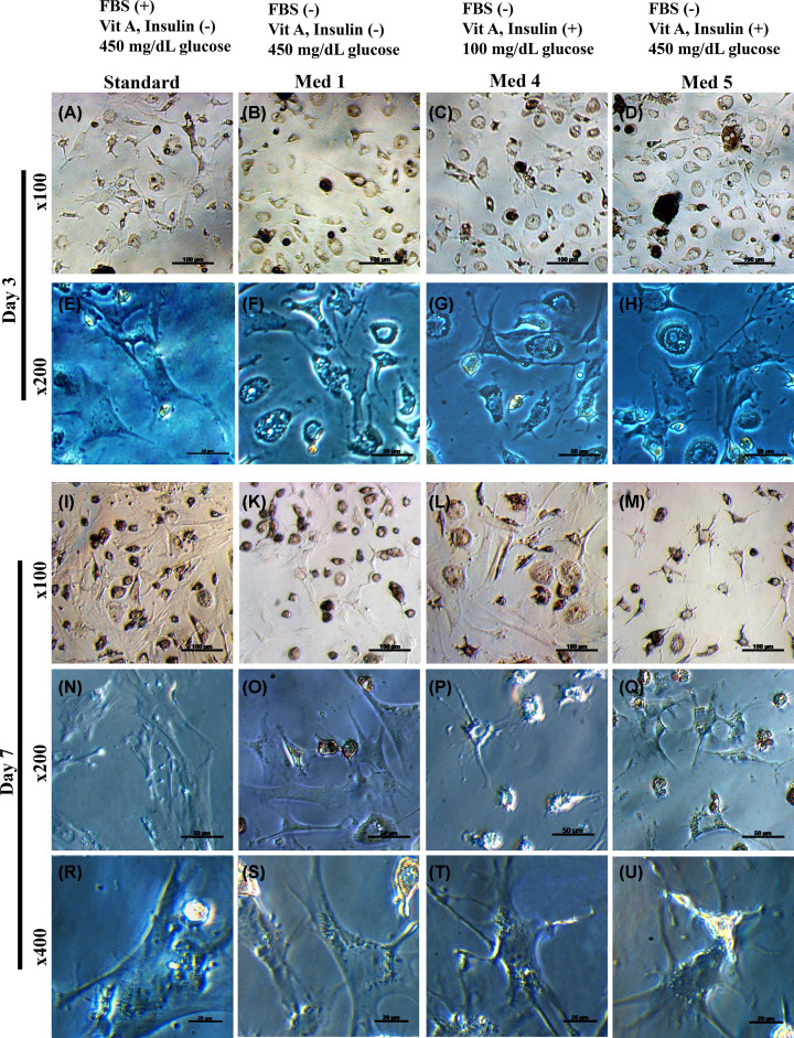
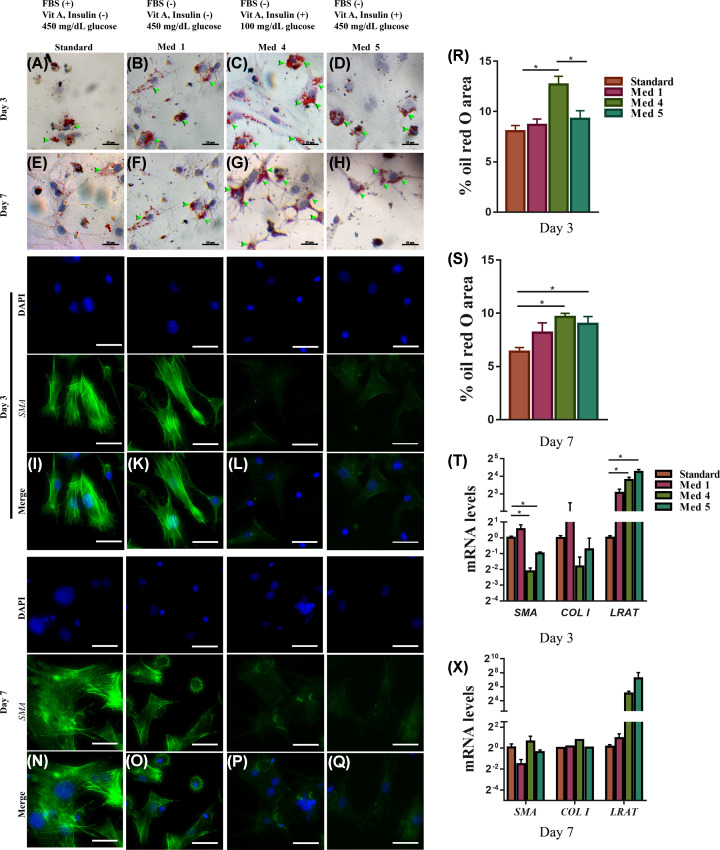
Liver fibrosis (LF) mortality rate is approximately 2 million per year. Irrespective of the etiology of LF, a key element in its development is the transition of hepatic stellate cells (HSCs) from a quiescent phenotype to a myofibroblast-like cell with the production of fibrotic proteins. It is necessary to define optimal isolation and culturing conditions for good HSCs yield and proper phenotype preservation for studying the activation of HSCs in vitro. In the present study, the optimal conditions of HSC isolation and culture were examined to maintain the HSC's undifferentiated phenotype. HSCs were isolated from Balb/c mice liver using Nycodenz, 8, 9.6, and 11%. The efficiency of the isolation procedure was evaluated by cell counting and purity determination by flow cytometry. Quiescent HSCs were cultured in test media supplemented with different combinations of fetal bovine serum (FBS), glutamine (GLN), vitamin A (vitA), insulin, and glucose. The cells were assessed at days 3 and 7 of culture by evaluating the morphology, proliferation using cell counting kit-8, lipid storage using Oil Red O (ORO) staining, expression of a-smooth muscle actin, collagen I, and lecithin-retinol acyltransferase by qRT-PCR and immunocytochemistry (ICC). The results showed that Nycodenz, at 9.6%, yielded the best purity and quantity of HSCs. Maintenance of HSC undifferentiated phenotype was achieved optimizing culturing conditions (serum-free Dulbecco's Modified Eagle's Medium (DMEM) supplemented with glucose (100 mg/dl), GLN (0.5 mM), vitA (100 μM), and insulin (50 ng/ml)) with a certain degree of proliferation allowing their perpetuation in culture. In conclusion, we have defined optimal conditions for HSCs isolation and culture.

Keywords: HSCs activation; Hepatic Stellate Cells; Nycodenz; Quiescent stellate cells; liver fibrosis.


Conflict of interest statement

The authors declare that there are no competing interests associated with the manuscript.

Figures

Figure 1
Figure 1. The flowchart of study design
(A) Flow chart of the isolation procedure of HSCs from mouse livers and (B) Experimental design of HSCs culture.
Figure 2
Figure 2. Evaluation of different Nycodenz concentrations on HSCs purification
The cell yield using Nycodenz concentrations at 8, 9.6, and 11% was evaluated by cell counting (A). Enrichment in HSCs cells was determined by cytofluorimetry (D) for desmin-positive cells as quantified in (B). The amount of contaminating cells (Kupffer and endothelial cells) was determined measuring the mRNA expression levels of clec4f and pecam1 genes, as markers of Kupffer and endothelial cells (C) after normalizing to the housekeeping gene Gapdh. Data are shown as means ± SEM, n=3, *P<0.05. Abbreviations: clec4f, C-type lectin domain family 4 member F; pecam1, platelet endothelial cell adhesion molecule-1.
Figure 3
Figure 3. HSCs characterization
Freshly isolated cells (I) and cells isolated at days 1 and 7 (II) were evaluated by phase-contrast microscopy (A,B,D,IK), by ORO dye staining for LDs (E,LN,R), by hemocytometer and Trypan Blue staining for viability (C), by flow cytometry for GFAP marker (H), by desmin immunofluorescence (F), GFAP (G) and a-SMA (O–Q) and mRNA levels of a-SMA, COL I and LRAT (S), western blot for a-SMA and collagen I (T). (III) HSCs proliferation evaluated by cell counting (U), by proliferation rate with CCK8 assay (V), by ki67 mRNA levels normalized to the housekeeping gene Gapdh (W), by cell cycle analysis (X). Data are shown as means ± SEM, n=3, *P<0.05, scale bar: 50 μm were not noted. Abbreviation: COL I, type I collagen.
Figure 4
Figure 4. Proliferation of HSCs in different culture media
HSCs cultured at day 3 (A,B) and day 7 (C,D) in standard medium and media 1–5 were evaluated for proliferation rate by the CCK8 assay and percentages of cells in the different phases of the cell cycles by cell cycle analysis (n=3), *P<0.05.
Figure 5
Figure 5. Changes in HSCs morphology by differences in culture media
Representative images of the morphology by light microscope of day 3 ((A–D) and (E–H) for ×100 and ×200 magnification, respectively) and day 7 HSCs cultured ((I–M), (N–Q), and (R–U) for ×100, ×200, and ×400 magnification, respectively) in standard medium and media 1–5.
Figure 6
Figure 6. Effects of different culture media on the activation of primary HSCs
Quiescent HSCs cultured under different test media (standard, 1–5) were examined for activation by LD content using ORO dye staining at day 3 (AD) and day 7 (E–H); by immunofluorescent staining for α-SMA day 3, (N–Q) and day 7 (I,K–M). Quantification of LDs accumulation at day 3 (R), and day 7 (S) were shown. mRNA levels of the activation-related genes (a-SMA, COL I) and quiescent-related gene (LRAT) at day 3 (T), and day 7 (X). Data are shown as means ± SEM, n=3, *P<0.05. The arrowheads show the ORO-stained area. Scale bar: 50 μm were not noted. Abbreviation: COL I, type I collagen.

References

    1. March S., Graupera M., Sarrias M.R. et al. . (2007) Identification and functional characterization of the hepatic stellate cell CD38 cell surface molecule. Am. J. Pathol. 170, 176–187 10.2353/ajpath.2007.051212 - DOI - PMC - PubMed
    1. Friedman S.L. (1993) The cellular basis of hepatic fibrosis–mechanisms and treatment strategies. N. Engl. J. Med. 328, 1828–1835 - PubMed
    1. Geerts A. (2001) History, heterogeneity, developmental biology, and functions of quiescent hepatic stellate cells. In Seminars in Liver Disease, Thieme Medical Publishers Inc., 333 Seventh Avenue, New, Copyright© 2001 - PubMed
    1. Blaner W.S., O'Byrne S.M., Wongsiriroj N. et al. . (2009) Hepatic stellate cell lipid droplets: a specialized lipid droplet for retinoid storage. Biochim. Biophys. Acta Mol. Cell Biol. Lipids 1791, 467–473 10.1016/j.bbalip.2008.11.001 - DOI - PMC - PubMed
    1. Gard A.L., White F.P. and Dutton G.R. (1985) Extra-neural glial fibrillary acidic protein (GFAP) immunoreactivity in perisinusoidal stellate cells of rat liver. J. Neuroimmunol. 8, 359–375 10.1016/S0165-5728(85)80073-4 - DOI - PubMed

Publication types

LinkOut - more resources