. 2020 Dec 1;3(12):e2030455.
doi: 10.1001/jamanetworkopen.2020.30455.

Assessment of Maternal and Neonatal SARS-CoV-2 Viral Load, Transplacental Antibody Transfer, and Placental Pathology in Pregnancies During the COVID-19 Pandemic

Affiliations

Assessment of Maternal and Neonatal SARS-CoV-2 Viral Load, Transplacental Antibody Transfer, and Placental Pathology in Pregnancies During the COVID-19 Pandemic

Andrea G Edlow et al. JAMA Netw Open. .

Abstract

Importance: Biological data are lacking with respect to risk of vertical transmission and mechanisms of fetoplacental protection in maternal severe acute respiratory syndrome coronavirus 2 (SARS-CoV-2) infection.

Objective: To quantify SARS-CoV-2 viral load in maternal and neonatal biofluids, transplacental passage of anti-SARS-CoV-2 antibody, and incidence of fetoplacental infection.

Design, setting, and participants: This cohort study was conducted among pregnant women presenting for care at 3 tertiary care centers in Boston, Massachusetts. Women with reverse transcription-polymerase chain reaction (RT-PCR) results positive for SARS-CoV-2 were recruited from April 2 to June 13, 2020, and follow-up occurred through July 10, 2020. Contemporaneous participants without SARS-CoV-2 infection were enrolled as a convenience sample from pregnant women with RT-PCR results negative for SARS-CoV-2.

Exposures: SARS-CoV-2 infection in pregnancy, defined by nasopharyngeal swab RT-PCR.

Main outcomes and measures: The main outcomes were SARS-CoV-2 viral load in maternal plasma or respiratory fluids and umbilical cord plasma, quantification of anti-SARS-CoV-2 antibodies in maternal and cord plasma, and presence of SARS-CoV-2 RNA in the placenta.

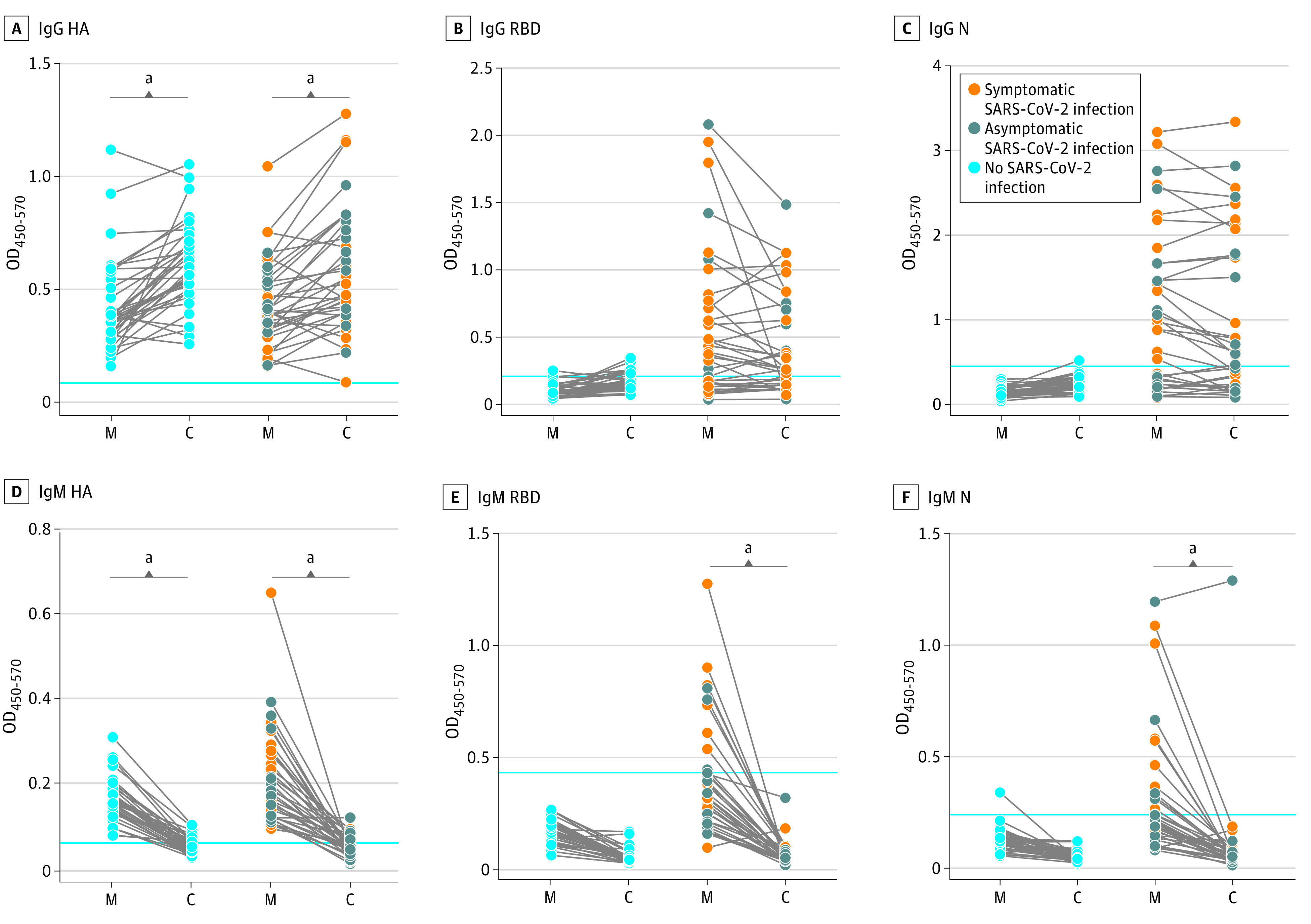
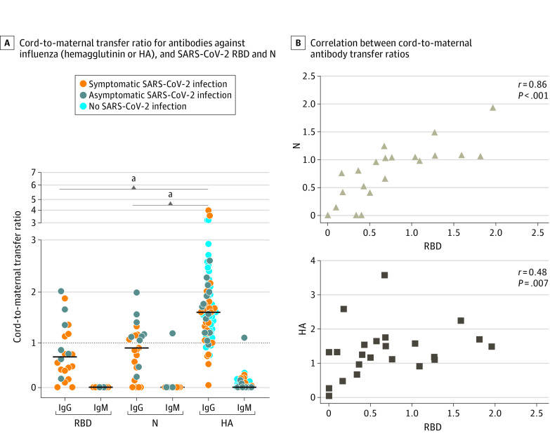
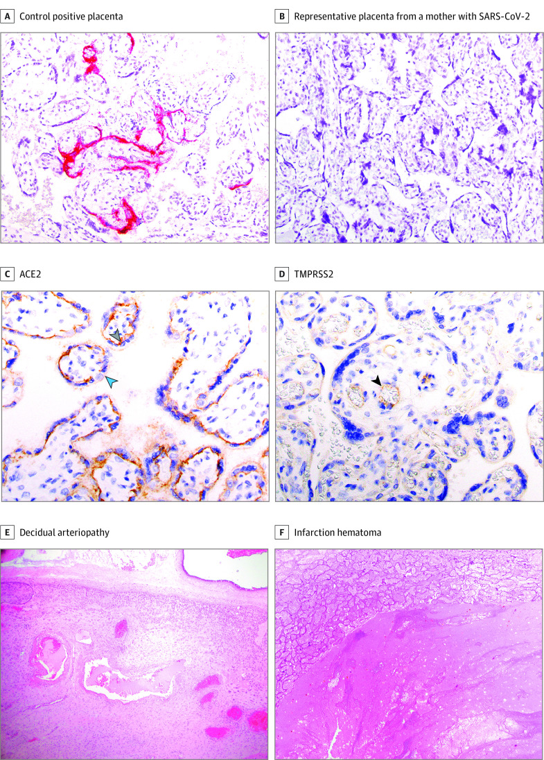
Results: Among 127 pregnant women enrolled, 64 with RT-PCR results positive for SARS-CoV-2 (mean [SD] age, 31.6 [5.6] years) and 63 with RT-PCR results negative for SARS-CoV-2 (mean [SD] age, 33.9 [5.4] years) provided samples for analysis. Of women with SARS-CoV-2 infection, 23 (36%) were asymptomatic, 22 (34%) had mild disease, 7 (11%) had moderate disease, 10 (16%) had severe disease, and 2 (3%) had critical disease. In viral load analyses among 107 women, there was no detectable viremia in maternal or cord blood and no evidence of vertical transmission. Among 77 neonates tested in whom SARS-CoV-2 antibodies were quantified in cord blood, 1 had detectable immunoglobuilin M to nucleocapsid. Among 88 placentas tested, SARS-CoV-2 RNA was not detected in any. In antibody analyses among 37 women with SARS-CoV-2 infection, anti-receptor binding domain immunoglobin G was detected in 24 women (65%) and anti-nucleocapsid was detected in 26 women (70%). Mother-to-neonate transfer of anti-SARS-CoV-2 antibodies was significantly lower than transfer of anti-influenza hemagglutinin A antibodies (mean [SD] cord-to-maternal ratio: anti-receptor binding domain immunoglobin G, 0.72 [0.57]; anti-nucleocapsid, 0.74 [0.44]; anti-influenza, 1.44 [0.80]; P < .001). Nonoverlapping placental expression of SARS-CoV-2 receptors angiotensin-converting enzyme 2 and transmembrane serine protease 2 was noted.

Conclusions and relevance: In this cohort study, there was no evidence of placental infection or definitive vertical transmission of SARS-CoV-2. Transplacental transfer of anti-SARS-CoV-2 antibodies was inefficient. Lack of viremia and reduced coexpression and colocalization of placental angiotensin-converting enzyme 2 and transmembrane serine protease 2 may serve as protective mechanisms against vertical transmission.


Conflict of interest statement

Conflict of Interest Disclosures: Dr Li reported serving as a consultant for Abbvie and Jan Biotech. Dr Boatin reported serving as a consultant for Microchips Biotech and as a scientific advisory board member for Reproductive Health Investors Alliance. Dr Gray reported receiving nonfinancial support from Illumina, and personal fees from Quest Diagnostics, BillionToOne, and Aetion outside the submitted work. Dr Fasano reported serving as a cofounder of and owning stock in Alba Therapeutics and serving on scientific advisory boards for NextCure and Viome outside the submitted work. Dr Schmidt reported receiving grants from the Bill and Melinda Gates Foundation, Defense Advanced Research Projects Agency (DARPA), Henry Jackson Foundation, amfAR, Ragon Institute, Massachusetts Consortium on Pathogen Readiness, Janssen, Gilead, Legend, Sanofi, Zentalis, Alkermes, and Intima; personal fees from SQZ Biotech; and having a patent for a SARS-CoV-2 vaccine licensed to Janssen. Dr Pepin reported owning stock in Gilead Sciences, BioNano Genomics, Biogen, Bluebird Bio, ImmunoGen, Pfizer, and Bristol-Myers Squibb. Dr Kaimal reported receiving grants from the National Institutes of Health outside the submitted work. Dr Roberts reported receiving author royalties from UpToDate and Cambridge University Press outside the submitted work. Dr Alter reported serving as a founder of Systems Seromyx. No other disclosures were reported.

Figures

Figure 1.
Figure 1.. Severe Acute Respiratory Syndrome Coronavirus 2 (SARS-CoV-2) Viral Load in Maternal and Neonatal Biofluids and Tissues by Maternal Disease Severity
The dot plot shows viral loads across samples by reverse transcription–polymerase chain reaction test results. Samples were analyzed in triplicate. No significant differences in viral load between any respiratory fluid were detected by Wilcoxon signed rank test.
Figure 2.
Figure 2.. Maternal-Cord Transplacental Antibody Transfer of Anti-Influenza Hemagglutinin A (HA) and Anti–Severe Acute Respiratory Syndrome Coronavirus 2 (SARS-CoV-2) Antibodies
The dot plots depict relative immunoglobuin (Ig) G or IgM titer against influenza HA, SARS-CoV-2 receptor binding domain (RBD), and SARS-CoV-2 nucleocapsid (N) present in maternal plasma (M) or matched umbilical cord blood (C). Data are represented as the optical density (OD) 450 value after background correction and are shown as the mean of 2 replicates. The blue lines represent the sum of the mean value of SARS-CoV-2 negative samples and 3 × the SD of those samples. Significance was determined by a Wilcoxon matched-pairs signed rank test. aP < .001.
Figure 3.
Figure 3.. Cord-to-Maternal Transplacental Antibody Transfer Ratios for Influenza and Severe Acute Respiratory Syndrome Coronavirus 2 (SARS-CoV-2)
A, Cord-to-maternal transfer ratio, calculated as (optical density [OD]450-570 cord)/(OD450-570 maternal) for antibodies against influenza hemagglutinin A (HA), and SARS-CoV-2 receptor binding domain (RBD) and nucleocapsid (N). Significance was assessed with a 1-way analysis of variance followed by Tukey post hoc testing to determine the source of significant differences in antibody transfer ratios among HA, RBD, and N. Ig indicates immunoglobin; black lines, means. B, Correlation between cord-to-maternal antibody transfer ratios for the indicated antibodies. Significance and ρ were determined by Spearman’s rank correlation. aP < .001.
Figure 4.
Figure 4.. Placental Findings in Maternal Severe Acute Respiratory Syndrome Coronavirus 2 (SARS-CoV-2) Infection
A and B: RNA in situ hybridization results at 20× original. Magenta red signal is visualized in the syncytiotrophoblast, with a complete lack of positivity in any stromal cells (including but not limited to Hofbauer cells) (A). C and D: SARS-CoV-2 receptor expression by immunohistochemistry at 40× original. C, Expression is restricted to the villous trophoblast with a polarity such that the highest expression is stromal side of the syncytiotrophoblast (gray arrow) with minimal to absent expression on the maternal vascular side (blue arrow). D, Weak expression limited to the villous endothelial cells (black arrow). Resident placental macrophages (Hofbauer cells) expressed neither angiotensin-converting enzyme 2 (ACE2) nor transmembrane serine protease 2 (TMPRSS2). E and F: Maternal vascular malperfusion (MVM) pathology. E, Hematoxylin and eosin stain at 4× original. F, Hematoxylin and eosin stain at 10× original.

Comment in

References

    1. Ellington S, Strid P, Tong VT, et al. . Characteristics of women of reproductive age with laboratory-confirmed SARS-CoV-2 infection by pregnancy status—United States, January 22-June 7, 2020. MMWR Morb Mortal Wkly Rep. 2020;69(25):769-775. doi:10.15585/mmwr.mm6925a1 - DOI - PMC - PubMed
    1. Khalil A, von Dadelszen P, Draycott T, Ugwumadu A, O’Brien P, Magee L. Change in the incidence of stillbirth and preterm delivery during the COVID-19 pandemic. JAMA. 2020;324(7):705-706. doi:10.1001/jama.2020.12746 - DOI - PMC - PubMed
    1. Takemoto MLS, Menezes MO, Andreucci CB, et al. . The tragedy of COVID-19 in Brazil: 124 maternal deaths and counting. Int J Gynaecol Obstet. 2020. doi:10.1002/ijgo.13300 - DOI - PMC - PubMed
    1. Liu H, Wang LL, Zhao SJ, Kwak-Kim J, Mor G, Liao AH. Why are pregnant women susceptible to COVID-19? an immunological viewpoint. J Reprod Immunol. 2020;139:103122. doi:10.1016/j.jri.2020.103122 - DOI - PMC - PubMed
    1. Dashraath P, Wong JLJ, Lim MXK, et al. . Coronavirus disease 2019 (COVID-19) pandemic and pregnancy. Am J Obstet Gynecol. 2020;222(6):521-531. doi:10.1016/j.ajog.2020.03.021 - DOI - PMC - PubMed

Publication types

MeSH terms