. 2020 Dec 22;4(24):6135-6147.
doi: 10.1182/bloodadvances.2020002456.

The evolution of factor XI and the kallikrein-kinin system

Affiliations

The evolution of factor XI and the kallikrein-kinin system

Michał B Ponczek et al. Blood Adv. .

Abstract

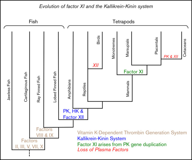
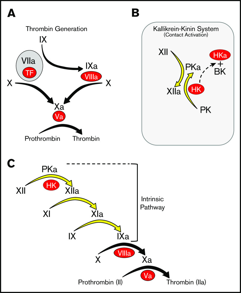
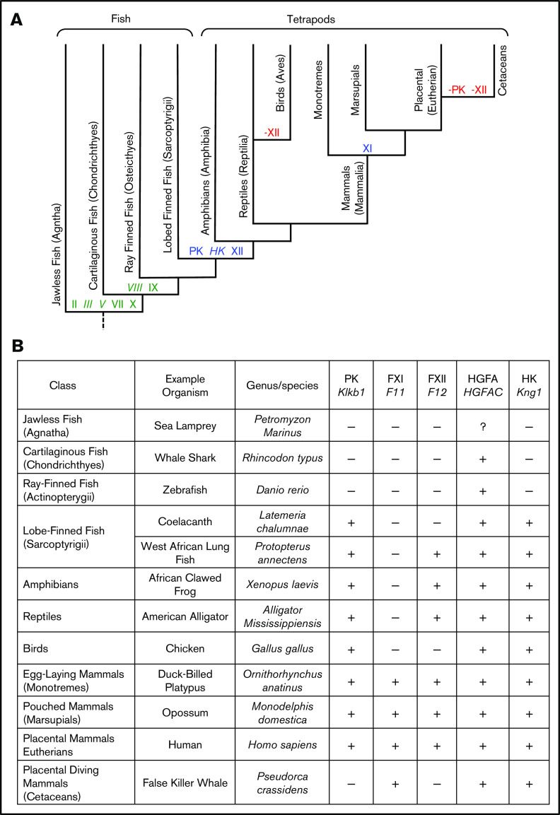
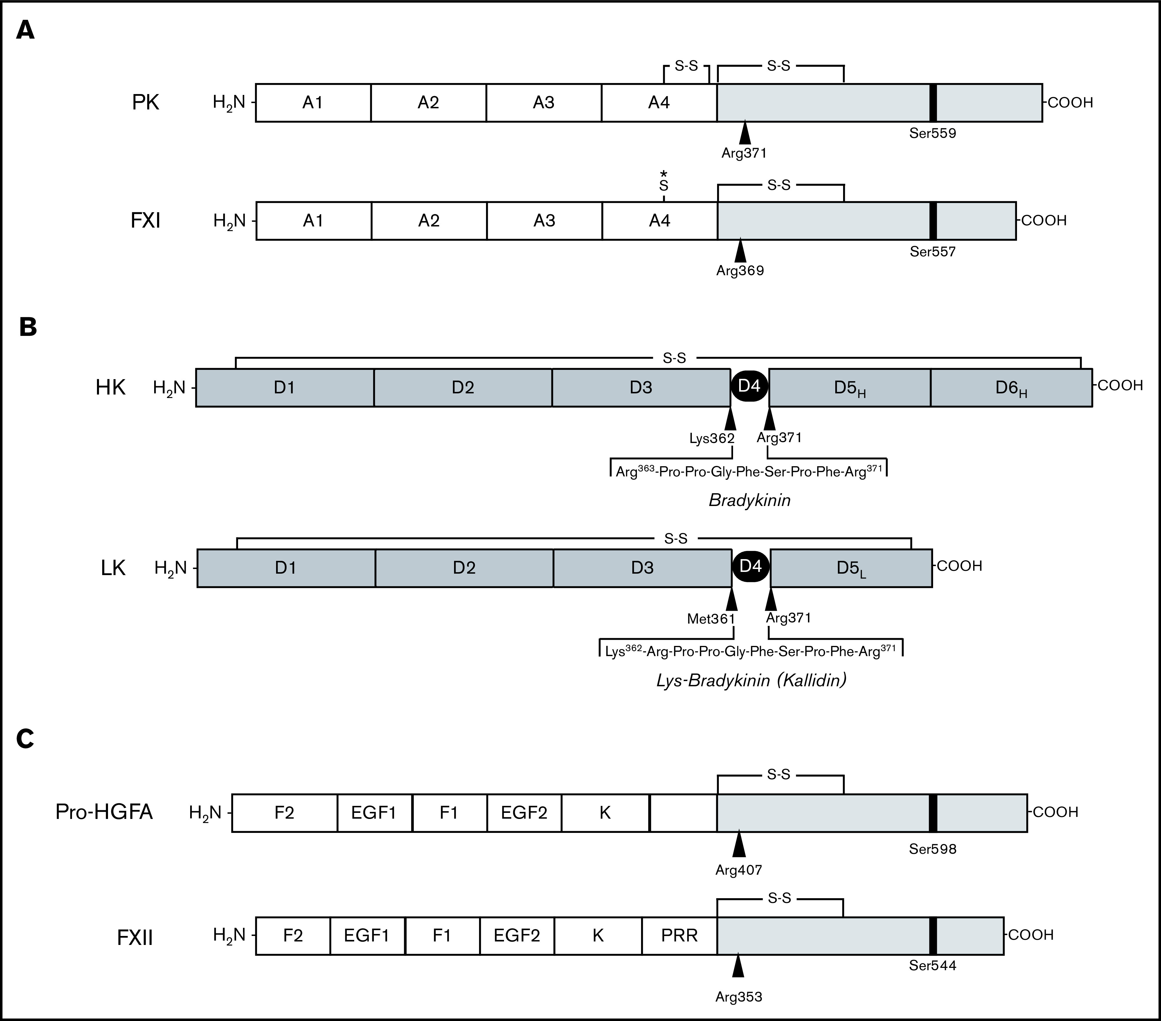
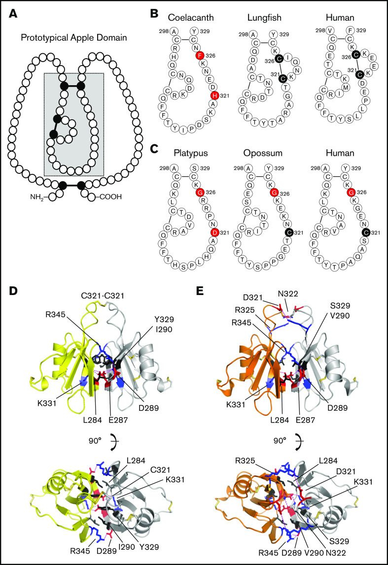
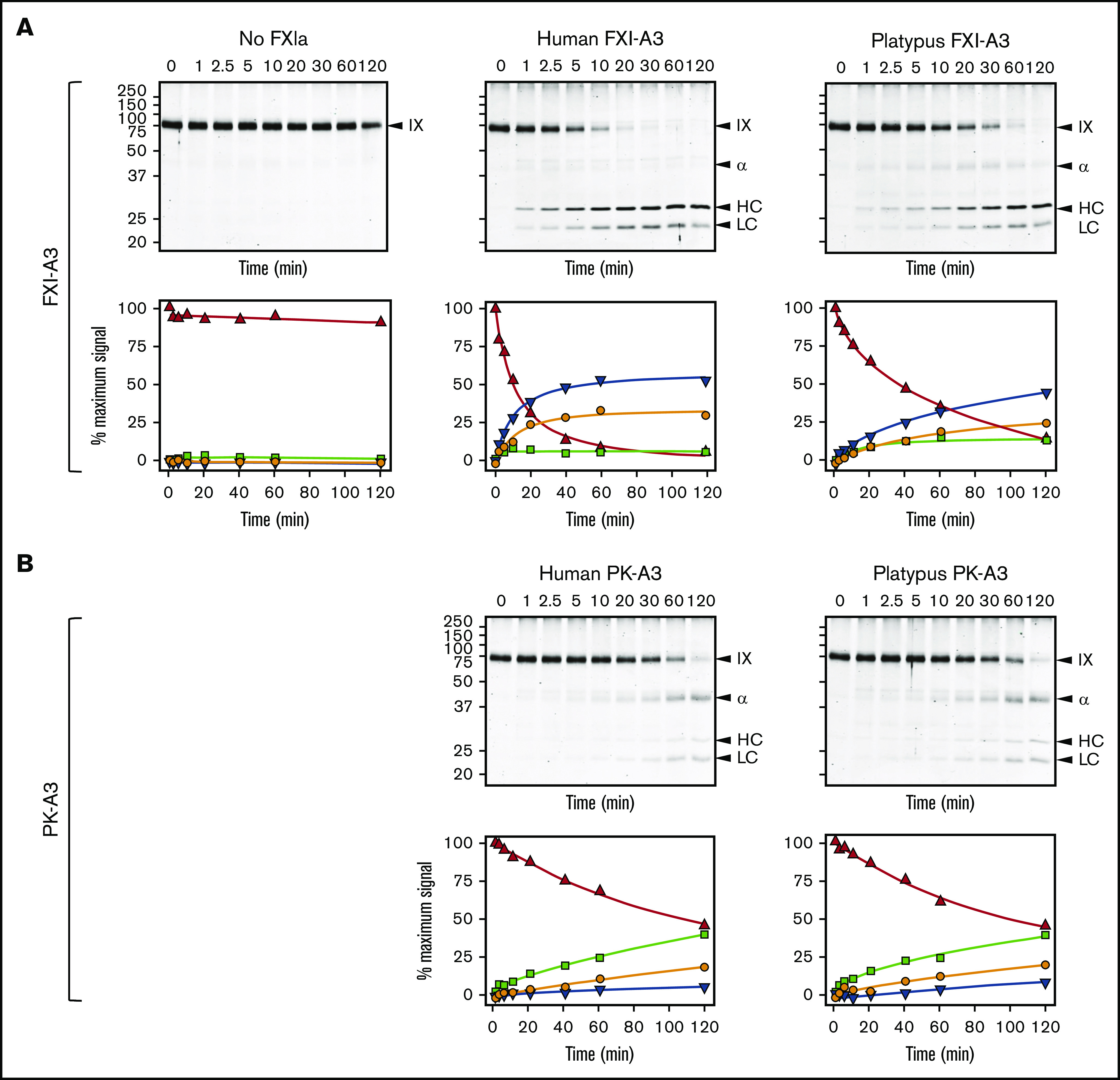
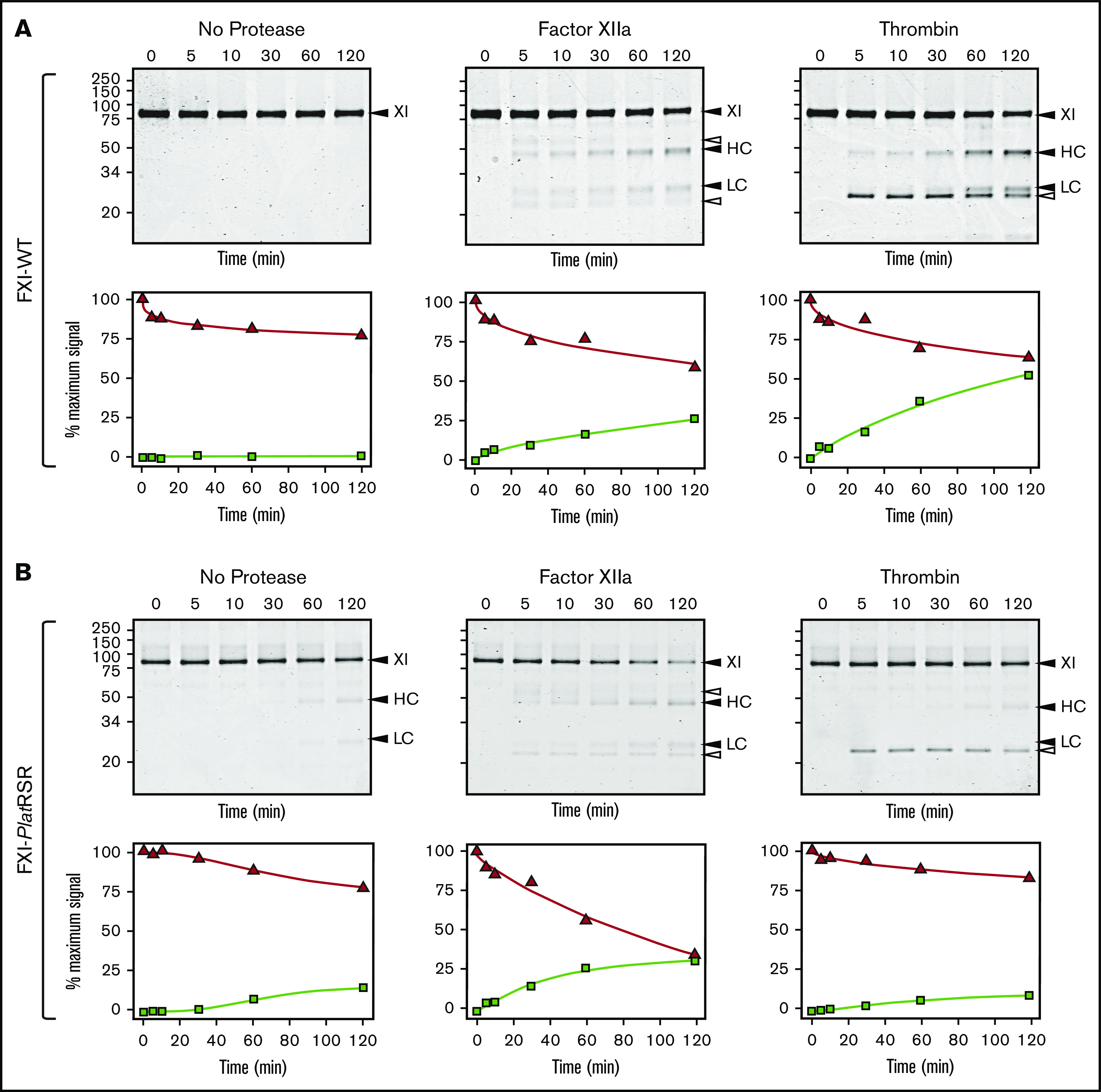
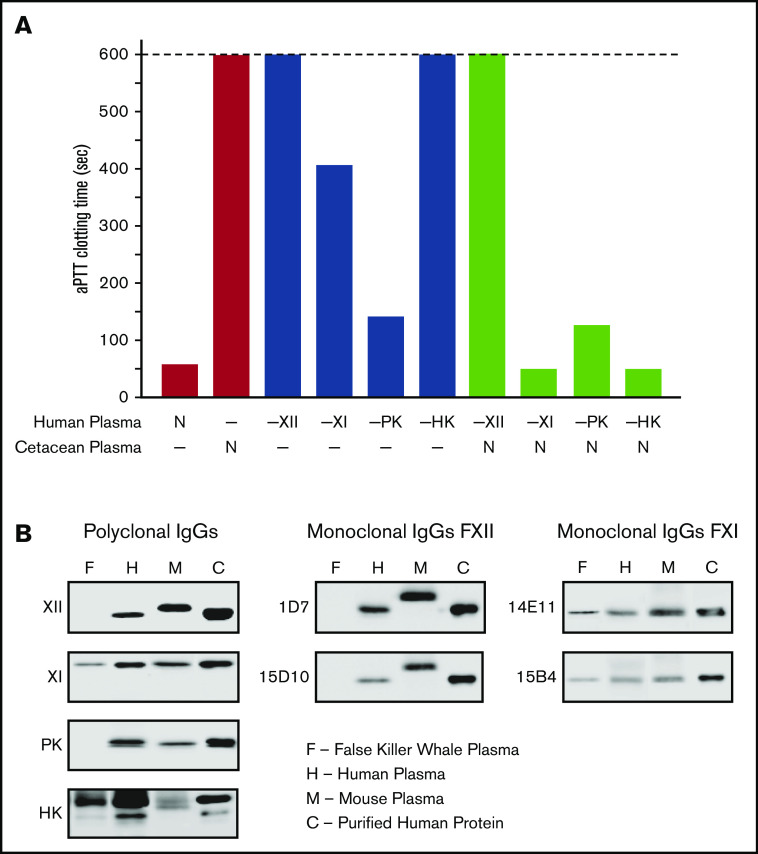
Factor XI (FXI) is the zymogen of a plasma protease (FXIa) that contributes to hemostasis by activating factor IX (FIX). In the original cascade model of coagulation, FXI is converted to FXIa by factor XIIa (FXIIa), a component, along with prekallikrein and high-molecular-weight kininogen (HK), of the plasma kallikrein-kinin system (KKS). More recent coagulation models emphasize thrombin as a FXI activator, bypassing the need for FXIIa and the KKS. We took an evolutionary approach to better understand the relationship of FXI to the KKS and thrombin generation. BLAST searches were conducted for FXI, FXII, prekallikrein, and HK using genomes for multiple vertebrate species. The analysis shows the KKS appeared in lobe-finned fish, the ancestors of all land vertebrates. FXI arose later from a duplication of the prekallikrein gene early in mammalian evolution. Features of FXI that facilitate efficient FIX activation are present in all living mammals, including primitive egg-laying monotremes, and may represent enhancement of FIX-activating activity inherent in prekallikrein. FXI activation by thrombin is a more recent acquisition, appearing in placental mammals. These findings suggest FXI activation by FXIIa may be more important to hemostasis in primitive mammals than in placental mammals. FXI activation by thrombin places FXI partially under control of the vitamin K-dependent coagulation mechanism, reducing the importance of the KKS in blood coagulation. This would explain why humans with FXI deficiency have a bleeding abnormality, whereas those lacking components of the KKS do not.


Conflict of interest statement

Conflict-of-interest disclosure: The authors declare no competing financial interests.

Figures

None
Graphical abstract
Figure 1.
Figure 1.
Models of thrombin generation. For all panels, plasma proteases are indicated in black lettering, with the activated forms indicated by a lower case “a.” Cofactors are indicated in red ovals. (A) Thrombin generation. Major protease activation reactions during thrombin generation are indicated by black arrows. Thrombin generation at a wound is typically initiated by a complex formed between the plasma protease factor VIIa and the cofactor TF. (B) The kallikrein-kinin system (KKS). On a surface (represented by the gray rectangle), factor XII (FXII), and PK undergo reciprocal activation to FXIIa and PKa. PKa cleaves HK, releasing BK. (C) The cascade-waterfall model of thrombin generation. In this model, the process is initiated by activation of FXII through the reactions shown in panel B. FXIIa then converts FXI to FXIa, which then activates FIX. The series of reactions indicated by the yellow arrows is referred to as the intrinsic pathway. Image adapted from Gailani and Gruber. BK, bradykinin; TF, tissue factor.
Figure 2.
Figure 2.
Evolution of vertebrate coagulation proteins. (A) Cladogram of vertebrate evolution. Vertebrates (animals with backbones) include fish and tetrapods (land vertebrates). The earliest tetrapods were amphibians, which arose from lobe-finned fish (sarcoptyrigii) ∼390 million years ago. Amphibians are ancestral to reptiles. Mammals descended from proto-mammalian synapsid reptiles, which diverged from sauropsid reptiles (ancestors of extant reptiles and birds) ∼250 to 300 million years ago. The most primitive mammals are egg-laying monotremes, which are represented today by the duck-billed platypus and a few species of spiny anteater (echidnas). Monotremes are the ancestors of pouched (marsupial) and placental (eutherian) mammals. Cetaceans are a group of aquatic placental mammals that include whales, dolphins, and porpoises. A more detailed presentation of the estimated times of divergence for certain vertebrates is presented in supplemental Figure 4. Green lettering indicates estimated points of origin of protein components of the vitamin K-dependent thrombin generation mechanism (factors II, III [tissue factor], V, VII, VIII, IX, and X). Points of origin of the kallikrein-kinin system (FXII, PK, and HK) and FXI are indicated in blue. Red lettering and a “–” sign indicate loss of the FXII or PK genes (–FXII, –PK). Plasma factors indicated in nonitalicized lettering are plasma proteases, whereas factors indicated in italics are nonenzymatic cofactors. (B) Representative vertebrate species. The animals listed in this table are used as representatives of their respective classes in the manuscript. The abbreviations for the 5 right-hand columns are PK, FXI, FXII, HGFA (pro-HGFA), and HK. The symbols in the columns indicate if a gene for the respective proteins was identified (+) or not identified (–) in genomic analyses. There is uncertainty on whether or not a gene for Pro-HGFA is present in jawless fish (?).
Figure 3.
Figure 3.
Proteins of the kallikrein-kinin system and factor XI. (A-B) Schematic diagrams of human protease precursors showing noncatalytic (white boxes) and catalytic (light gray boxes) domains. Positions of active site serine residues are indicated by black bars. Sites of proteolysis during activation are indicated by arrows. (A) Prekallikrein (PK) is a 93-kDa polypeptide that is cleaved after Arg371 to form plasma kallikrein (PKa). FXI is a homodimer of 80-kDa polypeptides. Each polypeptide is converted to FXIa by cleavage after Arg369. The noncatalytic portions of PK and FXI contain 4 apple domains, designated A1 to A4. The location of a Cys321-Cys326 intrachain disulfide bond in PK is indicated above A4. *The location of Cys321 in the A4 domain of FXI. In placental mammals and the opossum, Cys321 forms an interchain disulfide bond connecting the 2 subunits of the FXI dimer. (B) Human plasma kininogens come in high-molecular weight (HK) and low-molecular weight (LK) forms that are products of alternatively spliced messenger RNAs from the Kng1 gene. HK and LK have similar D1, D2, D3, and D4 domains, but different D5 domains (D5H and D5L, respectively). The D6 (D6H) domain is present only in HK and contains binding sites for PK and FXI. Cleavage sites for PKa in HK that release bradykinin are indicated by the black arrows. LK is cleaved at the sites indicated by black arrows by tissue kallikreins to release Lys-bradykinin (kallidin). (C) Pro-hepatocyte growth factor activator (pro-HGFA) is a 95-kDa polypeptide that is cleaved after Arg407 to form HGFA. FXII is an 80-kDa polypeptide that is converted to FXIIa by cleavage after Arg353. The Pro-HGFA and FXII noncatalytic domains are the fibronectin type 2 (F2), epidermal growth factor (EGF), fibronectin type 1 (F1), and kringle (K) domains. FXII also has a proline-rich region (PRR).
Figure 4.
Figure 4.
Prekallikrein and factor XI apple 4 domains. (A) Apple domain schematic. A typical apple (PAN) domain contains ∼90 amino acids (indicated by circles) and is constrained by 3 disulfide bonds (black circles and connecting bars). The region of the domain within the gray box is presented in detail in panels B and C. (B) PK-A4 residues 298 to 329. Amino acids from the PK-A4 domain highlighted by the gray box in panel A are shown for the coelacanth, West African lungfish, and human PK. Residues 321 and 326 are highlighted. In lungfish and human PK, these residues are cysteines that form a disulfide bond. In the coelacanth, they are histidine and phenylalanine. (C) FXI-A4 residues 298 to 329. Amino acids from the FXI-A4 domain highlighted by the gray box in panel A are shown for platypus, opossum, and human FXI. Residues 321 and 326 are highlighted. Residue 326 is glycine in all 3 species. In opossum and human FXI, residue 321 is a cysteine that forms the interchain disulfide bond connecting the subunits of the FXI dimer. (D) Topology diagrams of the human FXI dimer interface. Shown are 2 FXI-A4 domains (1 subunit is shown in yellow; the other in white) forming the FXI dimer interface. The Cys321-Cys321 interchain disulfide bond is shown at the top in orange. Hydrophobic residues Leu284, Ile290, and Tyr329 are shown in black, and salt bridges are formed between Lys331 (blue) and Glu287 (red) and Arg345 (blue) and Asp289 (red). The bottom image is rotated 90° relative to the top image. After Papagrigoriou et al. (E) Predicted FXI dimer interface for platypus FXI. Interactions are the same as those in panel D. The Cys321-Cys321 is not present in platypus FXI; instead, there is an additional salt bridge between Arg325 (blue) and Asp321 (red). The bottom image is rotated 90° relative to the top image.
Figure 5.
Figure 5.
FXIa activation of FIX. (A) FIX activation by FXIa with FXI-A3 domains. Human FIX (200 nM) was incubated at 37°C with vehicle (no FXIa), 2 nM human FXIa-WT (human FXI-A3), or 2 nM human FXIa with a platypus FXI-A3 domain replacing the human A3 domain (FXIa/PlatXIA3, platypus FXI-A3). At various times, samples were removed into reducing sample buffer, size-fractionated by SDS-PAGE and stained with Coomassie blue (top row). Positions of standards for FIX, the heavy chain of the intermediate α-FIX (α), the heavy chain of the final product FIXaβ (HC), and light chain of α-FIX and FIXaβ (LC) are shown on the right of each image; positions of molecular mass markers in kilodaltons are shown to the left of the images. Stained gels underwent densitometry scanning to generate the curves in the bottom row. Values for each band were compared with those for FIX at 0 minutes, which was assigned a value of 100%. Curves show the disappearance of FIX (Δ), and the appearance of the heavy chain of the intermediate α-FIX (□), the heavy chain of FIXaβ (∇), and the light chain of α-FIX and FIXaβ (○). (B) FIX activation by FXIa with PK-A3 domains. Human FIX was incubated as in panel A with 2 nM human FXI with a human PK-A3 domain replacing the FXI A3 domain (FXIa-PKA3, human PK-A3) or human FXI with a platypus PK-A3 domain replacing the FXI A3 domain (FXIa/PlatPKA3, platypus PK-A3). Time course experiments were run and analyzed as in panel A.
Figure 6.
Figure 6.
FXI activation. (A) FXI-WT activation. FXI-WT (200 nM) was incubated at 37°C with vehicle, or 40 nM FXIIa, or 140 nM thrombin in the presence of 4 μM polyphosphate. At various times, samples were removed into reducing sample buffer, size-fractionated by SDS-PAGE, and stained with Coomassie blue (top row). Positions of standards for FXI, and the heavy chain (HC) and light chain (LC) of FXIa are shown on the right of each image and positions of molecular mass markers in kilodaltons are shown to the left of the images. The white arrows indicate bands for FXIIa or thrombin that appear because of the high enzyme to substrate ratios in these reactions. Stained gels underwent densitometry scanning to generate the curves in the bottom row. Values for each band were compared with those for FXI at 0 minutes, which was assigned a value of 100%. Curves show the disappearance of FXI (Δ) and the appearance of the heavy chain of FXIa (□). (B) FXI-PlatRSR activation. FXI-PlatRSR was activated as in panel A with vehicle or with 40 nM FXIIa or 140 nM thrombin in the presence of 4 μM polyphosphate. Time course experiments were run and analyzed as in panel A.
Figure 7.
Figure 7.
Analysis of cetacean plasma. (A) Activated partial thromboplastin time studies. White bars show average aPTTs of pooled normal human plasma and plasma from a cetacean (a false killer whale). Gray bars show average aPTTs for plasmas from human patients lacking FXII (−XII), FXI (−XI), PK (−PK), or HK (−HK). Black bars indicate average aPTTs for mixtures of equal volumes of human factor-deficient plasmas and false killer whale plasma. Note the normal clotting times for mixtures with human plasma lacking FXI and HK. Bars represent averages of 3 runs for each plasma. (B) Plasma western blots. One-microliter samples of plasma from a false killer whale (F), a human (H), and a mouse (M) were size fractionated by SDS-PAGE and transferred to nitrocellulose. Protein controls (C) for human FXII (XII), FXI (XI), PK, or HK were included. Blots were developed with polyclonal IgGs to each human protein (left column) or monoclonal antibodies raised against mouse FXII (1D7 and 15D10, center column) or mouse FXI (14E11 and 15B4, right column). Note the absence of signals for FXII or PK, and the presence of FXI and HK, in plasma from the false killer whale.

References

    1. Jiang Y, Doolittle RF. The evolution of vertebrate blood coagulation as viewed from a comparison of puffer fish and sea squirt genomes. Proc Natl Acad Sci USA. 2003;100(13):7527-7532. - PMC - PubMed
    1. Doolittle RF, Jiang Y, Nand J. Genomic evidence for a simpler clotting scheme in jawless vertebrates. J Mol Evol. 2008;66(2):185-196. - PubMed
    1. Doolittle RF. Step-by-step evolution of vertebrate blood coagulation. Cold Spring Harb Symp Quant Biol. 2009;74(0):35-40. - PubMed
    1. Grover SP, Mackman N. Tissue factor: an essential mediator of hemostasis and trigger of thrombosis. Arterioscler Thromb Vasc Biol. 2018;38(4):709-725. - PubMed
    1. Gajsiewicz JM, Morrissey JH. Structure-function relationship of the interaction between tissue factor and factor VIIa. Semin Thromb Hemost. 2015;41(7):682-690. - PMC - PubMed

Publication types