. 2021 Feb 8;6(3):e136648.
doi: 10.1172/jci.insight.136648.

TCF-1 regulates HIV-specific CD8+ T cell expansion capacity

Affiliations

TCF-1 regulates HIV-specific CD8+ T cell expansion capacity

Rachel L Rutishauser et al. JCI Insight. .

Abstract

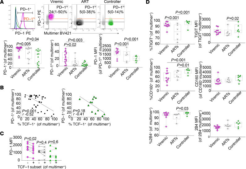
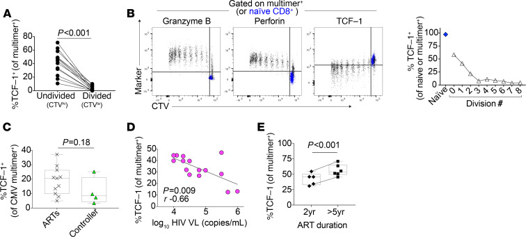
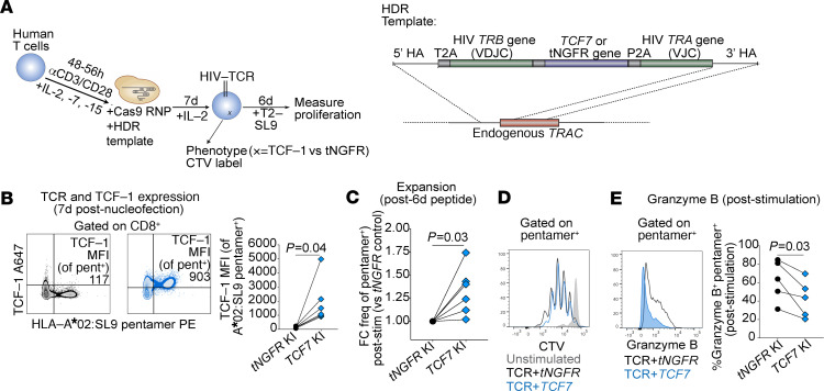
Although many HIV cure strategies seek to expand HIV-specific CD8+ T cells to control the virus, all are likely to fail if cellular exhaustion is not prevented. A loss in stem-like memory properties (i.e., the ability to proliferate and generate secondary effector cells) is a key feature of exhaustion; little is known, however, about how these properties are regulated in human virus-specific CD8+ T cells. We found that virus-specific CD8+ T cells from humans and nonhuman primates naturally controlling HIV/SIV infection express more of the transcription factor TCF-1 than noncontrollers. HIV-specific CD8+ T cell TCF-1 expression correlated with memory marker expression and expansion capacity and declined with antigenic stimulation. CRISPR-Cas9 editing of TCF-1 in human primary T cells demonstrated a direct role in regulating expansion capacity. Collectively, these data suggest that TCF-1 contributes to the regulation of the stem-like memory property of secondary expansion capacity of HIV-specific CD8+ T cells, and they provide a rationale for exploring the enhancement of this pathway in T cell-based therapeutic strategies for HIV.

Keywords: AIDS/HIV; Adaptive immunity; Immunology; T cells.


Conflict of interest statement

Conflict of interest: TLR and AM are cofounders of Arsenal Biosciences. TLR served as the CSO of Arsenal Biosciences from March to December 2019. AM is a cofounder of Spotlight Therapeutics and serves on the scientific advisory board of PACT Pharma, is an advisor to Trizell, and was a former advisor to Juno Therapeutics. The Marson Laboratory has received sponsored research support from Juno Therapeutics, Epinomics, and Sanofi and a gift from Gilead.

Figures

Figure 1
Figure 1. TCF-1 expression is elevated in HIV- and SIV-specific CD8+ T cells from controllers.
(A) Frequency of peripheral blood multimer+ HIV–specific CD8+ T cells. (B) Proliferation of HIV-specific CD8+ T cells in response to 6-day in vitro cognate peptide stimulation as measured by dilution of cell-trace violet (CTV). (C) Gating strategy (left: green, multimer+ from controller; gray, bulk CD8+ T cells) and distribution (right) of effector-memory phenotypes amongst multimer+ cells (TN, naive [gate includes, and likely primarily contains, CD95+ stem-cell memory cells, TSCM]; TCM, central memory; TTM, transitional memory; TEM, effector memory; TEMRA, effector memory-RA, separated by level of CD27 expression). (D) Gating (top left; TCF-1+ population gated based on CD8+ TN population from an HIV-uninfected participant, blue), representative flow plots (top right; median [range]), and summary data (bottom) showing TCF-1 expression in multimer+ HIV–specific CD8+ T cells from viremic (VIR; magenta), ART-suppressed (ARTs; black), and controller (C; green) individuals of all multimer specificities (left) and within the HIV Gag/HLA-A*02:SL9 multimer+ population (right). (E) TCF-1 expression in the SIV Gag/Mamu-A*01:CM9 multimer+ population from viremic and controller macaques. Phenotypes assessed by flow cytometry. FMO, fluorescence-minus-1 control. Box plots: median ± IQR. The human studies included data from a maximum of n = 13 viremic, 10 ART-suppressed, and 12 controller participants (as indicated in each figure), some with 2 multimer specificities. The macaque studies included n = 6 viremic and 4 controller animals. Linear mixed effects models to account for clustering within participants (A, C, D), Kruskal-Wallis followed by Dunn’s multiple comparison testing (B), Wilcoxon’s rank sum (E) were used.
Figure 2
Figure 2. High TCF-1 expression in HIV-specific CD8+ T cells is associated with a memory-like phenotype.
(A) Phenotype of HIV-specific multimer+ CD8+ T cells from viremic, ART-suppressed, or controller individuals (compared with naive CD8+ T cells from an HIV negative individual). (B and C) TCF-1 expression is higher in less differentiated naive and effector-memory subsets of bulk (B) and HIV-specific (C) CD8+ T cells. (D) Correlation between the expression of TCF-1 and other phenotypic markers in multimer+ CD8+ T cells (GranB, Granzyme B; black dots, all participants; green triangles, controllers only). (E) Expression of CD127, Granzyme B, and T-bet within TCF-1+ versus TCF-1 HIV-specific CD8+ T cells. These studies included data from a maximum of n = 13 viremic, 10 ART-suppressed, and 12 controller participants (as indicated in each figure), some with 2 multimer specificities. Linear mixed effects models to account for clustering within participants (A and B), Wilcoxon’s signed-rank test (C and E), Spearman’s correlation (D).
Figure 3
Figure 3. HIV-specific CD8+ T cells have lower expression of PD-1 but not other coinhibitory receptors.
(A) Expression of PD-1 in multimer+ HIV–specific multimer+ CD8+ T cells (annotated with median and range; MFI, median fluorescence intensity). (B) Correlation between PD-1 and TCF-1 expression in multimer+ HIV-specific CD8+ T cells (black dots, all participants; green triangles, controllers only). (C) Expression of PD-1 within TCF-1+ versus TCF-1 HIV-specific CD8+ T cells. (D) Expression of TIGIT, CD160, and 2B4 in multimer+ HIV–specific multimer+ CD8+ T cells. These studies included data from a maximum of n = 13 viremic, 10 ART-suppressed, and 12 controller participants (as indicated in each figure), some with 2 multimer specificities. Linear mixed effects models to account for clustering within participants (A, D), Wilcoxon’s signed-rank (C), and Spearman’s correlation (B).
Figure 4
Figure 4. TCF-1 expression is negatively correlated with antigen exposure.
(A) TCF-1 expression in HIV-specific CD8+ T cells after 6 days of in vitro peptide stimulation in divided versus undivided cells (n = 12 biological replicates). (B) TCF-1 expression in multimer+ CD8+ T cells (black) in each division peak after 6 days of in vitro peptide stimulation (naive CD8+ T cells shown in blue for reference); data are representative of pattern observed in n = 12 biological replicates. (C) TCF-1 expression in multimer+ CMV-specific CD8+ T cells from individuals with HIV (ART-suppressed [n = 12] versus controller [n = 4]). (D) Negative correlation between TCF-1 expression and plasma HIV viral load (VL; n = 15). (E) Expression of TCF-1 in HIV-specific CD8+ T cells in ART-suppressed individuals, depending on the duration of ART (n = 9 individuals). Wilcoxon’s signed-rank (A), Wilcoxon’s rank sum (C), Spearman’s correlation (D), and linear mixed effects models to account for clustering within participants (E).
Figure 5
Figure 5. TCF-1 KO impairs human CD8+ T cell proliferation.
(A) Correlation between TCF-1 expression in HIV-specific CD8+ T cells and the proportion that divide after 6-day in vitro peptide stimulation. (B) Correlation between TCF-1 expression in CMV-specific CD8+ T cells and their expansion after 6-day in vitro peptide stimulation (fold change [FC] in the frequency of CMV-specific CD8+ T cells [gated on total CD8+ T cells], stimulated versus unstimulated cells; n = 14). (C) CRISPR-Cas9–mediated deletion of TCF7 (Scr, scrambled guide RNA [gRNA]; RNP, ribonucleoprotein; n = 13 biological replicates). (D) TCF-1 protein downregulation in CD8+ T cells after TCF7 KO (red) compared with electroporation with Scr gRNA (gray). (E) CD8+ T cell phenotype after TCF7 KO. (F) Proportion of divided CD8+ T cells (%CTVlo) after 5-day stimulation with αCD3/CD28. (G) Correlation between the reduction in TCF-1 expression (MFI) and the reduction of CD8+ T cell proliferation (%CTVlo after stimulation). Spearman’s correlation (A, B, and G) and Wilcoxon’s signed-rank (D–F).
Figure 6
Figure 6. TCF-1 overexpression enhances HIV-specific CD8+ T cell expansion after peptide stimulation.
(A) Generation of HIV-specific T cell receptor (TCR) T cells using CRISPR-Cas9 knock-in (KI) of HIV-specific TCR and TCF7 (versus truncated Nerve Growth Factor Receptor [tNGFR]); T2, HLA-A*02-expressing T2 cell line; SL9, SLYNTVATL peptide. (B) TCF-1 protein expression after tNGFR (black) or TCF7 (blue) KI (left, representative flow plots; right, summary TCF-1 MFI from n = 6 biological replicates; lines connect control and TCF-1-expressing samples generated from the same donor). (C) Frequency of TCF7 KI TCR T cells after 6-day in vitro stimulation with SL9 peptide loaded on T2 cells (FC versus tNGFR KI; n = 6 biological replicates). (D) CTV tracings of TCR T cells (unstimulated or stimulated). (E) Granzyme B expression in TCF7 KI TCR T cells. Wilcoxon’s signed-rank (B, C, and E).

References

    1. Trautmann L, et al. Upregulation of PD-1 expression on HIV-specific CD8+ T cells leads to reversible immune dysfunction. Nat Med. 2006;12(10):1198–1202. doi: 10.1038/nm1482. - DOI - PubMed
    1. Day CL, et al. PD-1 expression on HIV-specific T cells is associated with T-cell exhaustion and disease progression. Nature. 2006;443(7109):350–354. doi: 10.1038/nature05115. - DOI - PubMed
    1. Kostense S, et al. High viral burden in the presence of major HIV-specific CD8(+) T cell expansions: evidence for impaired CTL effector function. Eur J Immunol. 2001;31(3):677–686. doi: 10.1002/1521-4141(200103)31:3<677::AID-IMMU677>3.0.CO;2-M. - DOI - PubMed
    1. McLane LM, et al. CD8 T cell exhaustion during chronic viral infection and cancer. Annu Rev Immunol. 2019;37(1):457–495. doi: 10.1146/annurev-immunol-041015-055318. - DOI - PubMed
    1. Borrow P, et al. Virus-specific CD8+ cytotoxic T-lymphocyte activity associated with control of viremia in primary human immunodeficiency virus type 1 infection. J Virol. 1994;68(9):6103–6110. doi: 10.1128/JVI.68.9.6103-6110.1994. - DOI - PMC - PubMed

Publication types

MeSH terms