Review
. 2020 Dec;10(12):200320.
doi: 10.1098/rsob.200320. Epub 2020 Dec 23.

HIV Rev-isited

Affiliations
Review

HIV Rev-isited

Catherine Toni-Sue Truman et al. Open Biol. 2020 Dec.

Abstract

The human immunodeficiency virus type 1 (HIV-1) proteome is expressed from alternatively spliced and unspliced genomic RNAs. However, HIV-1 RNAs that are not fully spliced are perceived by the host machinery as defective and are retained in the nucleus. During late infection, HIV-1 bypasses this regulatory mechanism by expression of the Rev protein from a fully spliced mRNA. Once imported into the nucleus, Rev mediates the export of unprocessed HIV-1 RNAs to the cytoplasm, leading to the production of the viral progeny. While regarded as a canonical RNA export factor, Rev has also been linked to HIV-1 RNA translation, stabilization, splicing and packaging. However, Rev's functions beyond RNA export have remained poorly understood. Here, we revisit this paradigmatic protein, reviewing recent data investigating its structure and function. We conclude by asking: what remains unknown about this enigmatic viral protein?

Keywords: HIV; Rev; human; immunodeficiency; virus.


Conflict of interest statement

We declare we have no competing interests.

Figures
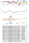
Figure 1.
Figure 1.
(a) Schematic of the HIV-1 genome, which is alternatively spliced. Start sites of open reading frames are numbered according to the NL4-3 HIV-1 sequence. (b) Though Rev is partially structured, it is predicted to be highly disordered at the amino acid level (data obtained from IUPred, where a score of greater than 0.4 indicates a high degree of disorder) [2]. Despite its disordered structure, Rev structures have been solved; regions indicated are labelled with Protein Data Base references and coloured for the ARM region (purple) and additional regions (gold). Post-translational modifications (PTMs) and amino acid variants obtained from UniProt are shown (main figure based on variant P04325).
Figure 2.
Figure 2.
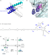
(a) The Rev N-terminal domain is composed of a helix–loop–helix motif and dimerizes in a hashtag motif. An arginine-rich RNA-binding motif is flanked by oligomerization motifs on both helices, which allow Rev to multimerize. NTD helices are stabilized by core hydrophobic interactions; some contributing residues are labelled (PDB: 6BSY). (b) A Rev dimer was crystallized in complex with the RRE; Rev binds stem IIB of the RRE using its arginine-rich motif, which makes contacts with the negatively charged RNA backbone (PDB: 4PMI [13]). (c) The structure of the 351-nucleotide Rev response element; regions are named using Roman numerals. (d) The canonical cycle of Rev exporting underspliced viral RNA to the cytoplasm. Rev binds the Rev response element on underspliced RNA at stem IIB, multimerizes and recruits CRM1 and RanGTP to cross the nuclear pore complex. In the cytoplasm, this complex can dissociate by RanGAP promoting RanGTP hydrolysis, freeing viral RNA. Rev can re-enter into the nucleus by binding importin-β.
Figure 3.
Figure 3.
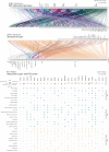
Almost 300 Rev–host protein interactions have been reported in the NCBI HIV-1 interaction database. Of these interactions, many cofactors appear to be nucleic acid binders, as shown by GO slim terms. In the second panel, the type of interaction reported is listed alongside the most reported proteins. Most of these cofactors are simply reported to interact with Rev, though some are known to be inhibitors or enhancers, etc. The third panel explores the interactions of 25 reported Rev cofactors, listing their known GO molecular functions, biological processes and cellular compartments in more detail.
Figure 4.
Figure 4.
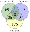
The overlap of host proteins identified to interact with Rev from three available datasets discovered using IP and MS. For Naji et al. [111], the top 250 candidates were considered as cited in the paper; for Jager et al. [114], proteins from both cell types were considered. For Arizala et al., reported proteins were normalized to HGNC IDs using the R package biomaRt [120] and pseudogenes/proteins which could not be mapped were filtered out.

References

    1. Laskey SB, Siliciano RF. 2014. A mechanistic theory to explain the efficacy of antiretroviral therapy. Nat. Rev. Microbiol. 12, 772 (10.1038/nrmicro3351) - DOI - PubMed
    1. Mészáros B, Erdós G, Dosztanyi Z.. 2018. IUPred2A: context-dependent prediction of protein disorder as a function of redox state and protein binding. Nucleic Acids Res. W329–W337. (10.1093/nar/gky384) - DOI - PMC - PubMed
    1. Katahira J. 2015. Nuclear export of messenger RNA. Genes 6, 163–184. (10.3390/genes6020163) - DOI - PMC - PubMed
    1. Luo M-j, Reed R. 1999. Splicing is required for rapid and efficient mRNA export in metazoans. Proc. Natl Acad. Sci. USA 96, 14937 (10.1073/pnas.96.26.14937) - DOI - PMC - PubMed
    1. Jacob AG, Smith CWJ. 2017. Intron retention as a component of regulated gene expression programs. Hum. Genet. 136, 1043–1057. (10.1007/s00439-017-1791-x) - DOI - PMC - PubMed

Publication types

LinkOut - more resources