. 2021 Feb:221:113165.
doi: 10.1016/j.ultramic.2020.113165. Epub 2020 Nov 15.

Robust scan synchronized force-fluorescence imaging

Affiliations

Robust scan synchronized force-fluorescence imaging

Patrick Schmidt et al. Ultramicroscopy. 2021 Feb.

Abstract

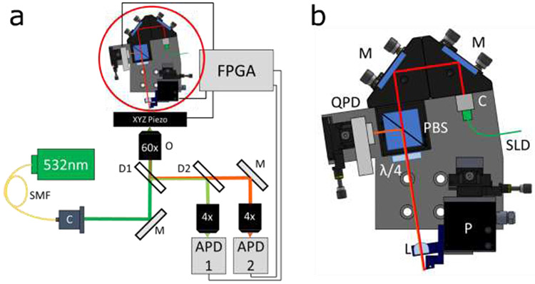
Simultaneous atomic force microscope (AFM) and sample scanning confocal fluorescence microscope measurements are widely used to obtain mechanistic and structural insights into protein dynamics in live cells. However, the absence of a robust technique to synchronously scan both AFM and confocal microscope piezo stages makes it difficult to visualize force-induced changes in fluorescent protein distribution in cells. To address this challenge, we have built an integrated AFM-confocal fluorescence microscope platform that implements a synchronous scanning method which eliminates image artifacts from piezo motion ramping, produces accurate pixel binning and enables the collection of a scanned image of a sample while applying force to a single point on the sample. As proof of principle, we use this instrument to monitor the redistribution of fluorescent E-cadherin, an essential transmembrane protein, in live cells, upon application of mechanical force.

Keywords: AFM-sample scanning confocal microscope; Integrated AFM–fluorescence microscope; Point scanning; Simultaneous force-fluorescence measurements; Synchronized scanning.


Figures

Figure 1:
Figure 1:. Instrumentation.
(a) Diagram of microscope, SMF- single mode fiber, O- objective, M- mirror, D1/D2- dichroic, C- collimator, FPGA- field programmable gate array, APD1/APD2-avalanche photodiode, (b) Diagram of custom AFM. M- mirror, L- lens, C- collimator, SLD-superluminescent diode, QPD- quadrant photodiode, PBS- polarizing cube beamsplitter, λ/4-quarter waveplate, P- 3 axis piezo.
Figure 2:
Figure 2:. Scanning method and synchronization.
(a) A 2D lateral scan is defined over a 10x10μm area with 10 scan lines. The scan definition is such that when the resulting data is formatted into a 2D image, each pixel value represents light collected from the center of that pixel. The scanning axis (X in this case) also extends before and after the scan area of interest to allow the triggering system which eliminates “warping” at the edges. Note that the range for each trigger extends slightly beyond the scan area, allowing a trigger “hand off’ between scan lines such that the trigger bounce does not signal the start of the next scan line collection. (b) Each piezo (sample and AFM) are programmed to internally record their positions and report the result at the end of the scan. To be sure the scan is not only spatially, but also temporally synced, the 2D scan is separated into each axis and plotted against time, X shown in (c) and Y shown in (d). The traces verify that the two piezo stages are synchronized both spatially and temporally.
Figure 3:
Figure 3:. Imaging force-induced redistribution of cell surface proteins.
To demonstrate the utility of the synchronized force fluorescence platform, we designed an experiment in which we collect fluorescence and backscatter images while applying a varying force on the apical surface of cells expressing DSRed-tagged E-cadherin. (a) Illustration of the experimental setup. The objective focus is set to the apical surface of the cell of interest (cell shown in red, beam waist in green), and the AFM cantilever with microsphere is positioned over the laser focus (or anywhere within the scan area the user chooses). (b) Backscatter and (c) fluorescence image results of a 50x50μm scan from APD 1 & 2, respectively (Figure 1). The triangular shape of the AFM cantilever as well as the silica microsphere sintered to the cantilever (along with a few smaller microspheres) are visible in (b). The fluorescently tagged E-cadherin largely reside at cell-cell junctions, making the edges of the cells bright as seen in (c). (d) Progression of the force applied to the cell by the AFM, and the resulting (e) backscatter and (f) fluorescent images (20x20μm scans). In row (e) we see the microsphere centered in the image, and as it presses into the cell the cantilever becomes brighter as it moves into the laser beam waist. In (f) we see a cluster of fluorescent E-cadherin on the apical surface of a cell. As higher force is applied, these cadherin are pushed out of the beam waist, and we see a dark spot form where the microsphere is pressing into the cell. Upon withdrawal of the AFM tip, we see the fluorescent E-cadherin return to the pressing site as the cell reforms its original shape. These images serve as good confirmation that our synchronized scanning method allows fluorescence imaging while applying force.

Similar articles

Cited by

References

    1. Kellermayer MSZ et al., Spatially and Temporally Synchronized Atomic Force and Total Internal Reflection Fluorescence Microscopy for Imaging and Manipulating Cells and Biomolecules. Biophys. J 91, 2665–2677 (2006). - PMC - PubMed
    1. Gumpp H et al., Triggering Enzymatic Activity with Force. Nano Lett. 9, 3290–3295 (2009). - PubMed
    1. Kassies R et al., Combined AFM and confocal fluorescence microscope for applications in bio-nanotechnology. Journal of microscopy 217, 109–116 (2005). - PubMed
    1. Meller K, Theiss C, Atomic force microscopy and confocal laser scanning microscopy on the cytoskeleton of permeabilised and embedded cells. Ultramicroscopy 106, 320–325 (2006). - PubMed
    1. Noy A, Huser TR, Combined force and photonic probe microscope with single molecule sensitivity. Rev. Sci. Instrum 74, 1217–1221 (2003).

Publication types

MeSH terms