. 2020 Dec 19;25(24):6025.
doi: 10.3390/molecules25246025.

Phenolic Constituents of Lamium album L. subsp. album Flowers: Anatomical, Histochemical, and Phytochemical Study

Affiliations

Phenolic Constituents of Lamium album L. subsp. album Flowers: Anatomical, Histochemical, and Phytochemical Study

Aneta Sulborska et al. Molecules. .

Abstract

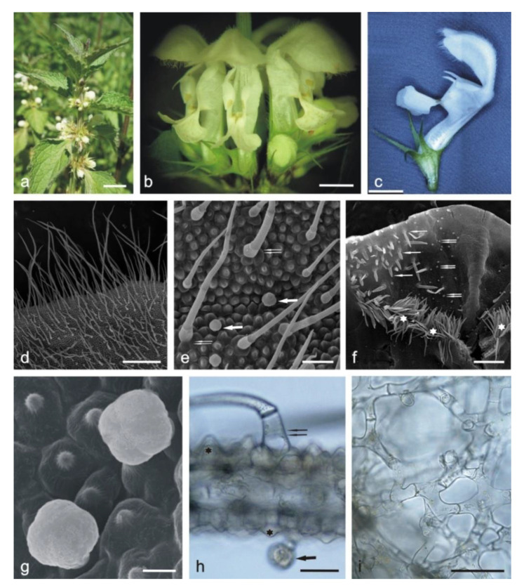
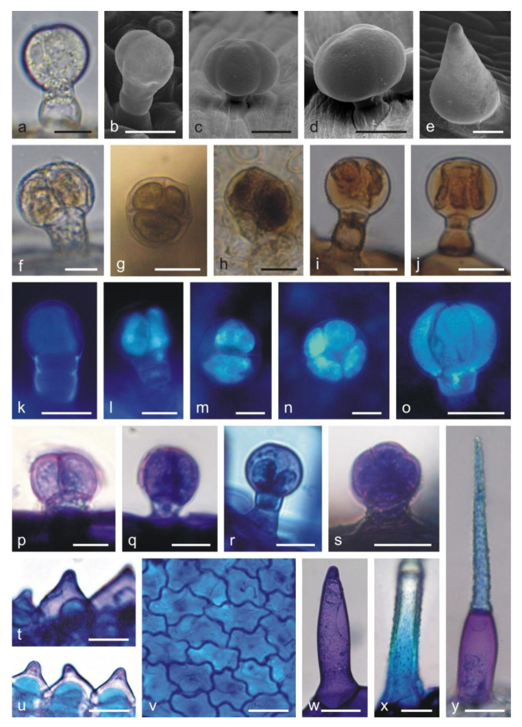
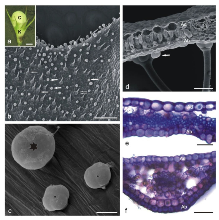
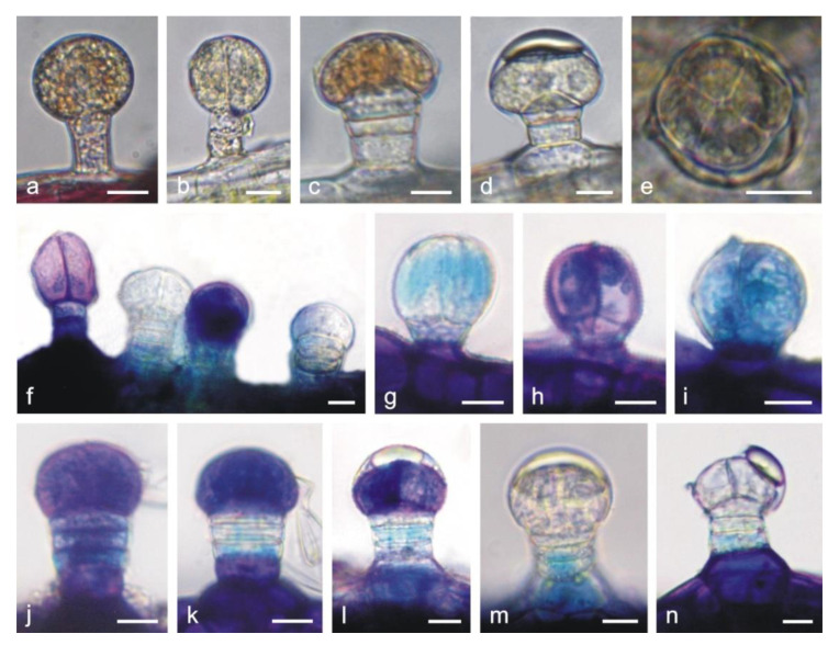
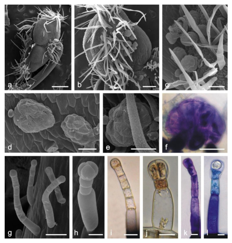
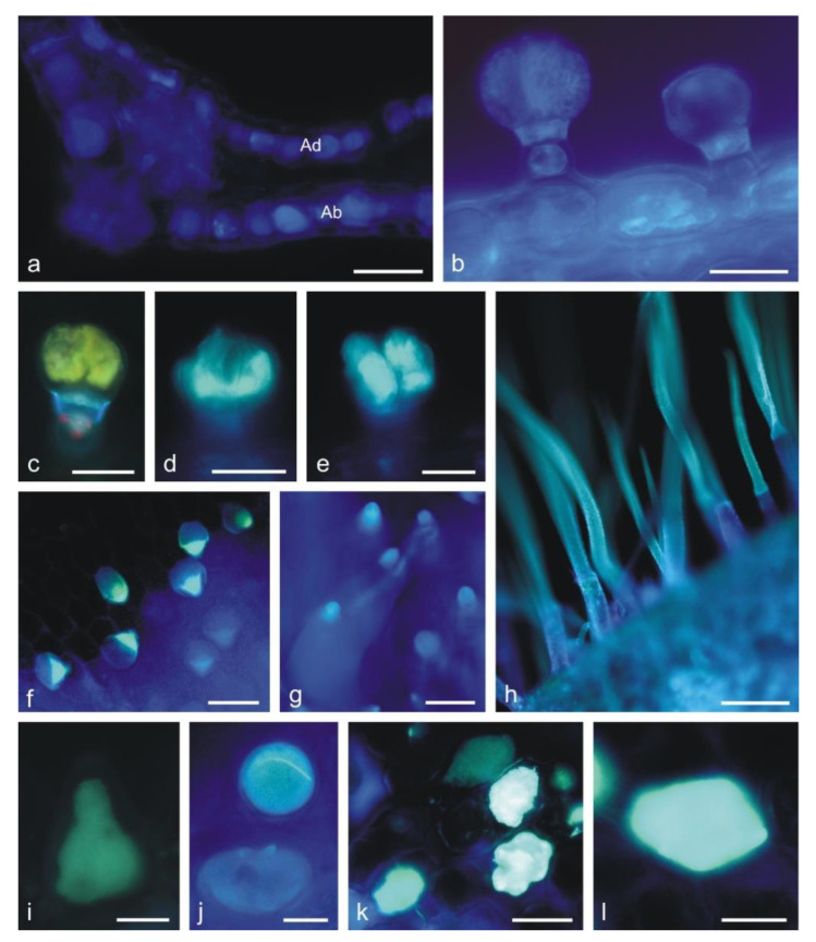
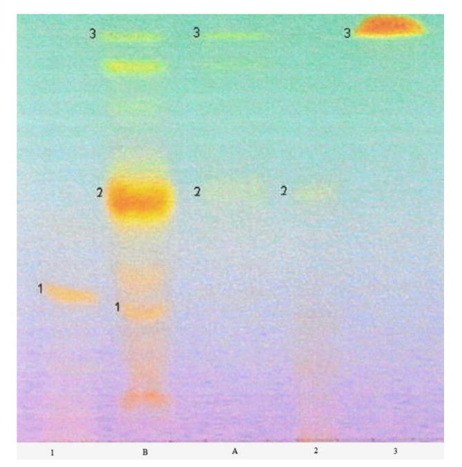
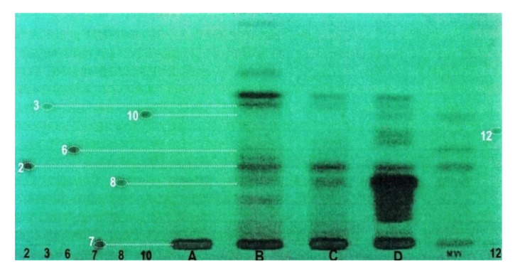
Flos Lamii albi has a high biological activity and is widely used in herbal medicine. The aim of the study was to characterize the secretory structures present in Lamium album subsp. album corolla and the location of phenolic compounds. Additionally, we carried out qualitative phytochemical analyses of flavonoids and phenolic acids. Light, fluorescence, and scanning electron microscopy were used to analyze the structure of the floral organs. The main classes of phenolic compounds and their localization were determined histochemically. Phytochemical analyses were performed with high-performance thin-layer chromatography (HPTLC) and high-performance liquid chromatography (HPLC). Six types of glandular trichomes were found which contained flavonoids, phenolic acids, and tannins. The phytochemical studies demonstrated the presence of caffeic, chlorogenic, ferulic, gallic, p-coumaric, protocatechuic, syringic, gentisic, and vanillic phenolic acids as well as rutoside, isoquercetin, and quercetin flavonoids. The corolla in L. album subsp. album has antioxidant properties due to the presence of various polyphenols, as shown by the histo- and phytochemical analyses. The distribution and morphology of trichomes and the content of phenolic compounds in the corolla have taxonomic, pharmacognostic, and practical importance, facilitating the identification of the raw material.

Keywords: flavonoids; phenolic acids; secretory trichomes; tannins; white nettle corolla.


Conflict of interest statement

The authors declare no conflict of interest.

Figures

Figure 1
Figure 1
Lamium album subsp. album flowers and fragments of the corolla. c—stereoscopic microscopy (SM), (dg)— scanning electron microscopy (SEM), (h,i)—light microscopy (LM). (a) Blooming plants. (b) Fragment of an inflorescence. (c) Lateral view of the flower. (d) Margin of the upper lip with long non-glandular trichomes. (e) Various types of trichomes and papillae on the abaxial surface of the upper lip: glandular trichomes (arrows), non-glandular trichomes (double arrows). (f) Fragment of the adaxial surface of the corolla tube with unevenly distributed non-glandular conical trichomes (double arrows), glandular trichomes (arrows) and a ring of flattened non-glandular trichomes (stars) located above the ovary. (g) Papillae and glandular trichomes with a 4-celled head on the adaxial surface of the upper lip. (h) Cross section of the upper lip with visible papillae (asterisks), glandular trichomes (arrow), and a non-glandular trichome (double arrow). (i) Loose arrangement of mesophyll cells in the cross section of the upper lip. Scale bars: 1 cm (a), 5 mm (b), 4 mm (c), 500 µm (d,f,i), 100 µm (e), 30 µm (h), 20 µm (g).
Figure 2
Figure 2
Types of trichomes and papillae on the L. album subsp. album corolla and the results of histochemical assays. (a,f) Control trichomes without staining (LM). (be) SEM images of trichomes. (gj,py) Histochemical reactions (LM). (ko) fluorescence microscopy (FM) images of trichomes. (a) Capitate trichome with a 1-celled head. (b) Capitate trichome with a 2-celled head. (c) Capitate trichome with a 4-celled head. (d) Peltate trichome. (e) Conical trichome. (f) Capitate trichome with a bicellular head and yellow content in the head cells. (g,h) Phenolic compounds stained black in the heads of trichomes after the application of ferric trichloride. (g) Capitate trichome with a 3-celled head. (h) Peltate trichome. (i,j) Brown color of tannins in trichomes stained with potassium dichromate. (ko) Blue autofluorescence in different capitate and peltate (o) trichomes indicating the presence of phenolic acids. (pr) Tannins in capitate trichomes stained blue after Toluidine blue O treatment. (s) Peltate trichome stained blue after Toluidine blue O treatment. (tu) Phenolic compounds in papillae stained blue after Toluidine blue O treatment. (v) Phenolic compounds visible in abaxial epidermis cells of the upper lip after Toluidine blue O treatment. (w) Non-glandular conical trichome from the corolla tube stained purple (pectins) after Toluidine blue O treatment. (x,y) Non-glandular trichomes containing phenolic compounds (blue) visible after Toluidine blue O treatment. Scale bars: 30 µm (c,d,h,i,q,r,w,y), 20 µm (a,b,f,g,j,k,o,p,s,t,u,v), 10 µm (e,l,m,n,x).
Figure 3
Figure 3
General habit and fragments of the L. album subsp. album calyx. (bd)—SEM; (e,f)—LM. (a) Flower bud with an open calyx. (b) Glandular (arrows) and non-glandular (double arrows) trichomes on the abaxial surface of the calyx. (c) Peltate trichome (large asterisk) and capitate trichomes (small asterisks) on the abaxial surface of a sepal nerve. (d) Cross section of a sepal with fragments of two non-glandular trichomes and a glandular trichome (arrow) on the abaxial surface. (e,f) Cross sections of sepals (Toluidine blue O staining). Abbreviations: K calyx, C corolla, Ad adaxial epidermis, Ab abaxial epidermis. Scale bars: 2 mm (a), 200 µm (b), 50 µm (df), 20 µm (c).
Figure 4
Figure 4
Glandular trichomes present on the L. album subsp. album calyx in LM. (ae) Trichomes from non-stained control preparations. (fn) Trichomes stained with Toluidine blue O. (a) Capitate trichome with 1-celled head. (b) Capitate trichome with 2-celled head. (ce) Peltate trichomes at various stages of development. (fi) Differently stained (blue and green) capitate trichomes after Toluidine blue O treatment indicating various contents of phenolic compounds. (jn) Peltate trichomes with differently stained head cells at different stages of development after Toluidine blue O treatment. In the older trichomes, the secretion is visible in the subcuticular space of the head (l,m) or on its surface with a ruptured cuticle (n). Scale bars: 10 µm (an).
Figure 5
Figure 5
Fragments of L. album subsp. album stamens with various types of trichomes. (ae,gh) SEM; (f,l)—LM. (ac) Long non-glandular trichomes and multicellular glandular trichomes anthers. (df) Multicellular glandular trichomes. (gl) Long-stalked capitate trichomes on filaments. (f,k,l) Tannins stained blue after Toluidine blue O treatment. (i,j) Tannins stained brown after potassium dichromate treatment. Scale bars: 500 µm (a), 200 µm (b), 100 µm (c), 50 µm (dg), 20 µm (hj), 10 µm (k,l).
Figure 6
Figure 6
Fragments of corolla petals L. album subsp. album imaged by a fluorescence microscope. (a,b) Light blue autofluorescence of phenolic acids (a) in epidermis cells in the cross section of the corolla tube, (b) in epidermis cells and capitate trichomes. (cl) Yellowish secondary fluorescence of flavonoids observed after the application of aluminum trichloride under the Cy 5 filter. (ce) Capitate trichomes containing flavonoids. (f,g) Different-length conical trichomes visible on the adaxial surface of the corolla tube with flavonoids in the apical part. (h) Non-glandular trichomes from the upper lip: flavonoid fluorescence in the cell walls. (i,j) Papillae on the upper lip containing flavonoids. (k,l) Epidermis cells from the upper lip with flavonoid content. Abbreviation: Ad adaxial epidermis, Ab abaxial epidermis. Scale bars: 100 µm (g,h), 50 µm (a), 30 µm (b,c,f,k), 20 µm (d,e,l), 10 µm (i,j).
Figure 7
Figure 7
Chromatogram of the separation of flavonoids in methanol extract from L. album subsp. album flowers with the multiple gradient development technique (MGD). The bands of the standards correspond with the bands of the compounds in the extract: 1—rutoside; 2—isoquercetin; 3—quercetin; A—ethyl acetate extract-excluded from the investigations; B—methanol extract. Visible light, derivatization with the use of a 1% methanolic solution of aluminum chloride. The mobile phase elution program is given in Table 2. Chromatographic plate HPTLC Si 60 F 254.
Figure 8
Figure 8
Separation of phenolic acids in methanol extracts of L. album subsp. album flowers. A—ethyl acetate extract-excluded from the investigations; B—methanol extract; C—acid hydrolysis; D—basic hydrolysis; MW—mixture of standards; 2—protocatechuic acid (3,4-dihydroxybenzoic); 3—vanillic acid (4-hydroxy-3-metoxybenzoic); 6—syringic acid (4-hydroxy-3,5-dimetoxybenzoic); 7—chlorogenic acid (depside of caffeic and quinic acids); 8—caffeic acid (3,4-dihydroxycinnamic); 10—p-coumaric acid (4-hydroxycinnamic); 12—ferulic acid (4-hydroxy-3-metoxycinnamic). Videoscaner Desaga (Germany). Stationary phase—chromatographic plate: Si 60 HPTLC F 254 (Merck). Elution program in Table 3. Wavelength 254 nm.
Figure 9
Figure 9
Separation of a mixture of phenolic acid standards in HPLC. Stationary phase—RP-18, mobile phase: water: methanol: formic acid, (75:25:0.5 v/v). Each compound is characterized by retention time in minutes (given above peaks): (1)—gallic acid (3,4,5-trihydroxybenzoic); (2)—protocatechuic acid (3,4-dihydroxybenzoic); (3)—gentisic acid (2,5-dihydroxybenzoic); (4)—vanillic acid (4-hydroxy-3-metoxybenzoic); (5)—caffeic acid (3,4-dihydroxycinnamic); (6)—syringic acid (4-hydroxy-3,5-dimetoxybenzoic); (7)—p-coumaric acid (4-hydroxycinamic); (8)—ferulic acid (4-hydroxy-3-metoxycinnamic); (9)—chlorogenic acid (depside of caffeic and quinic acids). Wavelength 254 nm.
Figure 10
Figure 10
Separation of phenolic acids in methanol extract after basic hydrolysis in HPLC. Stationary phase—RP-18; mobile phase: water: methanol: formic acid (75:25:0.5 v/v). (1)—gallic acid (3,4,5-trihydroxybenzoic); (2)—protocatechuic acid (3,4-dihydroxybenzoic); peak (3) is not identified; (4)—gentisic acid (2,5-dihydroxybenzoic); (5)—caffeic acid (3,4-dihydroxycinnamic) and vanillic acid (4-hydroxy-3-metoxybenzoic); (6)—syringic acid (4-hydroxy-3,5-dimetoxybenzoic); (7)—p-coumaric acid (4-hydroxycinamic); (8)—ferulic acid (4-hydroxy-3-metoxycinnamic); (9)—chlorogenic acid (depside of caffeic and quinic acids). Wavelength 254 nm.
Figure 11
Figure 11
Separation of phenolic acids after the acid hydrolysis of methanolic extract in HPLC. Stationary phase—RP-18; mobile phase: water: methanol: formic acid (75:25:0.5 v/v). (1)—protocatechuic acid (3,4-dihydroxybenzoic); (2)—caffeic acid (3,4-dihydroxycinnamic); (3)—p-coumaric acid (4-hydroxycinamic). Wavelength 254 nm.

Similar articles

Cited by

References

    1. Mabberley D.J. The Plant Book. 2nd ed. Cambridge University Press; Cambridge, UK: 1997.
    1. Yordanova Z.P., Zhiponova M.K., Iakimova E.T., Dimitrova M.A., Kapchina-Toteva V.M. Revealing the reviving secret of the white dead nettle (Lamium album L.) Phytochem. Rev. 2014;13:375–389. doi: 10.1007/s11101-014-9356-2. - DOI
    1. Dengler J., Berg C., Eisenberg M., Isermann M., Jansen F., Koska I., Löbel S., Manthey M., Pazolt J., Spangenberg A., et al. New descriptions and typifications of syntaxa within the project ‘Plant communities of Mecklenburg-Vorpommern and their vulnerability’—Part I. Feddes Repertorium Zeitschrift für Botanische Taxonomie und Geobotanik. 2003;114:587–631. doi: 10.1002/fedr.200311017. - DOI
    1. Metcalfe C.R., Chalk L. Anatomy of the Dicotyledons. Volume 2 Oxford Press; London, UK: 1972.
    1. Sulborska A., Dmitruk M., Konarska A., Weryszko-Chmielewska E. Adaptations of Lamium album L. flowers to pollination by Apoidea. Acta Sci. Pol. Hortorum Cultus. 2014;13:31–43.

MeSH terms

LinkOut - more resources