Review
. 2020 Dec 18;21(24):9669.
doi: 10.3390/ijms21249669.

Macrophage Proinflammatory Responses to Microorganisms and Transplanted Organs

Affiliations
Review

Macrophage Proinflammatory Responses to Microorganisms and Transplanted Organs

Malgorzata Kloc et al. Int J Mol Sci. .

Abstract

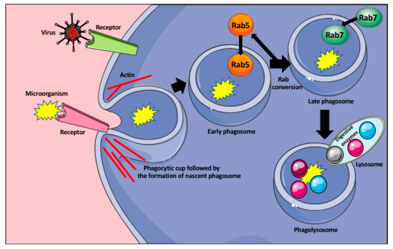
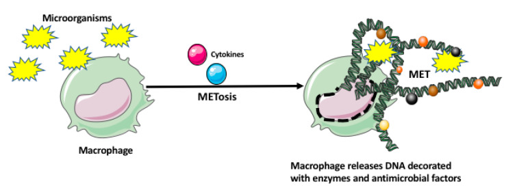
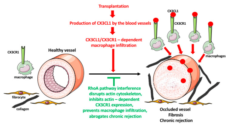
Tissue-resident macrophages and those conscripted from the blood/bone marrow are professional phagocytes. They play a role in tissue homeostasis, replacement, and healing, and are the first-line responders to microbial (viral, bacterial, and fungi) infections. Intrinsic ameboid-type motility allows non-resident macrophages to move to the site of inflammation or injury, where, in response to the inflammatory milieu they perform the anti-microbial and/or tissue repair functions. Depending on the need and the signaling from the surrounding tissue and other immune cells, macrophages acquire morphologically and functionally different phenotypes, which allow them to play either pro-inflammatory or anti-inflammatory functions. As such, the macrophages are also the major players in the rejection of the transplanted organs making an excellent target for the novel anti-rejection therapies in clinical transplantation. In this review, we describe some of the less covered aspects of macrophage response to microbial infection and organ transplantation.

Keywords: chronic rejection; infection; macrophage; transplantation.


Conflict of interest statement

The authors declare no conflict of interest.

Figures

Figure 1
Figure 1
Phagocytosis of microorganisms by macrophage. Recognition of the microorganism (bacteria, or virus) by the appropriate receptor induces the formation of the phagocytic cup that engulfs the microorganism and pinches off the membrane as a nascent phagosome. The formation of the phagocytic cup and detachment of the phagosome from the membrane are actin dependent. The nascent phagosome recruits small GTPase Rab5 that induce its remodeling and becomes the early phagosome. In the process of the Rab conversion, the early phagosome loses Rab5 and acquires Rab7 becoming the late phagosome. The Rab7 induces signaling pathways allowing the late phagosome to fuse with the lysosomes, containing various digestive enzymes, and become the phagolysosome that degrades the microorganism.
Figure 2
Figure 2
Macrophage extracellular trap (MET) formation by macrophage. The microorganisms and/or various cytokines may induce a unique cell death program in the macrophage, called METosis. The METosis causes breakage of the macrophage nuclear envelope and release of strands of DNA, which form the MET decorated with the enzymes and antimicrobial factors.
Figure 3
Figure 3
Role of macrophages in transplant rejection. Organ transplantation induces immune responses in the recipient. One of the responses is a massive production of the chemokine fractalkine (CX3CL1) by the endothelial cells of the graft blood vessels. The CX3CL1 recruits the monocytes/macrophages, which express the fractalkine receptor (CX3CR1), from the blood to the vicinity of the blood vessels. The macrophages induce an over-proliferation of the smooth muscle cells in the blood vessel wall, and fibroblast/fibrocyte to express a huge quantity of fibrotic factors such as collagen. These result in the occlusion of the blood vessel lumen, and graft tissue fibrosis, leading to chronic rejection of the transplant. The expression and recycling of the macrophage receptors are actin-dependent, and actin is regulated by the RhoA pathway. The interference (either RhoA deletion or pharmacologic inhibition) with the RhoA pathway disrupts the normal functioning of actin filaments and actin-dependent processes such as receptor expression and recycling. The lowered expression of CX3CR1 receptors makes macrophages less or nonresponsive to the fractalkine, prevents their infiltration into the graft, and inhibits chronic rejection.

References

    1. Liao X., Shen Y., Zhang R., Sugi K., Vasudevan N.T., Alaiti M.A., Sweet D.R., Zhou L., Qing Y., Gerson S.L., et al. Distinct roles of resident and nonresident macrophages in nonischemic cardiomyopathy. Proc. Natl. Acad. Sci. USA. 2018;115:E4661–E4669. doi: 10.1073/pnas.1720065115. - DOI - PMC - PubMed
    1. Allard B., Panariti A., Martin J.G. Alveolar Macrophages in the Resolution of Inflammation, Tissue Repair, and Tolerance to Infection. Front. Immunol. 2018;9:1777. doi: 10.3389/fimmu.2018.01777. - DOI - PMC - PubMed
    1. Apovian C.M., Apovian C.M. Macrophage functions in lean and obese adipose tissue. Metabolism. 2017;72:120–143. doi: 10.1016/j.metabol.2017.04.005. - DOI - PMC - PubMed
    1. Dixon L.J., Barnes M., Tang H., Pritchard M.T., Nagy L.E. Kupffer Cells in the Liver. Compr. Physiol. 2013;3:785–797. - PMC - PubMed
    1. Nagelkerke S.Q., Bruggeman C.W., Haan J.M.D., Mul E.P.J., Berg T.K.V.D., Van Bruggen R., Kuijpers T.W. Red pulp macrophages in the human spleen are a distinct cell population with a unique expression of Fc-γ receptors. Blood Adv. 2018;2:941–953. doi: 10.1182/bloodadvances.2017015008. - DOI - PMC - PubMed