Review
. 2021:42:473-518.
doi: 10.21775/cimb.042.473. Epub 2020 Dec 23.

Lyme Disease Pathogenesis

Affiliations
Review

Lyme Disease Pathogenesis

Jenifer Coburn et al. Curr Issues Mol Biol. 2021.

Abstract

Lyme disease Borrelia are obligately parasitic, tick- transmitted, invasive, persistent bacterial pathogens that cause disease in humans and non-reservoir vertebrates primarily through the induction of inflammation. During transmission from the infected tick, the bacteria undergo significant changes in gene expression, resulting in adaptation to the mammalian environment. The organisms multiply and spread locally and induce inflammatory responses that, in humans, result in clinical signs and symptoms. Borrelia virulence involves a multiplicity of mechanisms for dissemination and colonization of multiple tissues and evasion of host immune responses. Most of the tissue damage, which is seen in non-reservoir hosts, appears to result from host inflammatory reactions, despite the low numbers of bacteria in affected sites. This host response to the Lyme disease Borrelia can cause neurologic, cardiovascular, arthritic, and dermatologic manifestations during the disseminated and persistent stages of infection. The mechanisms by which a paucity of organisms (in comparison to many other infectious diseases) can cause varied and in some cases profound inflammation and symptoms remains mysterious but are the subjects of diverse ongoing investigations. In this review, we provide an overview of virulence mechanisms and determinants for which roles have been demonstrated in vivo, primarily in mouse models of infection.


Figures

Figure 1.
Figure 1.. Characteristics of select bacterial pathogens.
Schematic depiction of general properties of bacterial pathogens illustrated. Note that for many of the bacteria shown, the outcome of the interaction may depend on the host and even particular sites within the host. B. burgdorferi and T. pallidum are highly invasive and persistent, but non-toxigenic, while M. tuberculosis is usually less invasive but persistent and non-toxigenic; gut microbiota are persistent but neither invasive nor toxigenic. S. aureus is often a commensal that as opportunity arises can be invasive and toxigenic. L. interrogans is invasive and persistent, but unlike B. burgdorferi and T. pallidum, synthesizes lipopolysaccharide (LPS), a highly inflammatory glycolipid. B. pilosicoli causes persistent but not invasive infection, synthesizes LPS and hemolysins. V. cholerae and C. difficile are both toxigenic organisms, which cause disease largely attributable to toxin action; C. difficile is more persistent, at least in part, due to spore formation. The authors thank Valery Lozada-Fernandez, PhD candidate at the Medical College of Wisconsin, for generation of the 3D plot in this figure. Concept originally depicted in (Botkin et al., 2006).
Figure 2.
Figure 2.. Overview of Borrelia infection and Lyme disease in humans.
Schematic representation of the infection process (in black text) and common associated disease manifestations (in red text) in Lyme borreliosis. For detailed descriptions of Lyme disease clinical manifestations, stages, and associations with different Lyme spirochete species, we refer the reader to Radolf and Samuels (2021) and to Steere, A.C. (2001) “Lyme Disease” N Engl J Med; 345:115–125. Adapted from Coburn, J., Leong, J. and Chaconas, G. (2013). “Illuminating the roles of the Borrelia burgdorferi adhesins” Trends Microbiol 21(8): 372–379, with permission from Elsevier.
Figure 3.
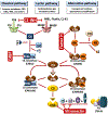
Figure 3.. Schematic representation of the human complement cascades.
Complement is activated by three pathways: the classical, lectin, and alternative pathway. In each pathway, a series of initial binding steps followed by proteolytic steps ultimately results in activation of the terminal pathway and lysis of the target cells. CRP, C-reactive protein; C1-INH, C1 esterase inhibitor; MBL, Mannose-binding lectin; MASP, MBL-associated serine protease; C4BP, C4b-binding protein; TCC, terminal complement complex. Parts of this figure are adapted from Kraiczy, P. (2016), “Travelling between two worlds: complement as a gatekeeper for an expanded host range of Lyme disease spirochetes” Vet. Sci., 3:12; doi:10.3390/vetsci3020012. Permission to reproduce under CC 4.0 (https://creativecommons.org/licenses/by/4.0).
Figure 4.
Figure 4.. Borrelia burgdorferi subversion of the complement system.
To evade complement, Lyme disease spirochetes produce outer surface lipoproteins that interfere with host complement by inhibiting complement activity directly or binding to host-derived regulators of complement activity (RCA). Several Borrelia inhibitors block the upstream initiation steps, including the CP-specific inhibitor BBK32, which binds to C1r and traps C1 in a zymogen state. OspC binds C4b and interferes with the activation of both the CP and LP. Lyme disease spirochetes also produce p43, which downregulates the CP and LP by recruiting the RCA called C4b-binding protein (C4BP). CspA and CspZ bind the negative regulators of the AP known as factor H (FH) and factor H-like protein 1 (FHL-1). ErpA and ErpP also bind to FH, but not to FHL-1. Borrelia produce at least four proteins that block the formation of the MAC complex. CspA binds C7 and C9 in a FH-independent manner and blocks C9 polymerization. Two homologous proteins from B. bavariensis, BGA66 and BGA71 also block C9 polymerization by binding to C7, C8, and C9. Finally, a surface protein that exhibits activity comparable to CD59 (i.e., CD59-like) was investigated, but the gene encoding this protein has not been identified. Proteins known to affect experimental infection by B. burgdorferi are outlined in blue, those that are important in transmission from Ixodes ticks are framed in green, and those whose functions in vivo are not defined are shown in red. PAMP: pathogen-associated molecular pattern; Ab: antibody; Ag: antigen; CL-11: collectin-11. This figure is reproduced from Skare J.T. and Garcia B.L. (2020) “Complement Evasion by Lyme Disease Spirochetes” Trends Microbiol. S0966-842X(20)30129-3. doi: 10.1016/j.tim.2020.05.004 with permission conveyed through Elsevier and Copyright Clearance Center.
Figure 5.
Figure 5.. Schematic of the vls antigenic variation locus of B. burgdorferi strain B31.
A, the vls expression locus (vlsE) with its promoter (P) is located 82 bp from the right covalently closed hairpin end of the linear plasmid lp28–1. To the left of the promoter and intergenic region (gray) are 15 silent cassettes carrying information corresponding to the variable region of vlsE and situated in the opposite orientation. B, the vlsE region is shown in greater detail, with the constant regions (CR) shown in yellow and the variable region, which corresponds to the information carried in the vls cassettes, shown in blue. The variable region is flanked by 17-bp direct repeats (DR)s. To the left of vlsE is its promoter, with the −10 and −35 sequences shown as green bars. Also shown by the bidirectional arrow is a 100-bp perfect inverted repeat (IR) that partially overlaps the promoter. C, an enlargement of the vlsE gene shows the product of multiple recombinational switching events that result in the copying of genetic information from the silent cassettes into the expression locus, producing a mosaic vlsE carrying information from a number of the silent cassettes. D, an IR found in the promoter region of the vlsE gene is shown in its normal linear configuration and as an extruded cruciform promoted by negative supercoiling or DNA unwinding from replication or transcription. This figure is republished from Chaconas, G., Castellanos, M. and Verhey, T. B. (2020). “Changing of the guard: How the Lyme disease spirochete subverts the host immune response” J Biol Chem 295(2): 301–313, with permission of The Journal of Biological Chemistry, © the American Society for Biochemistry and Molecular Biology, permission conveyed through Copyright Clearance Center, Inc.
Figure 6.
Figure 6.. Borrelia burgdorferi-host interactions that contribute to infection.
Shown are interactions between host molecules and B. burgdorferi virulence determinants that have been documented to influence infection in vivo in animal model(s). Many more await further characterization or have not yet been shown to make significant contributions in the mouse model of B. burgdorferi infection. Additional factors such as those involved in nutrient scavenging are not included here. Refer to Table 1 for a more complete summary of factors known to participate in B. burgdorferi survival in mice. Complement factors: C1r, C4b, Factor H (and related proteins). Adhesion substrates: GAG = glycosaminoglycan, Fn = fibronectin.

References

    1. Adams PP, Flores Avile C, Popitsch N, Bilusic I, Schroeder R, Lybecker M, and Jewett MW (2017). In vivo expression technology and 5’ end mapping of the Borrelia burgdorferi transcriptome identify novel RNAs expressed during mammalian infection. Nucleic Acids Research 45, 775–792. 10.1093/nar/gkw1180 - DOI - PMC - PubMed
    1. Aguirre JD, Clark HM, McIlvin M, Vazquez C, Palmere SL, Grab DJ, Seshu J, Hart PJ, Saito M, and Culotta VC (2013). A manganese-rich environment supports superoxide dismutase activity in a Lyme disease pathogen, Borrelia burgdorferi. The Journal of Biological Chemistry 288, 8468–8478. 10.1074/jbc.M112.433540 - DOI - PMC - PubMed
    1. Akins DR, Bourell KW, Caimano MJ, Norgard MV, and Radolf JD (1998). A new animal model for studying Lyme disease spirochetes in a mammalian host-adapted state. J Clin Invest 101, 2240–2250. 10.1172/JCI2325 - DOI - PMC - PubMed
    1. Alitalo A, Meri T, Lankinen H, Seppala I, Lahdenne P, Hefty PS, Akins D, and Meri S (2002). Complement inhibitor factor H binding to Lyme disease spirochetes is mediated by inducible expression of multiple plasmid-encoded outer surface protein E paralogs. J Immunol 169, 3847–3853. - PubMed
    1. Angel TE, Luft BJ, Yang X, Nicora CD, Camp DG 2nd, Jacobs JM, and Smith RD (2010). Proteome analysis of Borrelia burgdorferi response to environmental change. PLoS ONE 5, e13800. 10.1371/journal.pone.0013800 - DOI - PMC - PubMed

Substances