

Preferred Reporting Items for Root and Canal Anatomy in the Human Dentition (PROUD 2020) - A Systematic Review and a Proposal for a Standardized Protocol
- PMID: 33353923
- PMCID: PMC7881390
- DOI: 10.14744/eej.2020.88942
Preferred Reporting Items for Root and Canal Anatomy in the Human Dentition (PROUD 2020) - A Systematic Review and a Proposal for a Standardized Protocol
Abstract
Objective: Consistent reporting of publications in a given topic is essential. This systematic review aimed to identify and evaluate the reporting items in previous publications related to root canal anatomy in major Endodontic journals.
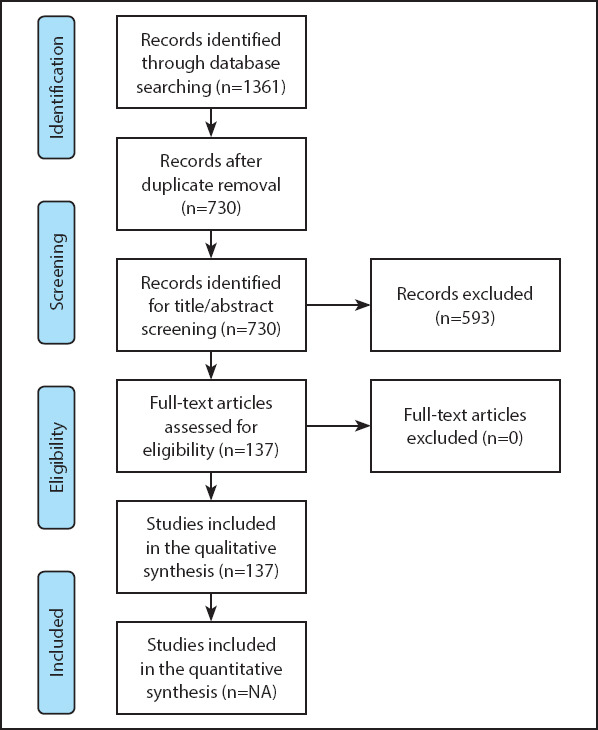
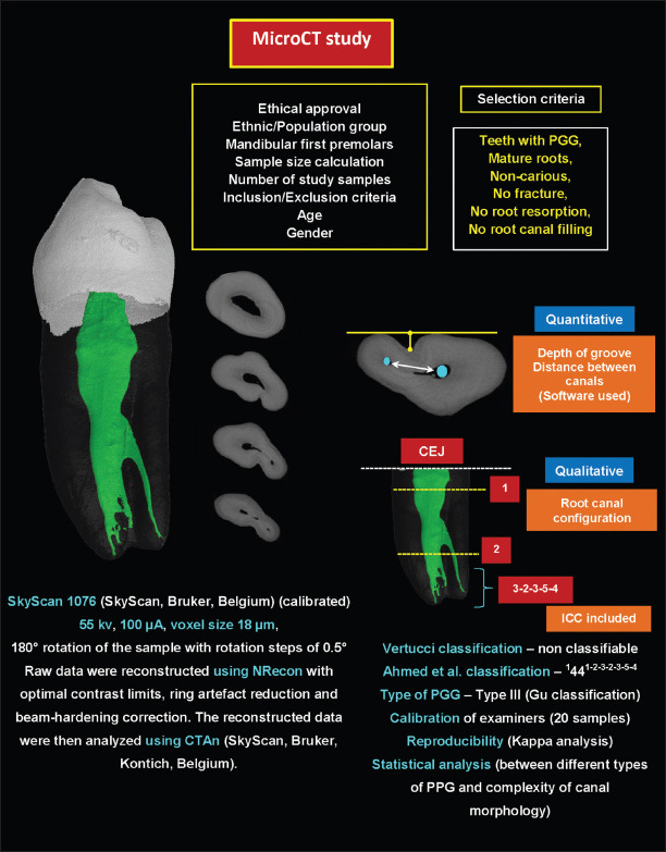
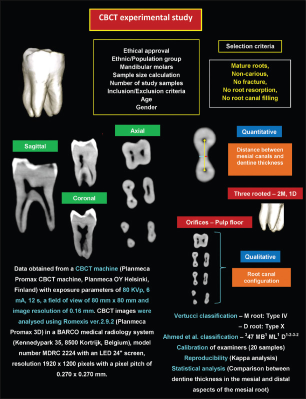
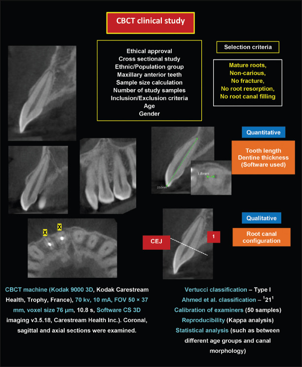
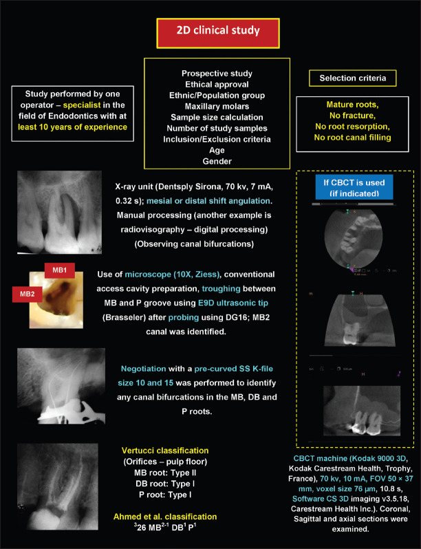
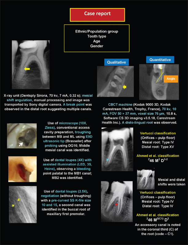
Methods: A systematic review was undertaken following the Preferred Reporting Items for Systematic Reviews and Meta-Analyses (PRISMA) guidelines. A comprehensive literature search was performed by 2 independent reviewers using a customized search strategy in major Endodontic journals through Scopus until November 2019. Studies investigating root and canal anatomy were included. The selected publications were divided into 7 categories according to the study design: micro-computed tomography (microCT) and cone-beam computed tomography (CBCT) experimental studies (extracted teeth), CBCT and 2D clinical studies, CBCT and 2D case reports in addition to others (i.e. staining and clearing method and root sectioning). The selected studies were evaluated according to three domains: 1) Criteria for study sample selection; 2) Criteria for methodological procedures and 3) Criteria for detection and evaluation.
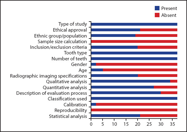
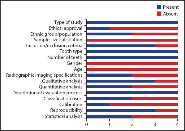
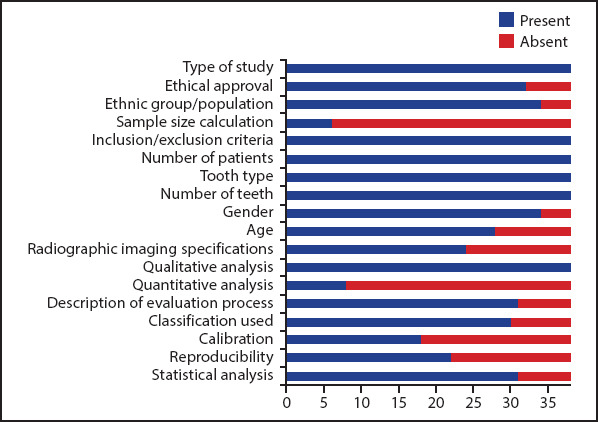
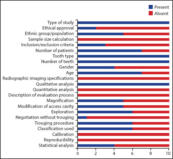
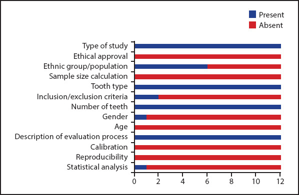
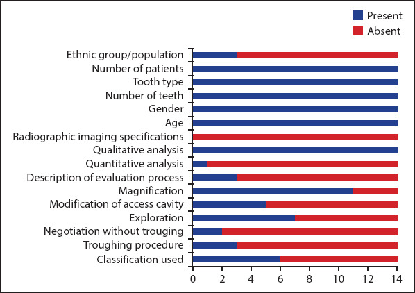
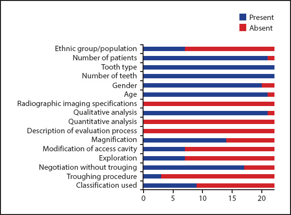
Results: After the removal of duplicated and irrelevant papers, 137 articles were included. Results showed that microCT studies reported accurately the tooth type, number of teeth, classifications used, qualitative and/or quantitative analysis (if required) and the evaluation process. However, sample size calculation, calibration, and reproducibility were not reported in the majority of microCT studies. CBCT clinical studies presented information for the type of study, inclusion/exclusion criteria, number of patients, tooth type, and number of teeth. However, the majority did not report sample size calculation and calibration of examiners. Radiographic exposure descriptions and classifications used were not reported adequately in CBCT and 2D case reports. Sample size calculation, calibration and reproducibility were not reported in staining and clearing method.
Conclusion: Despite accurate presentation of certain items, there is considerable inconsistent reporting of root and canal morphology regardless of the type of study and experimental procedure used. The PROUD checklist protocol presented in this systematic review aims to provide an accurate description of root canal anatomy in experimental, clinical, and case report publications.
Conflict of interest statement
Figures
Similar articles
-
A critical analysis of laboratory and clinical research methods to study root and canal anatomy.Int Endod J. 2022 Apr;55 Suppl 2:229-280. doi: 10.1111/iej.13702. Epub 2022 Mar 31. Int Endod J. 2022. PMID: 35124829 Review.
-
Application of a new system for classifying root and canal anatomy in studies involving micro-computed tomography and cone beam computed tomography: Explanation and elaboration.Int Endod J. 2021 Jul;54(7):1056-1082. doi: 10.1111/iej.13486. Epub 2021 Apr 18. Int Endod J. 2021. PMID: 33527452 Review.
-
Comparative accuracy of the Clearing Technique, CBCT and Micro-CT methods in studying the mesial root canal configuration of mandibular first molars.Int Endod J. 2017 Jan;50(1):90-96. doi: 10.1111/iej.12593. Epub 2016 Jan 19. Int Endod J. 2017. PMID: 26659613
-
Preferred Reporting Items for Epidemiologic Cross-sectional Studies on Root and Root Canal Anatomy Using Cone-beam Computed Tomographic Technology: A Systematized Assessment.J Endod. 2020 Jul;46(7):915-935. doi: 10.1016/j.joen.2020.03.020. Epub 2020 May 6. J Endod. 2020. PMID: 32387077
-
Cone-beam and micro-computed tomography for the assessment of root canal morphology: a systematic review.Braz Oral Res. 2020 Jun 19;34:e056. doi: 10.1590/1807-3107bor-2020.vol34.0056. eCollection 2020. Braz Oral Res. 2020. PMID: 32578799
Cited by
-
Comparative Evaluation of Mineral Trioxide Aggregate Obturation Using Four Different Techniques-A Laboratory Study.Materials (Basel). 2021 Jun 7;14(11):3126. doi: 10.3390/ma14113126. Materials (Basel). 2021. PMID: 34200233 Free PMC article.
-
Morphological assessment of the isthmus in mesial root canals of first mandibular molars.Acta Odontol Latinoam. 2023 Dec 31;36(3):163-168. doi: 10.54589/aol.36/3/163. Acta Odontol Latinoam. 2023. PMID: 38345278 Free PMC article.
-
Cone-beam Computed Tomography Analysis of the Root Canal Morphology of Mandibular Incisors Using Two Classification Systems in a Spanish Subpopulation: A Cross-Sectional Study.Eur Endod J. 2024 Mar 8;9(2):106 - 113. doi: 10.14744/eej.2023.10327. Epub 2024 Feb 21. Eur Endod J. 2024. PMID: 38380511 Free PMC article.
-
A microcomputed tomographic analysis of the morphological variabilities and incidence of extra canals in mandibular first molar teeth in an Egyptian subpopulation.Sci Rep. 2023 Jun 2;13(1):8985. doi: 10.1038/s41598-023-36005-7. Sci Rep. 2023. PMID: 37268728 Free PMC article.
-
Micro-computed tomography analysis and ex-vivo detection of six root canals in a four-rooted mandibular first premolar.BMC Oral Health. 2025 Apr 7;25(1):498. doi: 10.1186/s12903-025-05781-1. BMC Oral Health. 2025. PMID: 40197269 Free PMC article.
References
-
- Vertucci F. Root canal morphology and its relationship to endodontic procedures. Endod Topics. 2005;10(1):3–29.
-
- Martins JNR, Marques D, Mata A, Caramês J. Root and root canal morphology of the permanent dentition in a Caucasian population:a cone-beam computed tomography study. Int Endod J. 2017;50(11):1013–26. - PubMed
-
- Neelakantan P, Subbarao C, Subbarao CV, Ravindranath M. Root and canal morphology of mandibular second molars in an Indian population. J Endod. 2010;36(8):1319–22. - PubMed
-
- Versiani MA, Ordinola-Zapata R, Keleş A, Alcin H, Bramante CM, Pécora JD, et al. Middle mesial canals in mandibular first molars:A micro-CT study in different populations. Arch Oral Biol. 2016;61:130–7. - PubMed
-
- Morfis A, Sylaras SN, Georgopoulou M, Kernani M, Prountzos F. Study of the apices of human permanent teeth with the use of a scanning electron microscope. Oral Surg Oral Med Oral Pathol. 1994;77(2):172–6. - PubMed
Publication types
MeSH terms
LinkOut - more resources
Full Text Sources
