. 2020 Dec 18;6(51):eabd7197.
doi: 10.1126/sciadv.abd7197. Print 2020 Dec.

Digenic mutations in ALDH2 and ADH5 impair formaldehyde clearance and cause a multisystem disorder, AMeD syndrome

Affiliations

Digenic mutations in ALDH2 and ADH5 impair formaldehyde clearance and cause a multisystem disorder, AMeD syndrome

Yasuyoshi Oka et al. Sci Adv. .

Abstract

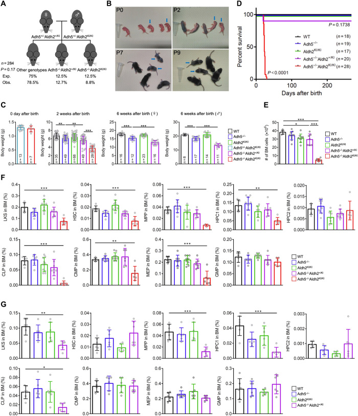
Rs671 in the aldehyde dehydrogenase 2 gene (ALDH2) is the cause of Asian alcohol flushing response after drinking. ALDH2 detoxifies endogenous aldehydes, which are the major source of DNA damage repaired by the Fanconi anemia pathway. Here, we show that the rs671 defective allele in combination with mutations in the alcohol dehydrogenase 5 gene, which encodes formaldehyde dehydrogenase (ADH5FDH ), causes a previously unidentified disorder, AMeD (aplastic anemia, mental retardation, and dwarfism) syndrome. Cellular studies revealed that a decrease in the formaldehyde tolerance underlies a loss of differentiation and proliferation capacity of hematopoietic stem cells. Moreover, Adh5-/-Aldh2 E506K/E506K double-deficient mice recapitulated key clinical features of AMeDS, showing short life span, dwarfism, and hematopoietic failure. Collectively, our results suggest that the combined deficiency of formaldehyde clearance mechanisms leads to the complex clinical features due to overload of formaldehyde-induced DNA damage, thereby saturation of DNA repair processes.


Figures

Fig. 1
Fig. 1. Identification of digenic variants in ADH5 and ALDH2 in patients with AMeDS.
(A) Pedigrees of AMeDS families 4 to 8. (B) Pathogenic variants identified in ADH5 and ALDH2. (C) Immunoblots of ADH5 and ALDH2 in primary fibroblasts from normal (1BR) and patients with AMeDS (N0608, N0611, and N0614). SMC3 is a loading control. (D) ADH5 transcript of normal (1BR) and AMeDS (N0608, N0611, and N0614) cells. The relative transcript levels analyzed by the ∆∆CT method are shown for triplicate experiments. (E) Cell viability after continuous 30 μM formaldehyde treatment. Results from triplicate experiments (means ± SD) are shown. **P < 0.01, two-tailed unpaired t test. (F) Immunoblots showing a reduced stability of ADH5 p.S75N identified in a healthy individual, NAG16714. Gene-edited hTERT-immortalized RPE1 (RPE1 hTERT) cells expressing the homozygous ADH5 p.S75N alleles (clones no. 10 and no. 52), and ∆ADH5 cells are examined. (G) Stable expression of the p.S75N mRNA. (H) Cell viability after continuous 40 μM formaldehyde treatment. Results from triplicate experiments (means ± SD) are shown. ***P < 0.001, two-tailed unpaired t test. (I) ADH5 p.S75N is unstable as with p.A278P. U2OS cells transfected with V5-tagged ADH5 WT (wild type), p.A278P, or p.S75N were harvested at the indicated times following cycloheximide (CHX) treatment. Cell lysates were immunoblotted with V5 and ACTB antibodies. (J) Quantification of ADH5-V5 levels in (I) by image analysis, normalized to ACTB levels. Means (± SD) from triplicate experiments are shown. *P < 0.05; one-way analysis of variance (ANOVA) with Tukey’s multiple comparisons test.
Fig. 2
Fig. 2. Formaldehyde is a possible endogenous aldehyde metabolized by both ADH5 and ALDH2.
(A) Formaldehyde treatment inhibits DNA replication in ADH5 and ALDH2 double-deficient cells. 5-ethynyl-2′-deoxyuridine (EdU) incorporation in WT, ∆ADH5, ALDH2E504K, or ∆ADH5 ALDH2E504K double-mutant U2OS cells after formaldehyde treatment is measured by fluorescence-activated cell sorting (FACS) analysis. Cells were incubated with indicated concentration of formaldehyde or 10 mM hydroxyurea (HU) as a positive control for 8 hours followed by EdU incorporation for 1 hour. Then, cells were fixed with 70% ethanol and analyzed by FACS for Alexa Fluor 488–labeled EdU and DNA stained with 7-aminoactinomycin D (7-AAD). Representative FACS images are shown. (B) Quantification of data in (A). Graph shows the percentage of EdU-positive cells. Means (± SD) from three independent experiments are shown. *P < 0.05, **P < 0.01, and ***P < 0.001, one-way ANOVA with Tukey’s multiple comparisons test. (C) Formaldehyde treatment induces DNA damage in cells from AMeDS-affected individuals. Immunoblots showing phospho-Ser139 histone H2AX (γH2AX), a DNA damage marker, and PARP1, an apoptosis marker in normal (1BR) and AMeDS (N0608 and N0611) cells. KU70 is a loading control. (D) EdU incorporation in normal and AMeDS cells after formaldehyde treatment measured by FACS analysis. Cells were incubated with indicated concentration of formaldehyde for 22 hours followed by EdU incorporation for 2 hours. Then, cells were fixed with 70% ethanol and analyzed by FACS for Alexa Fluor 488–labeled EdU and DNA stained with propidium iodide (PI). (E) Formaldehyde-induced DNA damage in AMeDS cells (N0611) is ameliorated with expression of either the wild-type ADH5 or ALDH2 cDNA. Green fluorescent protein as a mock control.
Fig. 3
Fig. 3. Deletion of ADH5 attenuates differentiation and proliferation capacity of CD34+ HSPCs harboring the ALDH2 rs671 allele.
(A) Schematic representation of CFU assay. CD34+ HSPCs are derived from umbilical cord blood of Japanese healthy donors. The numbers of HSPC pools with the designated ALDH2 rs671 alleles are shown. ADH5 was deleted in each HSPC pool by CRISPR-Cas9–based gene editing. (B) CFU assay of gene-edited CD34+ HSPCs was performed using a methylcellulose medium. Representative images are shown. Scale bar, 3 mm. (C) Total number of colonies after 14-day CFU assay of gene-edited CD34+ HSPCs. The number of colonies was normalized to untreated control. Statistical analysis was performed using one-way ANOVA with Tukey’s multiple comparisons test (***P < 0.001; ns, not significant). Lines represent median.
Fig. 4
Fig. 4. Loss of Adh5 function in combination with reduced Aldh2 activity recapitulates the phenotype of AMeDS in mice.
(A) Observed and expected frequencies of mice at 2 weeks of age from intercrossed of Adh5+/−Aldh2+/KI female mice with Adh5+/−Aldh2KI/KI male mice. Chi-square test shows no significant difference between observed and expected (P = 0.17). (B) Postnatal growth defects of Adh5−/−Aldh2KI/KI mice. Representative pictures are shown. Blue arrows indicate Adh5−/−Aldh2KI/KI mice. Photo credit: Yasuyoshi Oka, Nagoya University. (C) Body weights of individual mice at 0 days, 2 weeks, or 6 weeks of age. **P < 0.01 and ***P < 0.001, one-way ANOVA with Tukey’s multiple comparisons test. (D) Kaplan-Meier curves with log-rank (Mantel-Cox) test show a significant decrease in survival of Adh5−/−Aldh2KI/KI compared to the mice with other genotypes (P < 0.0001). (E) Quantification of nucleated bone marrow cells in bilateral femurs and tibias from 3-week-old mice is shown (means ± SD; n = at least 5 animals). *P < 0.05 and ***P < 0.001; one-way ANOVA with Tukey’s multiple comparisons test. (F and G) Quantification of hematopoietic subset: LKS (Linc-Kit+Sca-1+), HSC (Linc-Kit+Sca-1+CD150+CD48), MPP (Linc-Kit+Sca-1+CD150CD48), HPC1 (Linc-Kit+Sca-1+CD150CD48+), HPC2 (Linc-Kit+Sca-1+CD150+CD48+), CLP (Linc-KitlowSca-1lowCD127+CD135+), CMP (Linc-Kit+Sca-1CD34+CD16/32), MEP (Linc-Kit+Sca-1CD34CD16/32), and GMP (Linc-Kit+Sca-1CD34+CD16/32+) in individual mice at 3 weeks of age in (F) and at 8 to 9 months of age in (G). Means ± SD; n = at least 5 animals. *P < 0.05, **P < 0.01, and ***P < 0.001, one-way ANOVA with Tukey’s multiple comparisons test.

References

    1. Burgos-Barragan G., Wit N., Meiser J., Dingler F. A., Pietzke M., Mulderrig L., Pontel L. B., Rosado I. V., Brewer T. F., Cordell R. L., Monks P. S., Chang C. J., Vazquez A., Patel K. J., Mammals divert endogenous genotoxic formaldehyde into one-carbon metabolism. Nature 548, 549–554 (2017). - PMC - PubMed
    1. Walport L. J., Hopkinson R. J., Schofield C. J., Mechanisms of human histone and nucleic acid demethylases. Curr. Opin. Chem. Biol. 16, 525–534 (2012). - PubMed
    1. Harada S., Agarwal D. P., Goedde H. W., Aldehyde dehydrogenase deficiency as cause of facial flushing reaction to alcohol in Japanese. Lancet 2, 982 (1981). - PubMed
    1. Brooks P. J., Enoch M.-A., Goldman D., Li T.-K., Yokoyama A., The alcohol flushing response: An unrecognized risk factor for esophageal cancer from alcohol consumption. PLOS Med. 6, e1000050 (2009). - PMC - PubMed
    1. Yoshida A., Huang I. Y., Ikawa M., Molecular abnormality of an inactive aldehyde dehydrogenase variant commonly found in Orientals. Proc. Natl. Acad. Sci. U.S.A. 81, 258–261 (1984). - PMC - PubMed

Publication types