

Swept-source OCTA quantification of capillary closure predicts ETDRS severity staging of NPDR
- PMID: 33355147
- PMCID: PMC9046755
- DOI: 10.1136/bjophthalmol-2020-317890
Swept-source OCTA quantification of capillary closure predicts ETDRS severity staging of NPDR
Abstract
Purpose: To test whether a single or composite set of parameters evaluated with optical coherence tomography angiography (OCTA), representing retinal capillary closure, can predict non-proliferative diabetic retinopathy (NPDR) staging according to the gold standard ETDRS grading scheme.
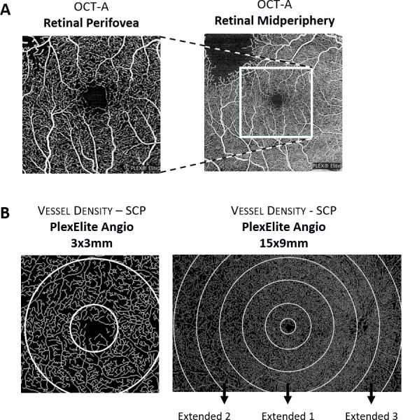
Methods: 105 patients with diabetes, either without retinopathy or with different degrees of retinopathy (NPDR up to ETDRS grade 53), were prospectively evaluated using swept-source OCTA (SS-OCTA, PlexElite, Carl Zeiss Meditec) with 15×9 mm and 3×3 mm angiography protocols. Seven-field photographs of the fundus were obtained for ETDRS staging. Eyes from age-matched healthy subjects were also imaged as control.
Results: In eyes of patients with type 2 diabetes without retinopathy or ETDRS levels 20 and 35, retinal capillary closure was in the macular area, with predominant alterations in the parafoveal retinal circulation (inner ring). Retinal capillary closure in ETDRS stages 43-53 becomes predominant in the retinal midperiphery with vessel density average values of 25.2±7.9 (p=0.001) in ETDRS 43 and 23.5±3.4 (p=0.001) in ETDRS 47-53, when evaluating extended areas of 15×9 protocol. Combination of acquisition protocols 3×3 mm and 15×9 mm, using SS-OCTA, allows discrimination between eyes with mild NPDR (ETDRS 10, 20, 35) and eyes with moderate-to-severe NPDR (ETDRS grades 43-53).
Conclusions: Retinal capillary closure, quantified by SS-OCTA, can identify NPDR severity progression. It is located mainly in the perifoveal retinal capillary circulation in the initial stages of NPDR, whereas the retinal midperiphery is predominantly affected in moderate-to-severe NPDR.
Keywords: diagnostic tests/Investigation; imaging; retina.
© Author(s) (or their employer(s)) 2022. Re-use permitted under CC BY-NC. No commercial re-use. See rights and permissions. Published by BMJ.
Conflict of interest statement
Competing interests: TS, ARS, IPM, LGM and MHM do not have financial disclosures. LHW, SK, LdS and MD: Carl Zeiss Meditec (E). JGC-V reports grants from Carl Zeiss Meditec and is consultant for Alimera Sciences, Allergan, Bayer, Gene Signal, Novartis, Pfizer, Precision Ocular, Roche, Sanofi-Aventis, Vifor Pharma, and Carl Zeiss Meditec.
Figures
References
Publication types
MeSH terms
LinkOut - more resources
Full Text Sources
Other Literature Sources
Medical