Development and validation of a deep learning system to screen vision-threatening conditions in high myopia using optical coherence tomography images
- PMID: 33355150
- PMCID: PMC9046742
- DOI: 10.1136/bjophthalmol-2020-317825
Development and validation of a deep learning system to screen vision-threatening conditions in high myopia using optical coherence tomography images
Abstract
Background/aims: To apply deep learning technology to develop an artificial intelligence (AI) system that can identify vision-threatening conditions in high myopia patients based on optical coherence tomography (OCT) macular images.
Methods: In this cross-sectional, prospective study, a total of 5505 qualified OCT macular images obtained from 1048 high myopia patients admitted to Zhongshan Ophthalmic Centre (ZOC) from 2012 to 2017 were selected for the development of the AI system. The independent test dataset included 412 images obtained from 91 high myopia patients recruited at ZOC from January 2019 to May 2019. We adopted the InceptionResnetV2 architecture to train four independent convolutional neural network (CNN) models to identify the following four vision-threatening conditions in high myopia: retinoschisis, macular hole, retinal detachment and pathological myopic choroidal neovascularisation. Focal Loss was used to address class imbalance, and optimal operating thresholds were determined according to the Youden Index.
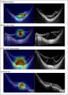
Results: In the independent test dataset, the areas under the receiver operating characteristic curves were high for all conditions (0.961 to 0.999). Our AI system achieved sensitivities equal to or even better than those of retina specialists as well as high specificities (greater than 90%). Moreover, our AI system provided a transparent and interpretable diagnosis with heatmaps.
Conclusions: We used OCT macular images for the development of CNN models to identify vision-threatening conditions in high myopia patients. Our models achieved reliable sensitivities and high specificities, comparable to those of retina specialists and may be applied for large-scale high myopia screening and patient follow-up.
Keywords: diagnostic tests/investigation; imaging; retina.
© Author(s) (or their employer(s)) 2022. Re-use permitted under CC BY-NC. No commercial re-use. See rights and permissions. Published by BMJ.
Conflict of interest statement
Competing interests: None declared.
Figures


References
Publication types
MeSH terms
LinkOut - more resources
Full Text Sources
