. 2021 Mar;9(3):279-290.
doi: 10.1158/2326-6066.CIR-20-0280. Epub 2020 Dec 22.

Antitumor Effects of CAR T Cells Redirected to the EDB Splice Variant of Fibronectin

Affiliations

Antitumor Effects of CAR T Cells Redirected to the EDB Splice Variant of Fibronectin

Jessica Wagner et al. Cancer Immunol Res. 2021 Mar.

Abstract

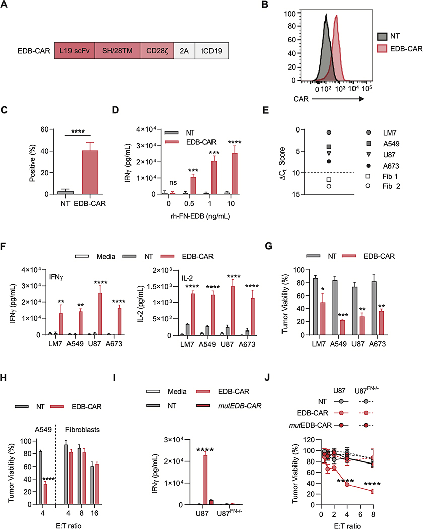
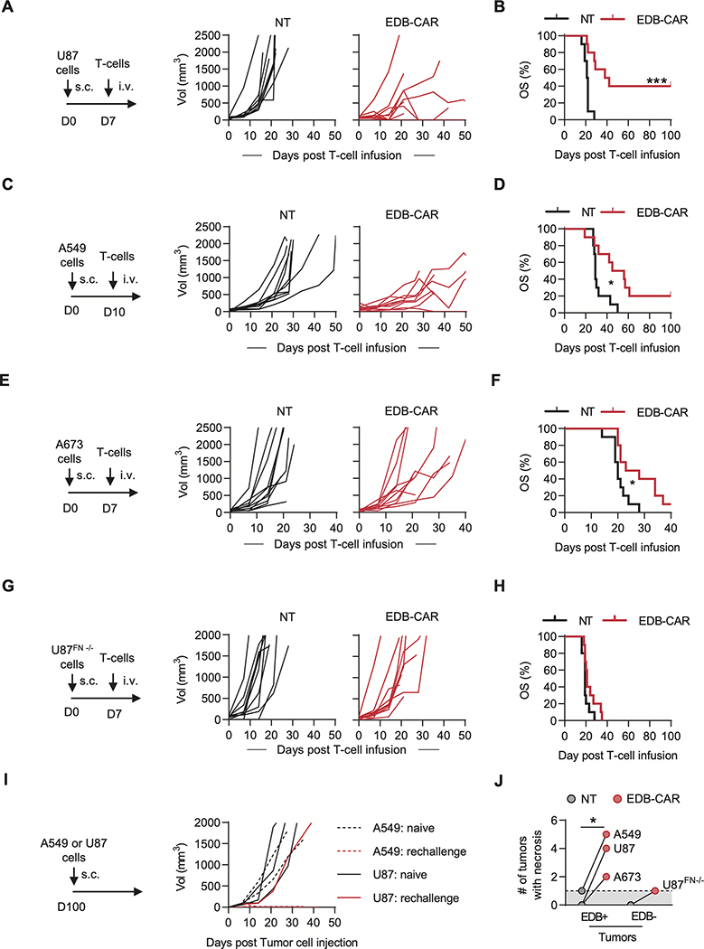
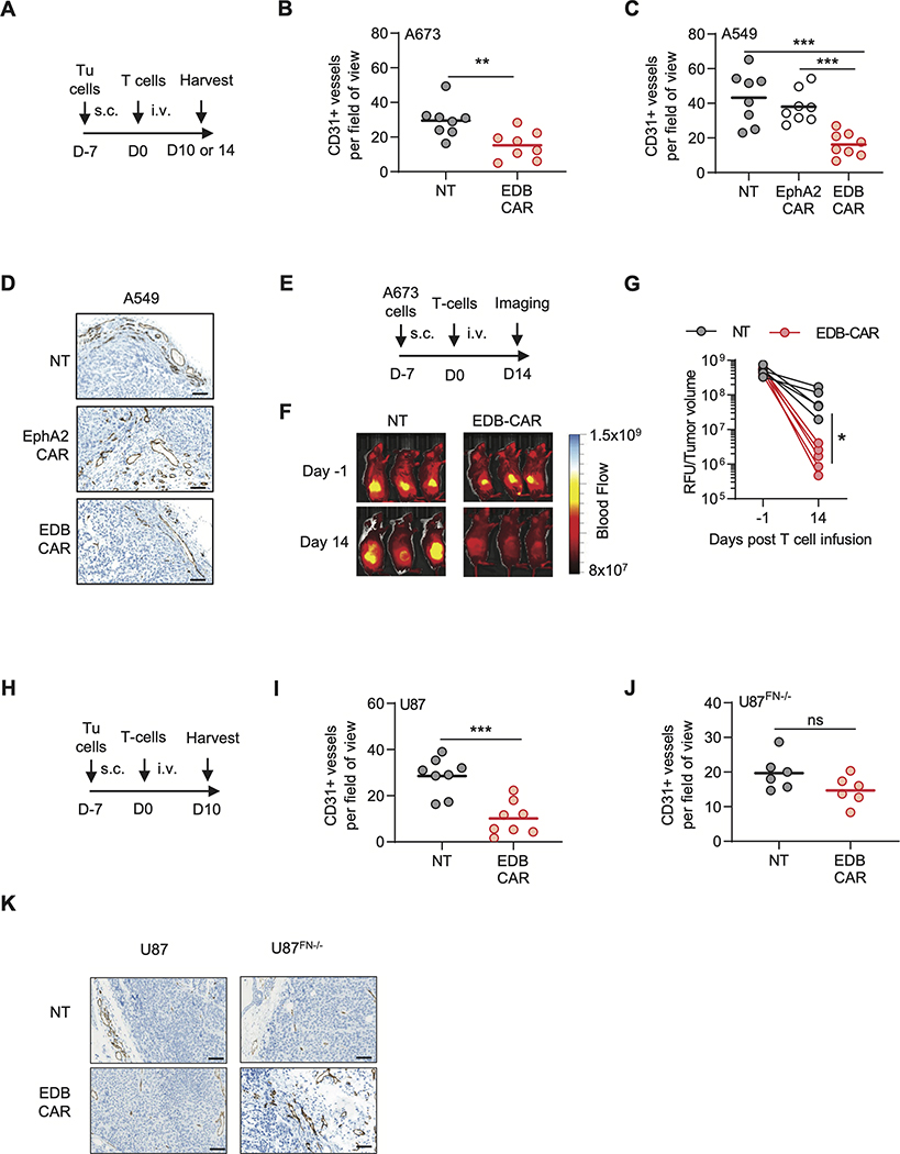
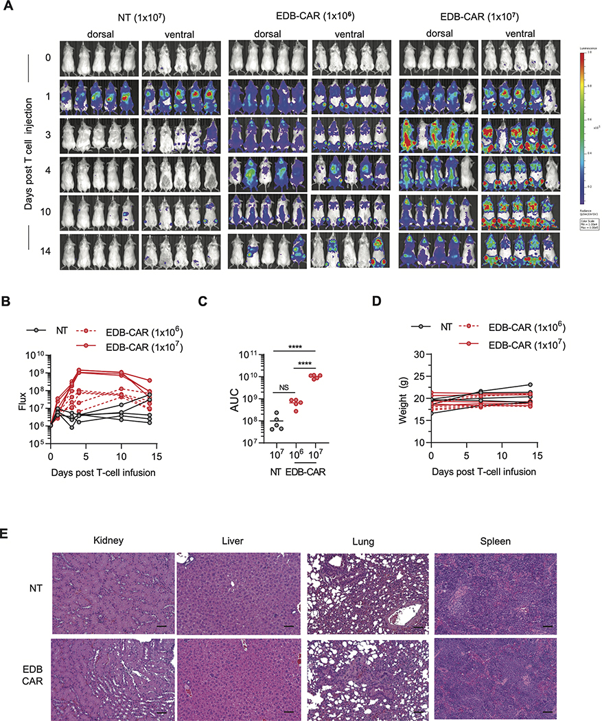
Chimeric antigen receptor (CAR) T-cell therapy has had limited success in early-phase clinical studies for solid tumors. Lack of efficacy is most likely multifactorial, including a limited array of targetable antigens. We reasoned that targeting the cancer-specific extra domain B (EDB) splice variant of fibronectin might overcome this limitation because it is abundantly secreted by cancer cells and adheres to their cell surface. In vitro, EDB-CAR T cells recognized and killed EDB-positive tumor cells. In vivo, 1 × 10⁶ EDB-CAR T cells had potent antitumor activity in both subcutaneous and systemic tumor xenograft models, resulting in a significant survival advantage in comparison with control mice. EDB-CAR T cells also targeted the tumor vasculature, as judged by IHC and imaging, and their antivascular activity was dependent on the secretion of EDB by tumor cells. Thus, targeting tumor-specific splice variants such as EDB with CAR T cells is feasible and has the potential to improve the efficacy of CAR T-cell therapy.


Conflict of interest statement

Conflict of interests: JW, GK, and SG have patent applications in the fields of T-cell and/or gene therapy for cancer. SG has a research collaboration with TESSA Therapeutics, is a DSMB member of Immatics, and on the scientific advisory board of Tidal.

Figures

Figure 1:
Figure 1:. Generation and functional characterization of EDB-CAR T cells.
(A) Scheme of retroviral vectors encoding the EDB-CAR, a 2A sequence, and truncated CD19 (tCD19); SH: short hinge; TM: transmembrane domain. (B-C) EDB-CAR expression was determined on T cells 7 days post-transduction by flow cytometric analysis. (B) Representative histogram (Black-filled line: NT T cells; Red-filled line: EDB-CAR transduced T cells) and (C) summary plot (n=7; Student’s t-test; ****p<0.0001). (D) NT and EDB-CAR T cells were incubated for 24 hours with increasing concentrations of rhFN-EDB-coated wells. IFNγ production in supernatants was determined by ELISA (n=4; two-way ANOVA; ***p<0.001; ****p<0.0001). (E) EDB expression of LM7, A673, A549, and U87 tumor cells, and two primary fibroblast cell lines (Fib 1, Fib 2) determined by RT-qPCR; dotted line represents the ΔCt score above which samples are considered positive. (F) NT and EDB-CAR T cells were incubated for 48 hours with EDB-positive tumor cells. IFNγ and IL2 production in supernatants was determined by ELISA (two-way ANOVA; ****p<0.0001). (G) Cytolytic activity of NT or EDB-CAR T cells at a E:T ratio of 4:1 against EDB-positive tumor cells (MTS assay; n=3; two-way ANOVA; *p<0.05; **p<0.01; ***p<0.001). (H) Cytolytic activity of NT or EDB-CAR T cells against primary fibroblasts (data of Fib 1 and Fib 2 was combined) at the indicated E:T ratios, with A549 serving as controls (n=3; two-way ANOVA; ****p<0.0001). (I) NT, EDB-CAR, or mutEDB-CAR T cells were incubated for 48 hours with U87 or U87FN−/− cells. IFNγ production in supernatants was determined by ELISA (n=3; two-way ANOVA; ****p<0.0001). (J) Cytolytic activity of NT, EDB-CAR, or mutEDB-CAR T cells against U87 or U87FN−/− cells (n=3; two-way ANOVA; ****p<0.0001). Mean+SEM is shown in panels.
Figure 2:
Figure 2:. EDB-CAR T cells have antitumor activity in vivo.
(A-H) 2×10⁶ of the indicated tumor cells were injected into NSG mice s.c.. On day (D)7 (U87, A673, U87FN−/−) or D10 (A549) post tumor cell injection, mice received a single i.v. dose of 1×10⁶ NT or EDB-CAR T cells. Tumor growth was monitored weekly by caliper measurement (n=10 mice/group; 5 mice/T-cell donor; log-rank test; *p<0.05; ***p<0.001). (A) Experimental scheme and tumor growth for the U87 model. (B) U87 model: Kaplan-Meier survival curve. (C) Experimental scheme and tumor growth for the A549 model. (D) A549 model: Kaplan-Meier survival curve. (E) Experimental scheme and tumor growth for the A673 model. (F) A673 model: Kaplan-Meier survival curve. (G) Experimental scheme and tumor growth for the U87FN−/− model. (H) U87FN−/− model: Kaplan-Meier survival curve. (I) NSG mice that survived long-term post initial A549 (n=2) or U87 (n=3) s.c. tumor cell injection and EDB-CAR T-cell therapy were re-challenged with a single s.c. dose of cognate tumor cells. Naïve mice served as controls. Tumor growth was monitored weekly by caliper measurement. (J) Macroscopic tumor necrosis of tumors <2,000 mm3 (Student’s t-test; *p<0.05) of the experiments shown in panels A-I; dots represent how many tumors had macroscopic necrosis out of 10 tumors per cell type; dotted line represent baseline (1 tumor with macroscopic necrosis).
Figure 3:
Figure 3:. EDB-CAR T cells target intratumoral CD31-positive endothelial cells.
(A-D) A673 or A549 s.c. tumor-bearing mice received a single dose of NT, EDB-CAR, or EphA2-CAR (A549 only) T cells. Tumors were processed on day (D)10 (A673) or 14 (A549) post T-cell injection and analyzed for CD31 expression (n=4 mice per group with each mouse bearing one tumor; dots represent average of three blind scores per tumor; bars represent average per cohort, two fields-of-view (FOV) per tumor). (B) CD31+ vessels/FOV in the A673 model; Student’s t-test; **p<0.01. (C) CD31+ vessels/FOV in the A549 model; one-way ANOVA; *p<0.05). (D) Representative IHC images for CD31 staining of A549 tumors. Images at 40x magnification; scale bar: 100μm. (E-G) A673 s.c. tumor-bearing NSG mice were imaged using Angiosense 750 on day −1 and day 14 post NT or EDB-CAR T-cell injection. (E) Experimental scheme. (F) Representative images. (G) Quantitation (relative light units (RLU) divided by tumor volume is plotted; n=5 mice/treatment group; Student’s t-test; *p<0.05). (H) Experimental scheme. s.c. tumor-bearing (I) U87 or (J) U87FN−/− NSG mice received a single i.v. dose of NT or EDB-CAR T cells. Tumor were processed on day 10 post T-cell injection and analyzed for CD31 expression (n=4 mouse/tumors per group except for U87FN−/−/NT T-cell group (n=3) with dots representing three blind score per FOV; bars represent average per cohort, 2 FOV per tumor). (K) Representative IHC images for CD31 for U87 and U87FN−/− cells. Images at 40x magnification; scale bar: 100μm.
Figure 4:
Figure 4:. EDB-CAR T cells transiently expand in non-tumor bearing mice post infusion.
Mice were injected with one i.v. dose of GFP.ffLuc-expressing EDB-CAR (1×10⁶ or 1×10⁷) or NT (1×10⁷) T cells (n=5 mice/group). (A) Bioluminescence images. (B) Summary of bioluminescence data. (C) Area under the curve (AUC) analysis; two-way ANOVA; ****p<0.001; n.s., not significant). (D) Mouse weight measured in grams (g). (E) Mice were euthanized on day 14, and kidneys, liver, lungs, and spleens were subjected for pathological analysis (5 mice/group). Representative stained hematoxylin and eosin (H&E) sections from each group are shown at 20x magnification; scale bar: 50 μm.

Similar articles

Cited by

References

    1. Grupp SA et al., Chimeric antigen receptor-modified T cells for acute lymphoid leukemia. N Engl J Med 368, 1509–1518 (2013). - PMC - PubMed
    1. Fry TJ et al., CD22-targeted CAR T cells induce remission in B-ALL that is naive or resistant to CD19-targeted CAR immunotherapy. Nat Med 24, 20–28 (2018). - PMC - PubMed
    1. Ramos CA et al., Clinical and immunological responses after CD30-specific chimeric antigen receptor-redirected lymphocytes. J Clin Invest 127, 3462–3471 (2017). - PMC - PubMed
    1. Kakarla S et al., Antitumor effects of chimeric receptor engineered human T cells directed to tumor stroma. Mol Ther 21, 1611–1620 (2013). - PMC - PubMed
    1. Martinez M, Moon EK, CAR T Cells for Solid Tumors: New Strategies for Finding, Infiltrating, and Surviving in the Tumor Microenvironment. Front Immunol 10, 128 (2019). - PMC - PubMed

Publication types

MeSH terms