Corneal confocal microscopy compared with quantitative sensory testing and nerve conduction for diagnosing and stratifying the severity of diabetic peripheral neuropathy
- PMID: 33355206
- PMCID: PMC7754626
- DOI: 10.1136/bmjdrc-2020-001801
Corneal confocal microscopy compared with quantitative sensory testing and nerve conduction for diagnosing and stratifying the severity of diabetic peripheral neuropathy
Abstract
Introduction: Diabetic neuropathy can be diagnosed and assessed using a number of techniques including corneal confocal microscopy (CCM).
Research design and methods: We have undertaken quantitative sensory testing, nerve conduction studies and CCM in 143 patients with type 1 and type 2 diabetes without neuropathy (n=51), mild neuropathy (n=47) and moderate to severe neuropathy (n=45) and age-matched controls (n=30).
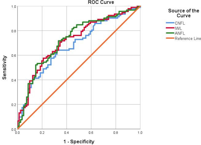
Results: Vibration perception threshold (p<0.0001), warm perception threshold (WPT) (p<0.001), sural nerve conduction velocity (SNCV) (p<0.001), corneal nerve fiber density (CNFD) (p<0.0001), corneal nerve branch density (CNBD) (p<0.0001), corneal nerve fiber length (CNFL) (p=0.002), inferior whorl length (IWL) (p=0.0001) and average nerve fiber length (ANFL) (p=0.0001) showed a progressive abnormality with increasing severity of diabetic neuropathy. Receiver operating characteristic curve analysis for the diagnosis of diabetic neuropathy showed comparable performance in relation to the area under the curve (AUC) but differing sensitivities and specificities for vibration perception threshold (AUC 0.79, sensitivity 55%, specificity 90%), WPT (AUC 0.67, sensitivity 50%, specificity 76%), cold perception threshold (AUC 0.64, sensitivity 80%, specificity 47%), SNCV (AUC 0.70, sensitivity 76%, specificity 54%), CNFD (AUC 0.71, sensitivity 58%, specificity 83%), CNBD (AUC 0.70, sensitivity 69%, specificity 65%), CNFL (AUC 0.68, sensitivity 64%, specificity 67%), IWL (AUC 0.72, sensitivity 70%, specificity 65%) and ANFL (AUC 0.72, sensitivity 71%, specificity 66%).
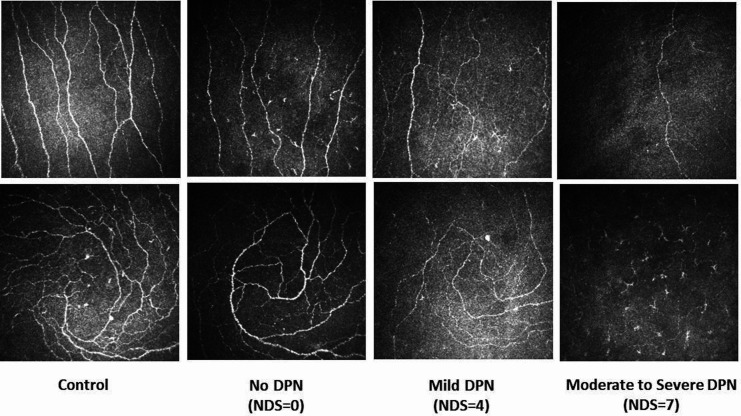
Conclusion: This study shows that CCM identifies early and progressive corneal nerve loss at the inferior whorl and central cornea and has comparable utility with quantitative sensory testing and nerve conduction in the diagnosis of diabetic neuropathy.
Keywords: cornea; diabetes complications; diabetic neuropathies; diagnosis.
© Author(s) (or their employer(s)) 2020. Re-use permitted under CC BY-NC. No commercial re-use. See rights and permissions. Published by BMJ.
Conflict of interest statement
Competing interests: None declared.
Figures