. 2021 May;11(5):1176-1191.
doi: 10.1158/2159-8290.CD-20-0790. Epub 2020 Dec 18.

DNA Polymerase and Mismatch Repair Exert Distinct Microsatellite Instability Signatures in Normal and Malignant Human Cells

Affiliations

DNA Polymerase and Mismatch Repair Exert Distinct Microsatellite Instability Signatures in Normal and Malignant Human Cells

Jiil Chung et al. Cancer Discov. 2021 May.

Abstract

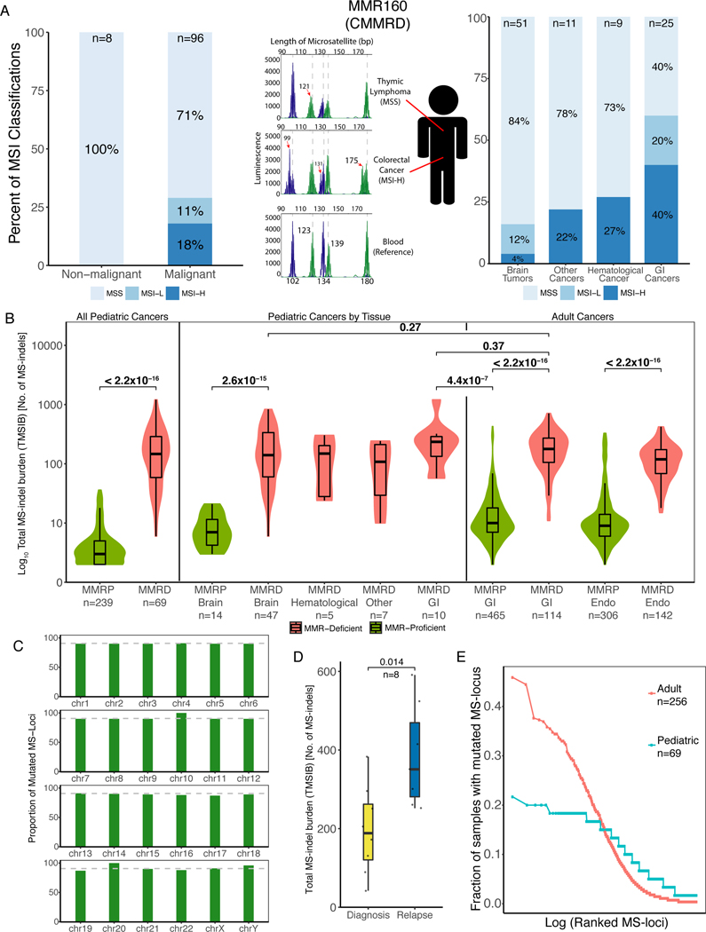
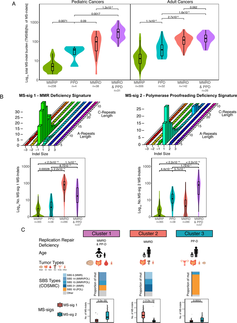
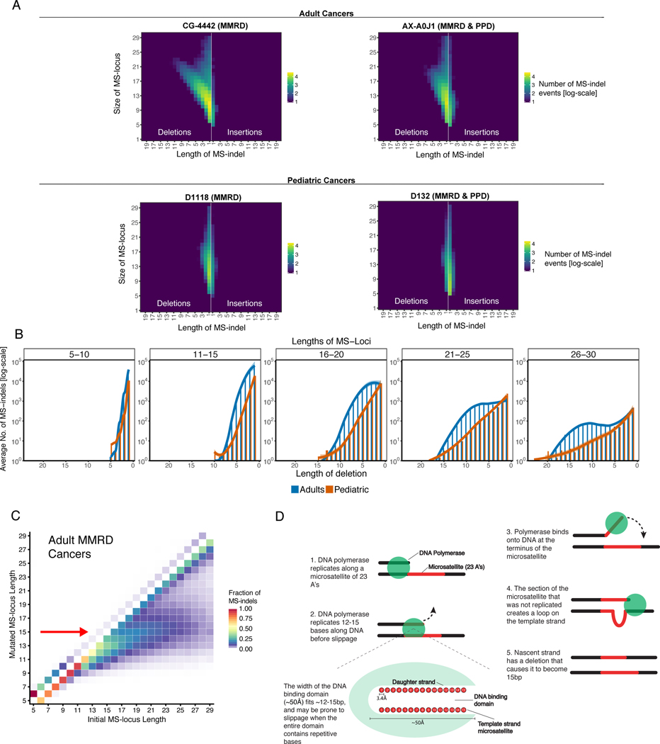
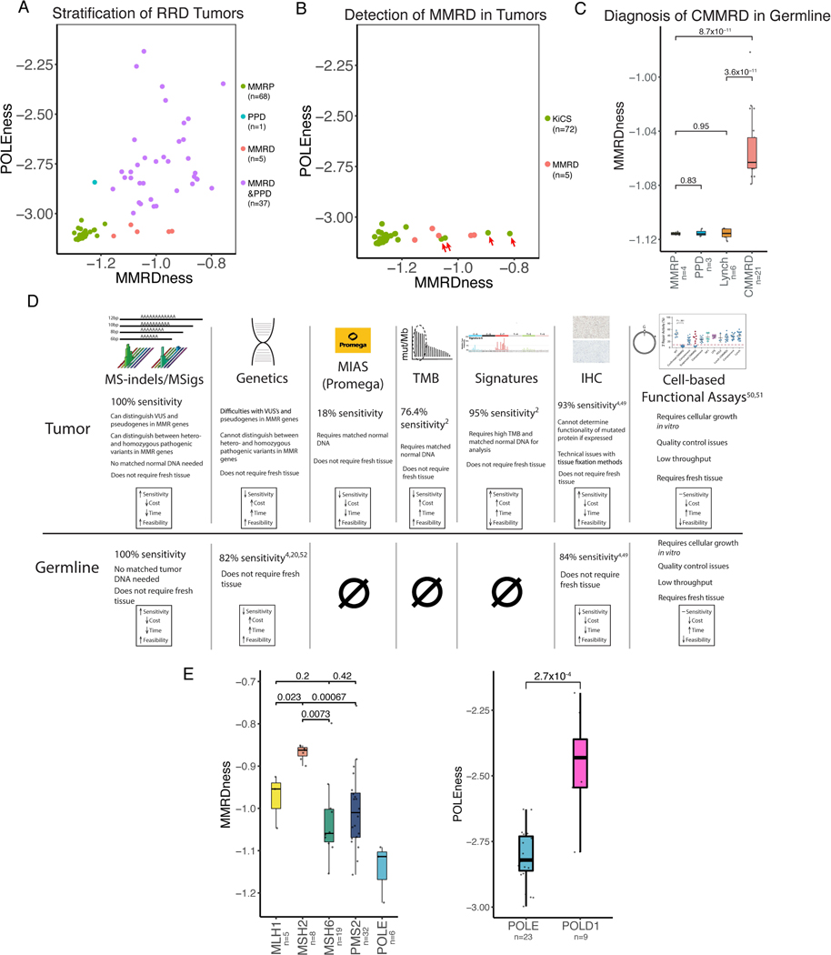
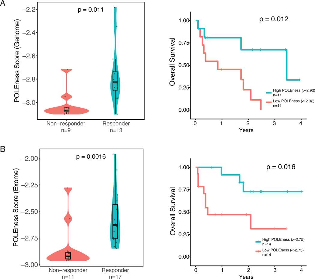
Although replication repair deficiency, either by mismatch repair deficiency (MMRD) and/or loss of DNA polymerase proofreading, can cause hypermutation in cancer, microsatellite instability (MSI) is considered a hallmark of MMRD alone. By genome-wide analysis of tumors with germline and somatic deficiencies in replication repair, we reveal a novel association between loss of polymerase proofreading and MSI, especially when both components are lost. Analysis of indels in microsatellites (MS-indels) identified five distinct signatures (MS-sigs). MMRD MS-sigs are dominated by multibase losses, whereas mutant-polymerase MS-sigs contain primarily single-base gains. MS deletions in MMRD tumors depend on the original size of the MS and converge to a preferred length, providing mechanistic insight. Finally, we demonstrate that MS-sigs can be a powerful clinical tool for managing individuals with germline MMRD and replication repair-deficient cancers, as they can detect the replication repair deficiency in normal cells and predict their response to immunotherapy. SIGNIFICANCE: Exome- and genome-wide MSI analysis reveals novel signatures that are uniquely attributed to mismatch repair and DNA polymerase. This provides new mechanistic insight into MS maintenance and can be applied clinically for diagnosis of replication repair deficiency and immunotherapy response prediction.This article is highlighted in the In This Issue feature, p. 995.


Conflict of interest statement

Conflicts of Interest:

G.G. is a founder, consultant and holds privately held equity in Scorpion Therapeutics. G.G. receives research funds from IBM and Pharmacyclics. G.G. is an inventor on patent applications related to MuTect, ABSOLUTE, MutSig and POLYSOLVER. Y.E.M and G.G. are inventors on patent applications related to MSMuTect and MSMutSig. G.G., Y.E.M., U.T., and J.C. are inventors on patent applications related to MSIdetect. Other authors declare no competing interests.

Figures

Figure 1.
Figure 1.. Microsatellites Accumulate MS-indels in germline MMRD Cancers
(A) Microsatellite Instability (MSI) status as measured by the Microsatellite Instability Analysis System (MIAS, Promega) of CMMRD samples. Left and right panels: The proportion of MSI high (MSI-H), MSI low (MSI-L) and microsatellite stable (MSS) samples in different tissue types with the number of samples tested (top). The middle section describes a single patient with constitutional mismatch repair deficiency (CMMRD), MMR160, who had a microsatellite stable (MSS) lymphoma and a microsatellite unstable (MSI-H) colorectal cancer. Histograms represent the results of the MIAS, with germline microsatellite lengths of the 5 tested loci (bottom plot) marked as dashed lines in all panels. GI = gastrointestinal. (B) Comparison of exome-wide total microsatellite indel (MS-indel) burden (TMSIB) between pediatric mismatch repair proficient (MMRP) and MMRD cancers, pediatric cancers by tissue type, and between pediatric and adult MMRD cancers (TMSIB in logarithmic scale). Median TMSIB for each group is written beside each box, and sample numbers are written below each group. Green represents MMRP, and red represents MMRD in children and adults. Statistical significance was calculated by Mann-Whitney U-test. Endo = Endometrial cancers. (C) Proportion of mutated MS-loci in each chromosome in the entire cohort. The average proportion (90.7%) is labelled and shown as a grey dotted line. The height of the bars in the indicate proportion, and similar heights indicate strong correlation of MS-indels to the number of MS-loci in that region of the chromosome. Supplementary Table S2 shows the similar proportions of MS-loci mutated in each chromosome. (D) Comparison of the number of MS-indels in diagnosis and relapsed pediatric RRD tumors (n=8). Statistical significance was calculated by the Paired-samples Wilcoxon test. (E) Analysis of recurrent indels in microsatellite loci (MS-loci), separated by adult MMRD cancers (red, n=256), and pediatric MMRD tumors (blue, n=69). Each point represents the fraction of tumors mutated for a given MS-locus (y-axis) sorted from most- to least-frequently altered (left to right on the x-axis).
Figure 2.
Figure 2.. MMRD and PPD tumors have distinct microsatellite indel signatures
(A) Comparison of TMSIB between RRD variants (MMRD, PPD, MMRD&PPD) to MMRP in pediatric brain and GI tumors (left) and adult endometrial tumors (right). Sample numbers are written below each group. P values calculated using Mann-Whitney U-test. (B) Graphical presentation of MS-sig1 (top left) and MS-sig2 (top right) based on indel size (x-axis) and microsatellite locus length, separated by A- or C- repeats (z-axis). The total number of MS-sig1 indels and MS-sig2 indels are compared between the RRD variants in boxplots (bottom left and bottom right). P values calculated using Mann-Whitney U-test. (C) Graphical representation of RRD subgroups(2) separated by age of tumor onset, most common tumor types, proportion of RRD-associated COSMIC SBS signatures, and MS-sigs 1 and 2. The size of each graphic corresponds to the proportion of the cancer observed clinically. MS-sigs correlate to the proportion of the COSMIC SBS signatures in each cluster. Statistical significance was calculated using the Mann-Whitney U-test.
Figure 3.
Figure 3.. Accumulation of large microsatellite deletions in MMRD cancers towards a preferred final length
(A) Examples of genomic MS-indel heatmaps of germline MMRD and MMRD & PPD cancers in adults (TCGA) and children. Pediatric cancers have an enrichment of single base MS-indels, while adult cancers accumulate insertions of one base and large (>3bp) deletions that increase in size (x-axis) with increasing locus length (y-axis). Each color represents the log10 count of MS-indel events. (B) The total number of MS-deletions in both pediatric and adults by locus length (each panel presents a range of 5 bases) and mutation size (x-axis). The number of deletions were averaged for each deletion length. (C) The fraction of MS-indels by initial (x-axis) and final/post-mutation (y-axis) lengths in adult tumors revealing the convergence of MS-loci to 15bp (indicated by red arrow). Fraction of indels was calculated by dividing the number of indels corresponding to each initial and mutated locus length by total number of MS-indels in each initial MS-locus length. (D) Schematic representation of the emergence of 15bp-long microsatellites as a targeted locus length in adult MMRD cancers. The polymerase is prone to slippage after replicating a tract of 15bp-repeats, resulting in a loop in the template strand upon rebinding. If unrepaired, the loop can result in a deletion that causes the nascent strand to become 15bp, irrespective of the original locus length.
Figure 4.
Figure 4.. Clinical applications of MS-sigs
(A) Two-dimensional plot separating all types of replication repair deficient (RRD) from wildtype (MMRP) cancers using whole genome sequencing. Green represents MMRP cancers, red represents mismatch repair deficient (MMRD) cancers, blue is polymerase proofreading deficient (PPD) cancers, and purple is combined MMRD and PPD cancers. Each dot represents an individual tumor. (B) Discovery of unexpected MMRD tumors using MS-sigs. Two-dimensional plot of presumed MMRP tumors from the Sickkids Cancer Sequencing (KiCS) program. KiCS samples shown in green, and MMRD tumors in red. Four tumors from the KiCS cohort that had increased MMRDness are highlighted by red arrows. These were later confirmed to be MMRD (Supplementary Table S6). (C) Detection of germline constitutional mismatch repair deficiency (CMMRD) using ultra low-passage WGS MMRDness scores. Blood samples from CMMRD and relevant control patients were used. Statistical significance was calculated using the Mann-Whitney U-test. (D) Comparison of MS-sigs to other clinically approved (CLIA) methods to diagnose MMRD in normal and malignant cells. Sensitivity was calculated by dividing the number of positive MMRD cases in each assay by the number of samples that were tested(,,,–65). (E) Genotype phenotype differences between different replication repair genes. Comparison of MMRDness between MMR gene mutations, and POLEness in POLEmut vs. POLD1mut cancers. P values were calculated using the Mann-Whitney U-test.
Figure 5.
Figure 5.. POLEness Score Predicts Response to Immunotherapy in RRD Cancers
(A) Genomic POLEness scores of 22 RRD cancers stratified by response to immune checkpoint inhibition. Overall survival of patients on immunotherapy separated by the median Genomic POLEness scores (median POLEness = −2.92, Methods). P values for comparing responders to non-responders were calculated using the Mann-Whitney U-test, and Kaplan-Meier curves were generated for survival with p values calculated using the Log Rank test. (B)Same as (A) using exomic POLEness scores from 28 RRD patients (median POLEness = −2.75, Methods).

Similar articles

Cited by

References

    1. Shlien A, Campbell BB, de Borja R, Alexandrov LB, Merico D, Wedge D, et al. Combined hereditary and somatic mutations of replication error repair genes result in rapid onset of ultra-hypermutated cancers. Nat Genet 2015;47(3):257–62 doi 10.1038/ng.3202. - DOI - PubMed
    1. Campbell BB, Light N, Fabrizio D, Zatzman M, Fuligni F, de Borja R, et al. Comprehensive Analysis of Hypermutation in Human Cancer. Cell 2017;171(5):1042–56 e10 doi 10.1016/j.cell.2017.09.048. - DOI - PMC - PubMed
    1. Kunkel TA, Erie DA. Eukaryotic Mismatch Repair in Relation to DNA Replication. Annu Rev Genet 2015;49:291–313 doi 10.1146/annurev-genet-112414-054722. - DOI - PMC - PubMed
    1. Tabori U, Hansford JR, Achatz MI, Kratz CP, Plon SE, Frebourg T, et al. Clinical Management and Tumor Surveillance Recommendations of Inherited Mismatch Repair Deficiency in Childhood. Clin Cancer Res 2017;23(11):e32–e7 doi 10.1158/1078-0432.Ccr-17-0574. - DOI - PubMed
    1. Kunkel TA. Evolving views of DNA replication (in)fidelity. Cold Spring Harbor symposia on quantitative biology 2009;74:91–101 doi 10.1101/sqb.2009.74.027. - DOI - PMC - PubMed

Publication types

Substances