Review
. 2020 Dec 21;13(12):dmm046920.
doi: 10.1242/dmm.046920.

Pulmonary neuroendocrine cells: physiology, tissue homeostasis and disease

Affiliations
Review

Pulmonary neuroendocrine cells: physiology, tissue homeostasis and disease

Masafumi Noguchi et al. Dis Model Mech. .

Abstract

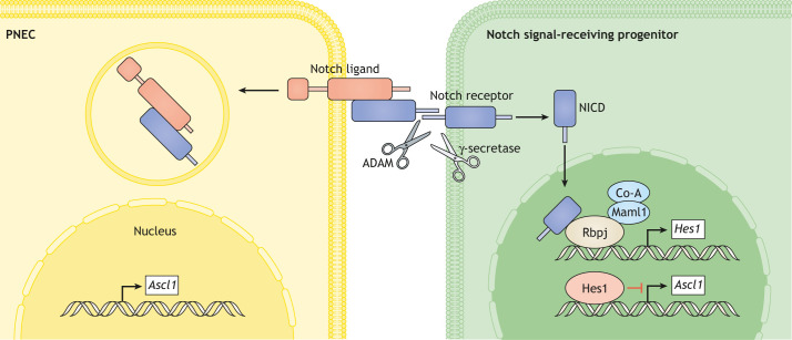
Mammalian lungs have the ability to recognize external environments by sensing different compounds in inhaled air. Pulmonary neuroendocrine cells (PNECs) are rare, multi-functional epithelial cells currently garnering attention as intrapulmonary sensors; PNECs can detect hypoxic conditions through chemoreception. Because PNEC overactivation has been reported in patients suffering from respiratory diseases - such as asthma, chronic obstructive pulmonary disease, bronchopulmonary dysplasia and other congenital diseases - an improved understanding of the fundamental characteristics of PNECs is becoming crucial in pulmonary biology and pathology. During the past decade, murine genetics and disease models revealed the involvement of PNECs in lung ventilation dynamics, mechanosensing and the type 2 immune responses. Single-cell RNA sequencing further unveiled heterogeneous gene expression profiles in the PNEC population and revealed that a small number of PNECs undergo reprogramming during regeneration. Aberrant large clusters of PNECs have been observed in neuroendocrine tumors, including small-cell lung cancer (SCLC). Modern innovation of imaging analyses has enabled the discovery of dynamic migratory behaviors of PNECs during airway development, perhaps relating to SCLC malignancy. This Review summarizes the findings from research on PNECs, along with novel knowledge about their function. In addition, it thoroughly addresses the relevant questions concerning the molecular pathology of pulmonary diseases and related therapeutic approaches.

Keywords: Development; Lung; Neuroendocrine; Regeneration; Respiratory diseases; Vagal nerves.


Conflict of interest statement

Competing interestsThe authors declare no competing or financial interests.

Figures

Fig. 1.
Fig. 1.
Schematic representation of pulmonary neuroendocrine cells (PNECs), neuroepithelial bodies (NEBs) and their innervation in the airway. In the mammalian lung, PNECs (yellow) localize at airway bifurcation sites (in the circled area and illustrated on the right), forming small clusters called NEBs. The NEB interacts with sensory nerve terminals, with myelinated afferent nerves (yellow and purple) branching and protruding into the NEB. The other sensory nerve (orange) comprises unmyelinated non-vagal immunoreactive nerve fibers originating from the dorsal root ganglia (DRG). Their axons enter the brain and transmit sensory information to the brainstem (green arrows). NEBs can sense CO2, air pressure, O2, H+ ions and nicotine, and activate reactions. ATP, adenosine triphosphate; CGRP, calcitonin gene-related peptide; GABA, gamma-aminobutyric acid; JG, jugular ganglion; NG, nodose ganglion; 5-HT, serotonin.
Fig. 2.
Fig. 2.
Notch-mediated cell–cell interaction. Notch is a type I transmembrane receptor that interacts with transmembrane ligands, such as Delta-like (Dll), on adjacent cells. Ligand binding leads to cleavage (by ADAM proteins and γ-secretase) and release of the Notch intracellular domain (NICD), which then moves to the nucleus to regulate transcriptional complexes containing the DNA-binding protein Rbpj. Hes1 is an Rbpj-dependent Notch target gene that encodes a transcription factor that suppresses the expression of Ascl1, a key determinant of PNEC fate. PNECs are a Dll-expressing cell type and their neighboring cells are often Notch active.
Fig. 3.
Fig. 3.
NEB development. Top left: branching airway. The circle indicates the area illustrated in the schematics below. Top right: developing airway of a fetal mouse at E14.5 [blue, 4′,6-diamidino-2-phenylindole (DAPI); green, laminin of basal membrane; magenta, Ret-expressing PNECs]. The development of an NEB is a stepwise process. Step 1: PNECs differentiate in the developing airway. Step 2: many PNECs appear while keeping distance between each other by Notch-mediated lateral inhibition (see Fig. 2). Step 3: PNECs detach from neighboring cells and migrate to the airway bifurcation site. Step 4: PNECs form clusters at the bifurcation site. Step 5: a primitive NEB microenvironment develops. Shown on the right are snapshots from time-lapse imaging of the developing murine airway (red, PNECs; blue, epithelial cells; arrows indicate migrating PNECs). These images are reproduced and modified from Noguchi et al. (2015) under the terms of the CC-BY 4.0 license.
Fig. 4.
Fig. 4.
The NEB microenvironment can foster two regeneration modes. NEB-mediated epithelial regeneration in the naphthalene injury-repair model occurs in two modes: (A) the variant club cells (vClub)/SSEA-1+, peri-pNEB, N1ICD+, CC10 cells (SPNC)/uroplakin-3a+ club cells (U-CC), which reside next to PNECs, act as transient-amplifying cells after injury; (B) the rare PNECs (PNECstem), which reserve stem cell potential, contribute to tissue regeneration after injury.
Fig. 5.
Fig. 5.
PNEC hyperplasia in lung diseases. (A) PNEC hyperplasia is an abnormal expansion of PNECs and is associated with several lung diseases. (B) Pulmonary emphysema occurs upon alveolar septal destruction in asthma, chronic obstructive pulmonary disease (COPD) and bronchopulmonary dysplasia (BPD). (C) Schematic representation of inadequate closure of the pleuroperitoneal membrane in congenital diaphragmatic hernia (CDH) patients (arrow). (D) Chest high-resolution computed tomography in neuroendocrine hyperplasia of infancy (NEHI); characteristic ground-glass opacities can be observed (arrows). This panel was reproduced and modified from Popler et al. (2010). This image is not published under the terms of the CC-BY license of this article. For permission to reuse, please see Popler et al. (2010).
Fig. 6.
Fig. 6.
Generation of a high-resolution 3D image of NEBs by two-photon microscopy. (A) The isolated fetal mouse cranial lung lobe is cleared with CUBIC, a hydrophilic tissue-clearing reagent. The cleared specimen is placed in a custom chamber and the whole lobe is imaged with a two-photon microscope using a ×25 objective lens. (B) Geometric computational analysis of the high-resolution 3D image reveals the stereotypic distribution of nodal NEBs (green/cyan). The 3D structure of the entire airway epithelium is visualized (blue). The central lines of the bronchial lumen structure (purple) are drawn using IMARIS filament tracing. The panel is reproduced and modified from Noguchi et al. (2015) under the terms of the CC-BY 4.0 license.

References

    1. Adriaensen D. and Timmermans J. P (2004). Purinergic signalling in the lung: important in asthma and COPD? Curr. Opin. Pharmacol. 4, 207-214. 10.1016/j.coph.2004.01.010 - DOI - PubMed
    1. Adriaensen D., Timmermans J. P., Brouns I., Berthoud H. R., Neuhuber W. L. and Scheuermann D. W (1998). Pulmonary intraepithelial vagal nodose afferent nerve terminals are confined to neuroepithelial bodies: an anterograde tracing and confocal microscopy study in adult rats. Cell Tissue Res. 293, 395-405. 10.1007/s004410051131 - DOI - PubMed
    1. Adriaensen D., Brouns I., Pintelon I., De Proost I. and Timmermans J. P (2006). Evidence for a role of neuroepithelial bodies as complex airway sensors: comparison with smooth muscle-associated airway receptors. J. Appl. Physiol. 101, 960-970. 10.1152/japplphysiol.00267.2006 - DOI - PubMed
    1. Agarwala S. and Tamplin O. J (2018). Neural crossroads in the hematopoietic stem cell niche. Trends Cell Biol., 28, 987-998. 10.1016/j.tcb.2018.05.003 - DOI - PMC - PubMed
    1. Aguayo S. M., Kane M. A., King T. E. J. R., Schwarz M. I., Grauer L. and Miller Y. E. (1989). Increased levels of bombesin-like peptides in the lower respiratory tract of asymptomatic cigarette smokers. J. Clin. Invest. 84, 1105-1113. 10.1172/JCI114273 - DOI - PMC - PubMed

Publication types