Review
. 2021 Jan;70(1):17-26.
doi: 10.2337/dbi19-0033.

Deciphering the Complex Communication Networks That Orchestrate Pancreatic Islet Function

Affiliations
Review

Deciphering the Complex Communication Networks That Orchestrate Pancreatic Islet Function

Jonathan Weitz et al. Diabetes. 2021 Jan.

Abstract

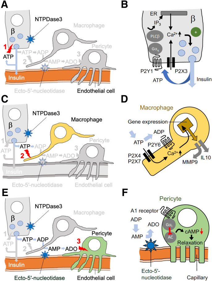
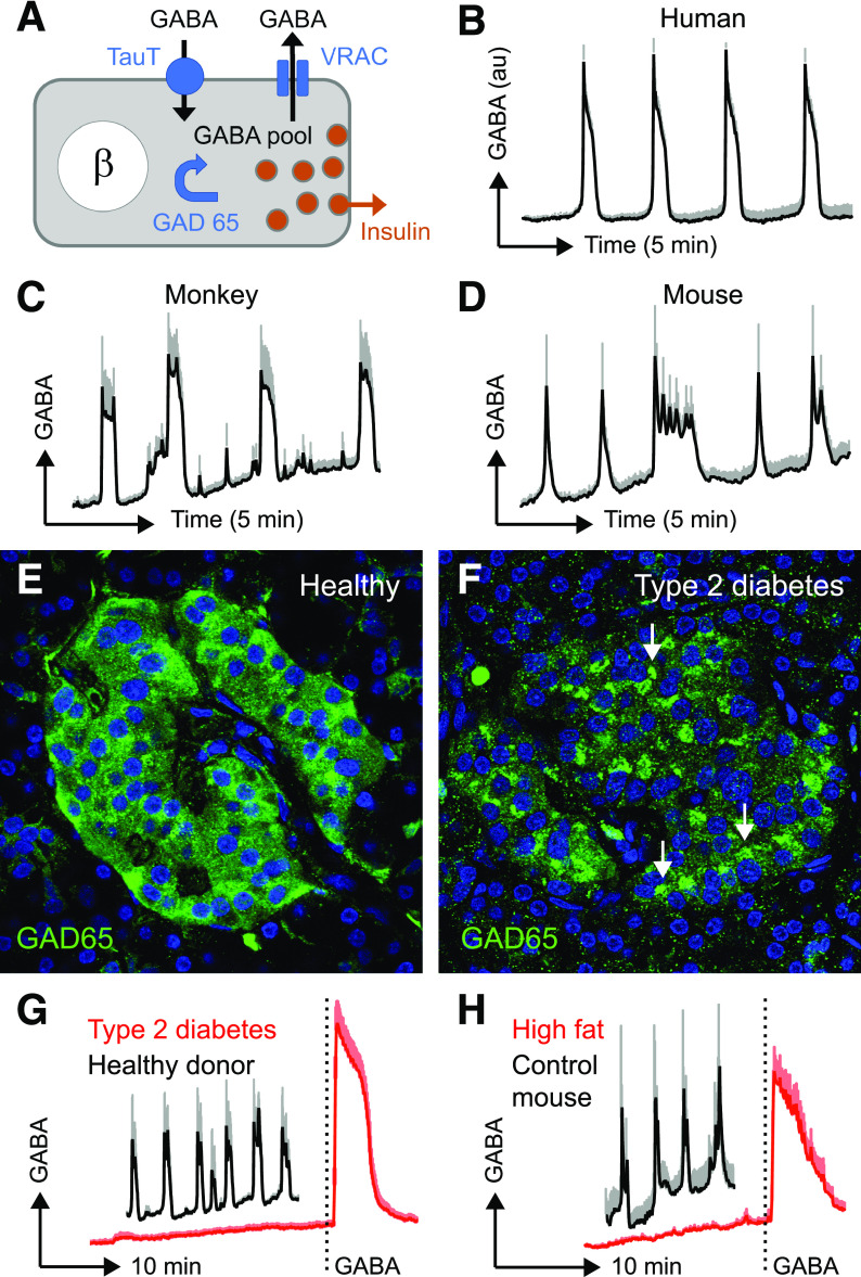
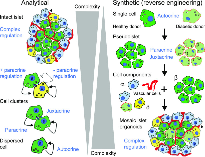
Pancreatic islets are clusters of hormone-secreting endocrine cells that rely on intricate cell-cell communication mechanisms for proper function. The importance of multicellular cooperation in islet cell physiology was first noted nearly 30 years ago in seminal studies showing that hormone secretion from endocrine cell types is diminished when these cells are dispersed. These studies showed that reestablishing cellular contacts in so-called pseudoislets caused endocrine cells to regain hormone secretory function. This not only demonstrated that cooperation between islet cells is highly synergistic but also gave birth to the field of pancreatic islet organoids. Here we review recent advances related to the mechanisms of islet cell cross talk. We first describe new developments that revise current notions about purinergic and GABA signaling in islets. Then we comment on novel multicellular imaging studies that are revealing emergent properties of islet communication networks. We finish by highlighting and discussing recent synthetic approaches that use islet organoids of varied cellular composition to interrogate intraislet signaling mechanisms. This reverse engineering of islets not only will shed light on the mechanisms of intraislet signaling and define communication networks but also may guide efforts aimed at restoring islet function and β-cell mass in diabetes.


Figures

Figure 1
Figure 1
The fate of extracellular ATP in the islet. A: ATP is released together with insulin from β-cells. As soon as it leaves the granule, ATP acts on purinergic receptors on β-cells to potentiate insulin secretion, thus establishing a positive-feedback loop (1). B: Transduction mechanisms and effects of ATP activation of β-cells. C: ATP further activates macrophages (2). ATP is hydrolyzed by NTPDase3, producing ADP (and AMP), which can also activate macrophages. D: Transduction mechanisms and effects of purinergic activation of resident macrophages. E: On its way through the interstitial space, AMP encounters ectonucleotidases on endothelial cells, which produces adenosine (ADO). Adenosine inhibits vascular pericytes (3), allowing capillaries to dilate. This increases local blood flow. F: Transduction mechanisms and effects of purinergic inhibition of islet pericytes.
Figure 2
Figure 2
β-cells secrete the paracrine signal GABA constitutively and in a pulsatile manner. A: Cartoon depicting the mechanisms of GABA synthesis, transport across the membrane, and efflux from β-cells. The GABA-synthetizing enzyme GAD65 increases the cytoplasmic pool of GABA. This GABA leaves the cell via the VRAC. GABA is recaptured by the taurine transporter TauT. BD: Detection of GABA by cytosolic Ca2+ flux in GABAB receptor–expressing biosensor cells shows that GABA secretion is periodic in human (B), monkey (Macaca fascicularis) (C), and mouse (D) islets. E and F: Confocal images of pancreatic sections from a donor without diabetes (E) and a donor with type 2 diabetes (F) show a redistribution of the enzyme during diabetes (arrows). G and H: GABA secretion measured as in BD is impaired in human type 2 diabetes (G) as well as in mice fed a high-fat diet (H). For the original study on the mechanisms of GABA secretion, see Menegaz et al. (24). All experiments were performed at 3 mmol/L glucose concentration. au, arbitrary units.
Figure 3
Figure 3
Illustration of analytical and synthetic strategies to decode complex cell-cell interactions in the pancreatic islet. Left: In analytical approaches the islet is broken down, either literally or using imaging tools, to dissect out individual signaling components (autocrine, paracrine, and juxtacrine). Right: In synthetic approaches, the islet is reconstructed from individual components. This reverse engineering reestablishes step-by-step the different signaling mechanisms. The advantages and limitations of these strategies are discussed in the text. Two light-green diabetic β-cells are included in the cell components in the pseudoislet below. The red squiggly line denotes vascular cells.

Similar articles

Cited by

References

    1. Halban PA, Wollheim CB, Blondel B, Meda P, Niesor EN, Mintz DH. The possible importance of contact between pancreatic islet cells for the control of insulin release. Endocrinology 1982;111:86–94 - PubMed
    1. Weir GC, Halban PA, Meda P, Wollheim CB, Orci L, Renold AE. Dispersed adult rat pancreatic islet cells in culture: A, B, and D cell function. Metabolism 1984;33:447–453 - PubMed
    1. Pipeleers D, in’t Veld PI, Maes E, Van De Winkel M. Glucose-induced insulin release depends on functional cooperation between islet cells. Proc Natl Acad Sci U S A 1982;79:7322–7325 - PMC - PubMed
    1. Hauge-Evans AC, Squires PE, Persaud SJ, Jones PM. Pancreatic beta-cell-to-beta-cell interactions are required for integrated responses to nutrient stimuli: enhanced Ca2+ and insulin secretory responses of MIN6 pseudoislets. Diabetes 1999;48:1402–1408 - PubMed
    1. Lammert E, Thorn P. The role of the islet niche on beta cell structure and function. J Mol Biol 2020;432:1407–1418 - PubMed

Publication types

MeSH terms