

Flexural wave-based soft attractor walls for trapping microparticles and cells
- PMID: 33355319
- PMCID: PMC7612665
- DOI: 10.1039/d0lc00865f
Flexural wave-based soft attractor walls for trapping microparticles and cells
Abstract
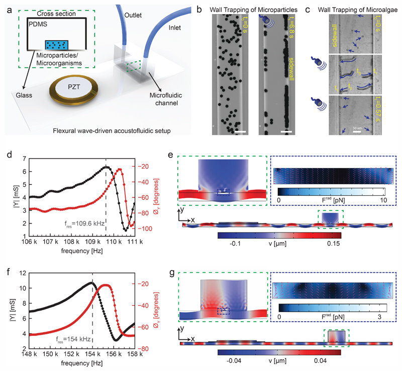
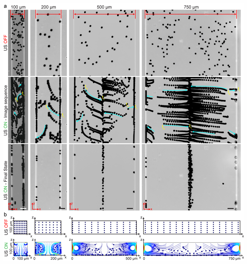
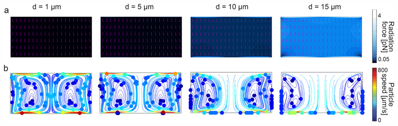
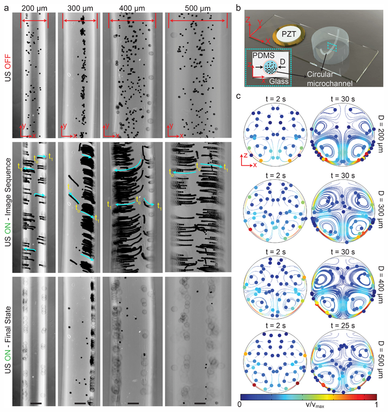
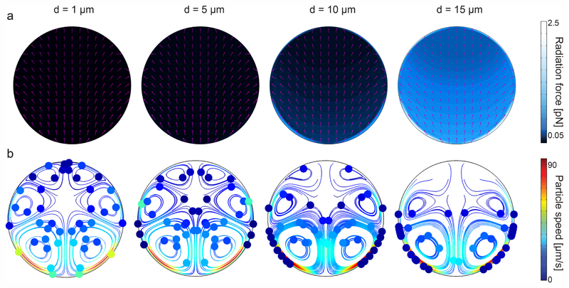
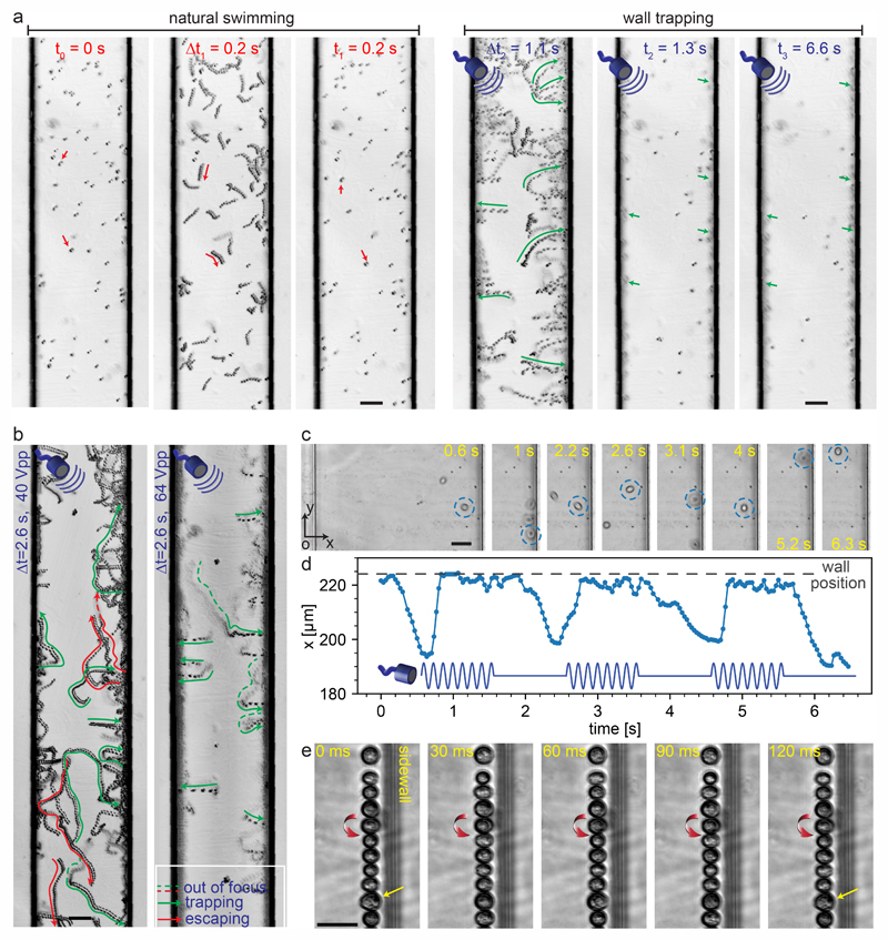
Acoustic manipulation of microparticles and cells, called acoustophoresis, inside microfluidic systems has significant potential in biomedical applications. In particular, using acoustic radiation force to push microscopic objects toward the wall surfaces has an important role in enhancing immunoassays, particle sensors, and recently microrobotics. In this paper, we report a flexural-wave based acoustofluidic system for trapping micron-sized particles and cells at the soft wall boundaries. By exciting a standard microscope glass slide (1 mm thick) at its resonance frequencies <200 kHz, we show the wall-trapping action in sub-millimeter-size rectangular and circular cross-sectional channels. For such low-frequency excitation, the acoustic wavelength can range from 10-150 times the microchannel width, enabling a wide design space for choosing the channel width and position on the substrate. Using the system-level acousto-structural simulations, we confirm the acoustophoretic motion of particles near the walls, which is governed by the competing acoustic radiation and streaming forces. Finally, we investigate the performance of the wall-trapping acoustofluidic setup in attracting the motile cells, such as Chlamydomonas reinhardtii microalgae, toward the soft boundaries. Furthermore, the rotation of microalgae at the sidewalls and trap-escape events under pulsed ultrasound are demonstrated. The flexural-wave driven acoustofluidic system described here provides a biocompatible, versatile, and label-free approach to attract particles and cells toward the soft walls.
Conflict of interest statement
The authors declare no conflict of interest.
Figures
Similar articles
-
Concentration of Microparticles Using Flexural Acoustic Wave in Sessile Droplets.Sensors (Basel). 2022 Feb 8;22(3):1269. doi: 10.3390/s22031269. Sensors (Basel). 2022. PMID: 35162014 Free PMC article.
-
Manipulation with sound and vibration: A review on the micromanipulation system based on sub-MHz acoustic waves.Ultrason Sonochem. 2023 Jun;96:106441. doi: 10.1016/j.ultsonch.2023.106441. Epub 2023 May 13. Ultrason Sonochem. 2023. PMID: 37216791 Free PMC article. Review.
-
Effect of microchannel protrusion on the bulk acoustic wave-induced acoustofluidics: numerical investigation.Biomed Microdevices. 2021 Dec 29;24(1):7. doi: 10.1007/s10544-021-00608-6. Biomed Microdevices. 2021. PMID: 34964071
-
Residue-free acoustofluidic manipulation of microparticles via removal of microchannel anechoic corner.Ultrason Sonochem. 2022 Sep;89:106161. doi: 10.1016/j.ultsonch.2022.106161. Epub 2022 Sep 6. Ultrason Sonochem. 2022. PMID: 36088893 Free PMC article.
-
Acoustofluidics 24: theory and experimental measurements of acoustic interaction force.Lab Chip. 2022 Sep 13;22(18):3290-3313. doi: 10.1039/d2lc00447j. Lab Chip. 2022. PMID: 35969199 Review.
Cited by
-
Acoustofluidic medium exchange for preparation of electrocompetent bacteria using channel wall trapping.Lab Chip. 2021 Nov 9;21(22):4487-4497. doi: 10.1039/d1lc00406a. Lab Chip. 2021. PMID: 34668506 Free PMC article.
-
Hemodynamics Challenges for the Navigation of Medical Microbots for the Treatment of CVDs.Materials (Basel). 2021 Dec 2;14(23):7402. doi: 10.3390/ma14237402. Materials (Basel). 2021. PMID: 34885556 Free PMC article. Review.
-
Concentration of Microparticles Using Flexural Acoustic Wave in Sessile Droplets.Sensors (Basel). 2022 Feb 8;22(3):1269. doi: 10.3390/s22031269. Sensors (Basel). 2022. PMID: 35162014 Free PMC article.
-
Application of Lab-on-Chip for Detection of Microbial Nucleic Acid in Food and Environment.Front Microbiol. 2021 Nov 4;12:765375. doi: 10.3389/fmicb.2021.765375. eCollection 2021. Front Microbiol. 2021. PMID: 34803990 Free PMC article. Review.
-
Manipulation with sound and vibration: A review on the micromanipulation system based on sub-MHz acoustic waves.Ultrason Sonochem. 2023 Jun;96:106441. doi: 10.1016/j.ultsonch.2023.106441. Epub 2023 May 13. Ultrason Sonochem. 2023. PMID: 37216791 Free PMC article. Review.
References
Publication types
MeSH terms
Grants and funding
LinkOut - more resources
Full Text Sources
Other Literature Sources