. 2021 Feb;23(2):152.
doi: 10.3892/mmr.2020.11791. Epub 2020 Dec 23.

PMCA4 gene expression is regulated by the androgen receptor in the mouse testis during spermatogenesis

Affiliations

PMCA4 gene expression is regulated by the androgen receptor in the mouse testis during spermatogenesis

Rui Sun et al. Mol Med Rep. 2021 Feb.

Abstract

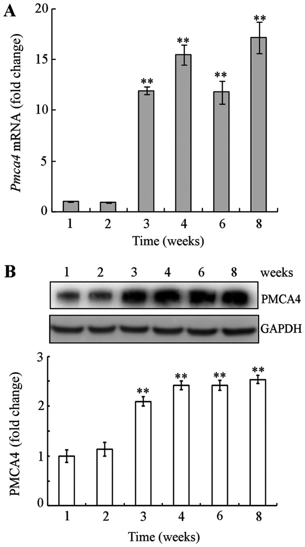
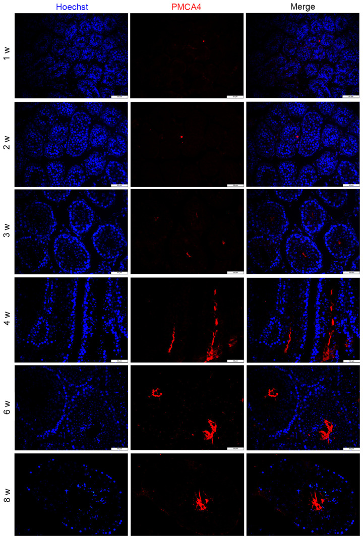
The present study aimed to investigate the expression of ATPase Ca++ transporting plasma membrane 4 (PMCA4) in mouse testis and to determine its role in spermatogenesis. Reverse transcription‑quantitative PCR, western blotting and immunofluorescence were performed to evaluate the expression levels of PMCA4 in mouse testes at various weeks postnatal in wild type mice, and in testes from Sertoli cell‑specific androgen receptor knockout and androgen receptor knockout (ARKO) mice. Luciferase assay, androgen receptor (AR) overexpression and AR antagonist experiments were used to confirm that AR regulated the expression of PMCA4. The results demonstrated that PMCA4 was highly expressed in mouse testes at 3‑8 weeks postnatal. PMCA4 expression levels in ARKO mouse testes were decreased compared with wild type. In addition, activation of AR by testosterone administration resulted in an increase in the activity of the PMCA4 promoter. Cells transfected with an AR‑overexpressing plasmid exhibited increased expression levels of the PMCA4 protein. Finally, the increase in PMCA4 protein levels induced by testosterone was prevented by pre‑treatment with the AR antagonist flutamide. The present results confirmed that PMCA4 was upregulated during mouse testis development and that PMCA4 mRNA and protein expression levels were regulated by androgens and AR. The present findings suggest that PMCA4 may be involved in the regulation of spermatogenesis.

Keywords: ATPase Ca++ transporting plasma membrane 4; spermatogenesis; testis; androgen receptor.


Figures

Figure 1.
Figure 1.
Expression of PMCA4 in different mouse tissues. Western blot analysis of PMCA4 protein expression in various tissues form adult C57BL/6 mice. GAPDH was used as in internal control. PMCA4, ATPase Ca++ transporting plasma membrane 4.
Figure 2.
Figure 2.
Temporal expression of PMCA4 during mouse testis postnatal development. (A) mRNA expression levels were evaluated at 1, 2, 3, 4, 6 and 8 weeks postnatal. mRNA levels were normalized to GAPDH and plotted relative to those of the 1-week-old mice. (B) Protein expression levels were detected by western blotting, with GAPDH used an internal control. Data were analyzed by one-way ANOVA followed by post hoc Bonferroni's test and are presented as mean ± standard error of the mean (n=4). **P<0.01 vs. 1 week. PMCA4, ATPase Ca++ transporting plasma membrane 4; w, week.
Figure 3.
Figure 3.
PMCA4 expression is decreased in testes from ARKO mice. (A) mRNA and (B) protein expression levels of PMCA4 were evaluated in WT, SCARKO and ARKO mouse testes. Data are presented as the mean ± standard error of the mean relative to the WT group (n=4). **P<0.01 vs. WT. PMCA4, ATPase Ca++ transporting plasma membrane 4; AR, androgen receptor; WT, wild type; SCARKO, Sertoli cell-specific AR knockout; ARKO, AR knockout.
Figure 4.
Figure 4.
Immunofluorescence staining for PMCA4 in mouse testis at different postnatal weeks. Representative microscopy images show PMCA4 staining (red) and nuclear staining (Hoechst 33342; blue) (n=4). Scale bar, 50 µm. PMCA4, ATPase Ca++ transporting plasma membrane 4; w, week.
Figure 5.
Figure 5.
Immunofluorescence staining for PMCA4 in mouse testis from wild type and ARKO mice, and in wild type mouse sperm. Representative microscopy images show PMCA4 staining (red) and nuclear staining (Hoechst 33342; blue) (n=4). Scale bar, 50 µm. PMCA4, ATPase Ca++ transporting plasma membrane 4; ARKO, AR knockout.
Figure 6.
Figure 6.
PMCA4 promoter activity is increased by testosterone treatment in TM4 cells in vitro. Dual-luciferase assays were used to detect the effects of testosterone on the PMCA4 promoter activity in TM4 cells. Cells were transfected with empty vector as a control. Results are plotted as fold-change of testosterone-treated relative to vehicle-treated. Data are presented as the mean ± standard error of the mean (n=3). *P<0.05 vs. PMCA4 vehicle control. PMCA4, ATPase Ca++ transporting plasma membrane 4; AR, androgen receptor.
Figure 7.
Figure 7.
PMCA4 expression is upregulated by androgens and AR in TM4 cells. (A) TM4 cells were transfected with either empty vector or pcDNA-AR, and subsequently the AR and PMCA4 protein expression levels were evaluated via western blotting. GAPDH was used as an internal control. Data are presented as the mean ± standard error of the mean (n=3). **P<0.01 vs. empty vector group. (B) Cells were pre-treated with flutamide for 6 h, prior to 10 nM testosterone administration for 24 h. AR and PMCA4 protein expression levels were then evaluated by western blotting. Testosterone increased PMCA4 and AR protein expression, and this increase was blocked by the AR antagonist flutamide. Data are presented as the mean ± standard error of the mean (n=3). The statistical significance of the differences between groups was determined by one-way ANOVA followed by all pairwise Holm-Sidak test. *P<0.05 and **P<0.01, with comparisons shown in brackets. PMCA4, ATPase Ca++ transporting plasma membrane 4; AR, androgen receptor; NC, negative control for Flu; C, control for T and ethanol; T, testosterone; Flu, flutamide.

Similar articles

Cited by

References

    1. Walker WH. Testosterone signaling and the regulation of spermatogenesis. Spermatogenesis. 2011;1:116–120. doi: 10.4161/spmg.1.2.16956. - DOI - PMC - PubMed
    1. Yong EL, Lim LS, Wang Q, Mifsud A, Lim J, Ong YC, Sim KS. Androgen receptor polymorphisms and mutations in male infertility. J Endocrinol Invest. 2000;23:573–577. doi: 10.1007/BF03343778. - DOI - PubMed
    1. Tahmasbpour E, Balasubramanian D, Agarwal A. A multi-faceted approach to understanding male infertility: Gene mutations, molecular defects and assisted reproductive techniques (ART) J Assist Reprod Genet. 2014;31:1115–1137. doi: 10.1007/s10815-014-0280-6. - DOI - PMC - PubMed
    1. O'Hara L, Smith LB. Androgen receptor roles in spermatogenesis and infertility. Best Pract Res Clin Endocrinol Metab. 2015;29:595–605. doi: 10.1016/j.beem.2015.04.006. - DOI - PubMed
    1. De Gendt K, Swinnen JV, Saunders PT, Schoonjans L, Dewerchin M, Devos A, Tan K, Atanassova N, Claessens F, Lécureuil C, et al. A Sertoli cell-selective knockout of the androgen receptor causes spermatogenic arrest in meiosis. Proc Natl Acad Sci USA. 2004;101:1327–1332. doi: 10.1073/pnas.0308114100. - DOI - PMC - PubMed