. 2021 Jan 13;13(1):277-286.
doi: 10.1021/acsami.0c20088. Epub 2020 Dec 23.

Hyaluronan-Conjugated Carbon Quantum Dots for Bioimaging Use

Affiliations

Hyaluronan-Conjugated Carbon Quantum Dots for Bioimaging Use

Bedia Begüm Karakoçak et al. ACS Appl Mater Interfaces. .

Abstract

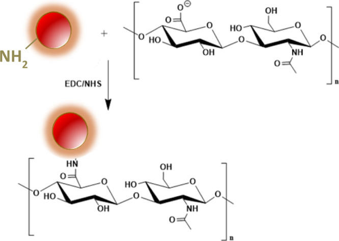
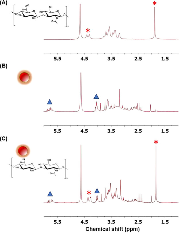
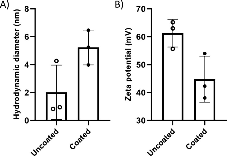
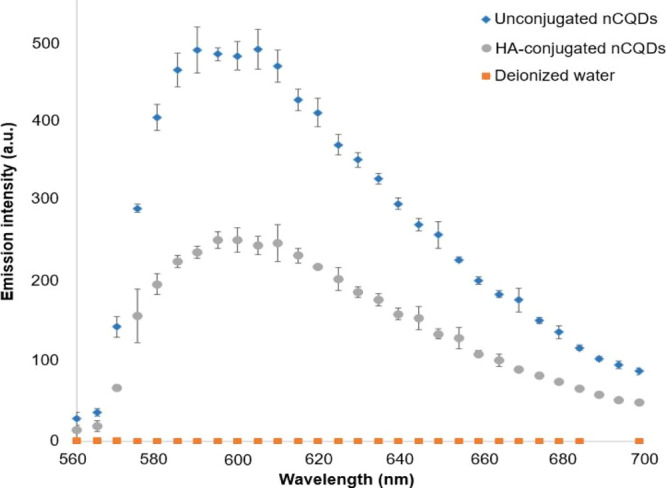
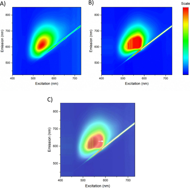
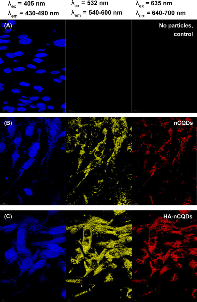
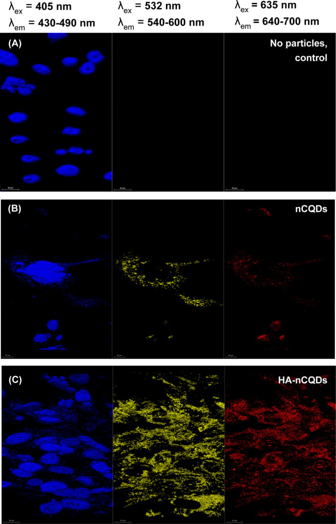
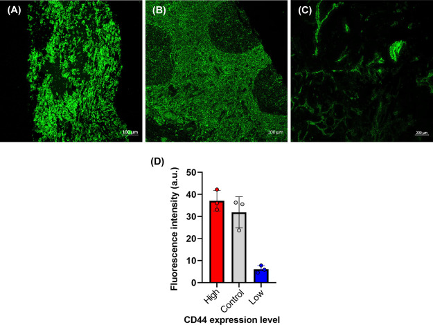
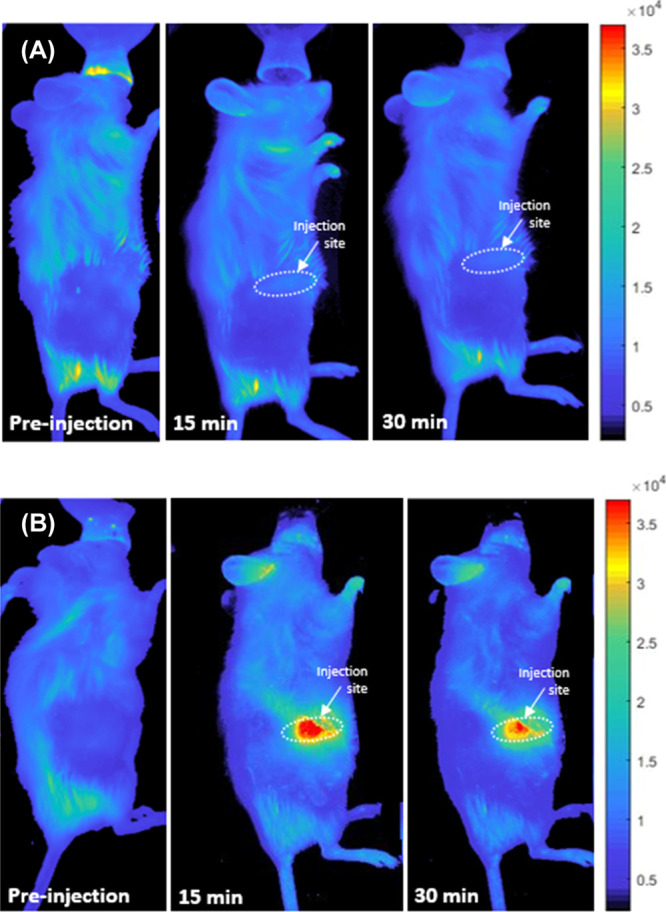
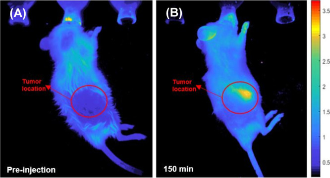
This work demonstrates the application of hyaluronan-conjugated nitrogen-doped carbon quantum dots (HA-nCQDs) for bioimaging of tumor cells and illustrates their potential use as carriers in targeted drug delivery. Quantum dots are challenging to deliver with specificity, which hinders their application. To facilitate targeted internalization by cancer cells, hyaluronic acid, a natural ligand of CD44 receptors, was covalently grafted on nCQDs. The HA-nCQD conjugate was synthesized by carbodiimide coupling of the amine moieties on nCQDs and the carboxylic acids on HA chains. Conjugated HA-nCQD retained sufficient fluorescence, although with 30% lower quantum efficiency than the original nCQDs. Confocal microscopy showed enhanced internalization of HA-nCQDs, facilitated by CD44 receptors. To demonstrate the specificity of HA-nCQDs toward human tumor cells, patient-derived breast cancer tissue with high-CD44 expression was implanted in adult mice. The tumors were allowed to grow up to 200-250 mm3 prior to the injection of HA-nCQDs. With either local or systemic injection, we achieved a high level of tumor specificity judged by a strong signal-to-noise ratio between the tumor and the surrounding tissue in vivo. Overall, the results show that HA-nCQDs can be used for imaging of CD44-specific tumors in preclinical models of human cancer and potentially used as carriers for targeted drug delivery into CD44-rich cells.

Keywords: CD44 receptors; bioimaging; breast cancer; cancer; carbon quantum dots; hyaluronic acid; tumor imaging.


Conflict of interest statement

The authors declare no competing financial interest.

Figures

Figure 1
Figure 1
Reaction scheme of conjugating nCQD with hyaluronic acid.
Figure 2
Figure 2
1H NMR spectra of (A) HA, (B) nCQDs, and (C) HA-nCQD conjugate confirming the presence of HA and nCQD peaks in the HA-nCQD conjugate.
Figure 3
Figure 3
DLS analysis of nCQDs consisting of (A) hydrodynamic particle diameter and (B) zeta potential measurements. With HA coating, although the zeta potential of the nCQDs has decreased, 44.8 mV is still considered a high enough potential for particle stability. No aggregates or precipitates were observed in the solution after HA conjugation to nCQDs.
Figure 4
Figure 4
PL emission spectra of unconjugated nCQDs with citric acid to aliphatic diamines ratio of 2.0, and their HA-nCQD conjugates. λexcitation = 540 nm.
Figure 5
Figure 5
3D fluorescence excitation-emission spectroscopy of (A) nCQDs, from our previous study, and (B) HA-nCQDs. (C) Overlay of (A,B), indicating a redshift (marked with white arrow) in the emission characteristics of the HA-nCQDs. Although the excitability and the range for emission characteristics did not significantly differ between the two CQDs, the broader nonhomogenous shape of the emission peak in HA-nCQDs indicates heterogeneity among the emitters.
Figure 6
Figure 6
Confocal microscopy images of ARPE-19 cells exposed to 0.6 mg/mL nCQDs, with and without HA conjugation. The cell nuclei were stained with DAPI. The scale bars are 10 μm.
Figure 7
Figure 7
Confocal microscopy images of CHO cells exposed to 0.6 mg/mL nCQDs, with and without HA conjugation. The cell nuclei were stained with DAPI. The scale bars are 10 μm.
Figure 8
Figure 8
Tissue screening for CD44 expression. (A) High-CD44 expressing TMA sample. (B) Tonsil (Control for CD44 expression). (C) Low-CD44-expressing TMA sample. (D) Fluorescence intensity comparison of the CD44 expression of all three tissues. Scale bars in confocal images are 100 μm for (A) and (B) and 200 μm for (C).
Figure 9
Figure 9
In vivo fluorescent images of mice injected with WHIM4 tumor cells after the tumor has grown up to 226.8 mm3 in volume. The images were obtained before and after subcutaneous injection of (A) nCQDs and (B) HA-nCQDs.
Figure 10
Figure 10
In vivo fluorescent images of mice bearing patient-derived WHIM4 tumor cells (up to 226.8 mm3 in volume) after intravenous injections of HA-nCQDs. The HA-nCQDs were detectable at the tumor site at 150 min postinjection.

Similar articles

Cited by

References

    1. Cherukula K.; Lekshmi K. M.; Uthaman S.; Cho K.; Cho C.-S.; Park I.-K. Multifunctional Inorganic Nanoparticles: Recent Progress in Thermal Therapy and Imaging. Nanomaterials 2016, 6, 76.10.3390/nano6040076. - DOI - PMC - PubMed
    1. Cattaneo A. G.; Gornati R.; Sabbioni E.; Chiriva-Internati M.; Cobos E.; Jenkins M. R.; Bernardini G. Nanotechnology and human health: risks and benefits. J. Appl. Toxicol. 2010, 30, 730–744. 10.1002/jat.1609. - DOI - PubMed
    1. Gutiérrez Millán C.; Colino Gandarillas C. I.; Sayalero Marinero M. L.; Lanao J. M. Cell-based drug-delivery platforms. Ther. Deliv. 2012, 3, 25–41. 10.4155/tde.11.141. - DOI - PubMed
    1. Hola K.; Zhang Y.; Wang Y.; Giannelis E. P.; Zboril R.; Rogach A. L. Carbon dots-Emerging light emitters for bioimaging, cancer therapy and optoelectronics. Nano Today 2014, 9, 590–603. 10.1016/j.nantod.2014.09.004. - DOI
    1. Nair B. G.; Varghese S. H.; Nair R.; Yoshida Y.; Maekawa T.; Kumar D. S. Nanotechnology platforms; an innovative approach to brain tumor therapy. Med. Chem. 2011, 7, 488–503. 10.2174/157340611796799113. - DOI - PubMed

MeSH terms