. 2021 Jan 26;93(3):1369-1376.
doi: 10.1021/acs.analchem.0c02966. Epub 2020 Dec 23.

High-Temporal-Resolution smFISH Method for Gene Expression Studies in Caenorhabditis elegans Embryos

Affiliations

High-Temporal-Resolution smFISH Method for Gene Expression Studies in Caenorhabditis elegans Embryos

Seleipiri Charles et al. Anal Chem. .

Abstract

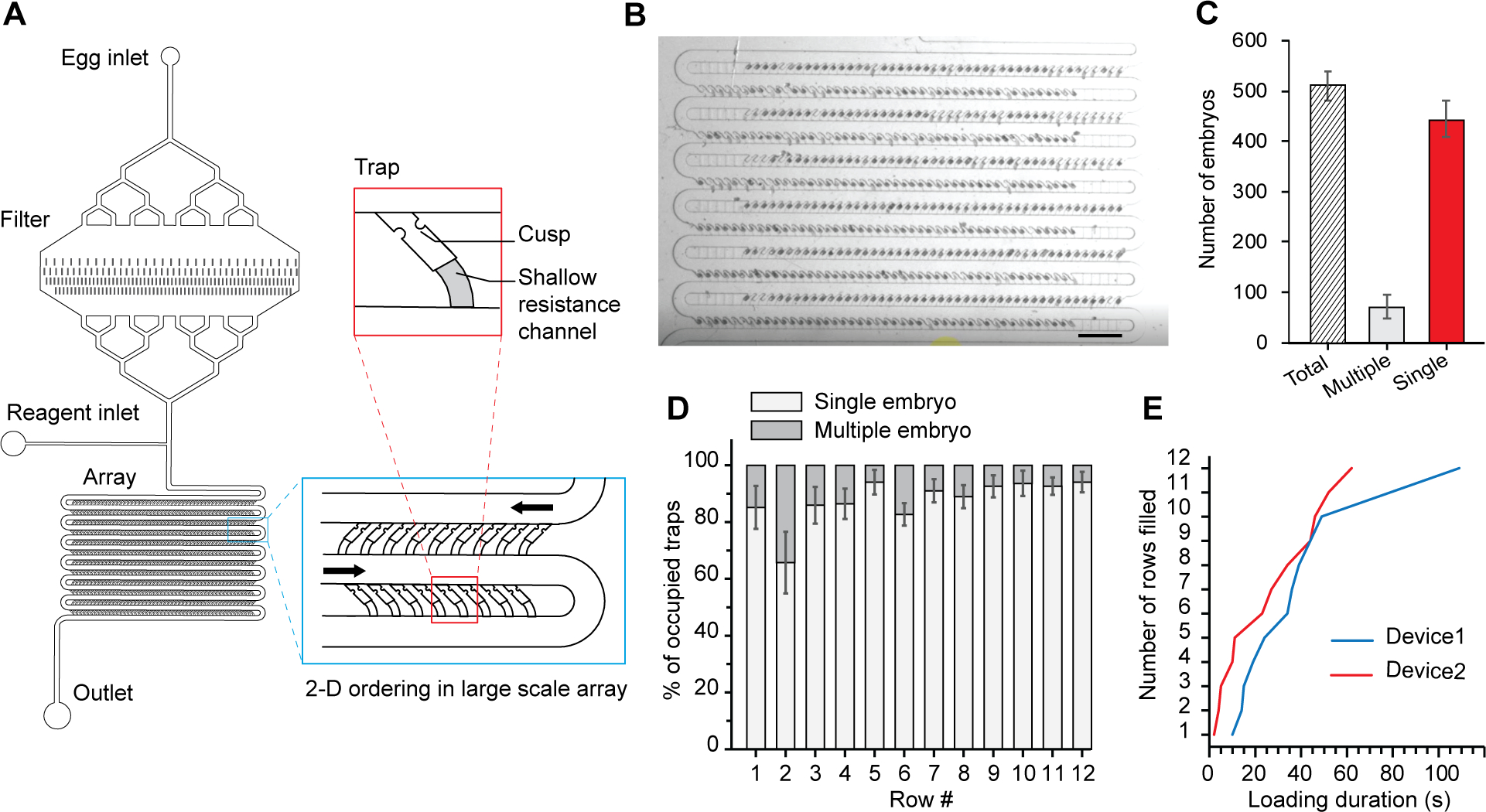
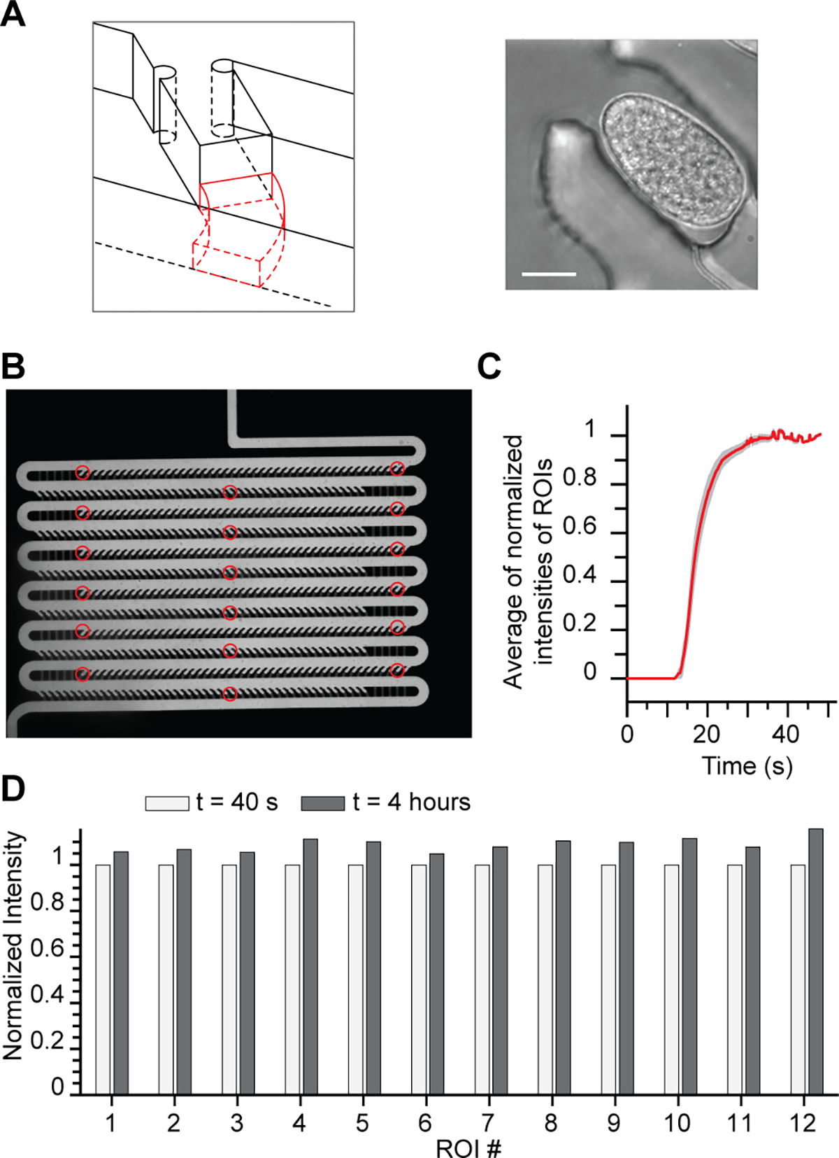
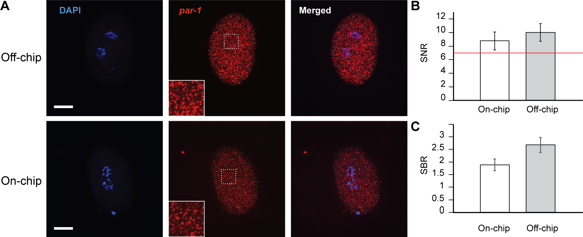
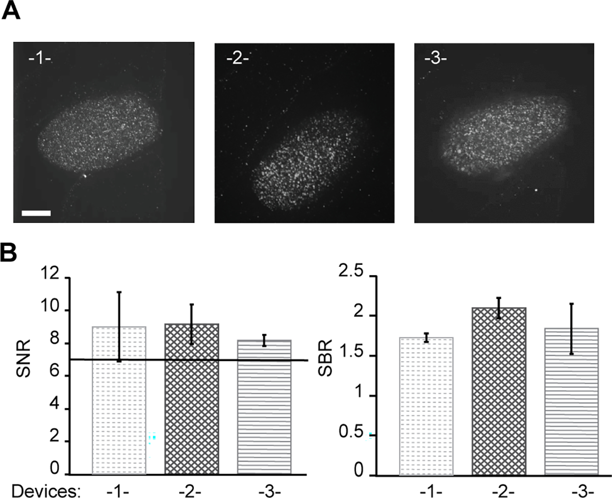
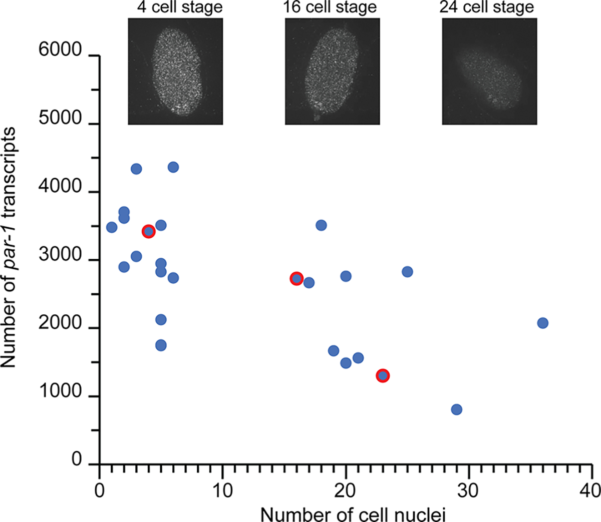
Recent development in fluorescence-based molecular tools has contributed significantly to developmental studies, including embryogenesis. Many of these tools rely on multiple steps of sample manipulation, so obtaining large sample sizes presents a major challenge as it can be labor-intensive and time-consuming. However, large sample sizes are required to uncover critical aspects of embryogenesis, for example, subtle phenotypic differences or gene expression dynamics. This problem is particularly relevant for single-molecule fluorescence in situ hybridization (smFISH) studies in Caenorhabditis elegans embryogenesis. Microfluidics can help address this issue by allowing a large number of samples and parallelization of experiments. However, performing efficient reagent exchange on chip for large numbers of embryos remains a bottleneck. Here, we present a microfluidic pipeline for large-scale smFISH imaging of C. elegans embryos with minimized labor. We designed embryo traps and engineered a protocol allowing for efficient chemical exchange for hundreds of C. elegans embryos simultaneously. Furthermore, the device design and small footprint optimize imaging throughput by facilitating spatial registration and enabling minimal user input. We conducted the smFISH protocol on chip and demonstrated that image quality is preserved. With one device replacing the equivalent of 10 glass slides of embryos mounted manually, our microfluidic approach greatly increases throughput. Finally, to highlight the capability of our platform to perform longitudinal studies with high temporal resolution, we conducted a temporal analysis of par-1 gene expression in early C. elegans embryos. The method demonstrated here paves the way for systematic high-temporal-resolution studies that will benefit large-scale RNAi and drug screens and in systems beyond C. elegans embryos.


Conflict of interest statement

The authors declare no conflict of interest.

Figures

Figure 1.
Figure 1.
Pipeline for studying gene expression during embryogenesis with high temporal resolution. (A) Timeline of early stage development of C. elegans embryos. (B) Schematic highlighting advantages of our integrated microfluidic pipeline vs traditional off-chip method for smFISH imaging: parallelization of experiments and automated imaging.
Figure 2.
Figure 2.
Microfluidic device design and loading characterization. (A) Schematic illustrating the device design and features incorporated to enable robust and reproducible hydrodynamic loading of hundreds of eggs (total trap number: 600) with Insert detailing the relevant geometrical dimensions; main channel (100 μm), trap size (35 μm) and resistance channel (12 μm). (B) Image of microfluidic array showing single embryos loaded in device in AP direction. (C) Quantification of loading occupancy in device. (D) Homogeneity of egg loading throughout the array (c-d: n=4 devices) Error bars represent SD. (E) Loading duration across 2 different devices.
Figure 3.
Figure 3.
Device features enable efficient reagent exchange necessary for executing smFISH protocol. (A) Schematic of resistance channel geometry designed to maintain embryo positioning during smFISH protocol (left). Representative image of embryo trapped using the resistance channel geometry (right, scalebar: 15 μm). (B) Image of device filled with FITC-dextran. ROIs analyzed are indicated by the red circles. (C) Plot describing the transition regimes for different ROI throughout the array: delivery reaches rapidly permanent regime after 30 seconds. Device was filled with wash buffer and exchanged with FITC-dextran in hybridization buffer to mimic the hybridization step of the smFISH experiment. (D) Bar graph of average fluorescence intensity of the different ROIs in the device at the beginning (40 s) and end (4 hr) of the mock hybridization experiment.
Figure 4.
Figure 4.
Comparison of on-chip smFISH protocol with traditional off-chip method. (A) Representative images of eggs stained off-chip (top) and on-chip (bottom) with the smFISH protocol. Blue indicates DAPI and identifies nuclei by staining chromosomes. Red puncta indicate individual par-1 molecules with zoom-in views in inserts. Scale bars are 15 μm. (B-C) Quantification of image quality between off-chip and on-chip experiments using SNR and SBR of the puncta for each condition. The red line indicates the minimum acceptable SNR for smFISH quantification (SNR = 7). Error bars represent SD, n = 20 embryos.
Figure 5.
Figure 5.
Integration of multiple arrays on single substrate for high-throughput confocal imaging. (A) Representative smFISH images of embryos from each device. Red puncta indicate individual par-1 molecules. Scale bar is 15 μm. (B) Quantification of image quality using SNR and SBR of puncta in each separate device. Error bars represent SD, n = 20 embryos.
Figure 6.
Figure 6.
Time-resolved analysis of par-1 gene expression changes during early embryogenesis showing the transcript count versus number of cell nuclei (n = 25 embryos). Inserts show representative smFISH images of embryos at different developmental stages corresponding to the red-circled data points.

Similar articles

Cited by

References

    1. Wood WB The Nematode Caenorhabditis Elegans; Cold Spring Harbor Laboratory: New York, 1988.
    1. Bryson-Richardson R; Berger S; Currie P Atlas of Zebrafish Development; Academic Press: San Diego, 2012.
    1. Bate M; Martinez Arias A The Development of Drosophila melanogaster; Cold Spring Harbor Laboratory: New York, 1993.
    1. Raj A; Rifkin SA; Andersen E; van Oudenaarden A Variability in gene expression underlies incomplete penetrance. Nature 2010, 463, 913–918. - PMC - PubMed
    1. Kistler KE; Trcek T; Hurd TR; Chen R; Liang F-X; Sall J; Kato M; Lehmann R Phase transitioned nuclear Oskar promotes cell division of Drosophila primordial germ cells. Elife 2018, 7, e37949. - PMC - PubMed

Publication types

MeSH terms