

Characterization of Retinal Microvascular and Choroidal Structural Changes in Parkinson Disease
- PMID: 33355613
- PMCID: PMC7758829
- DOI: 10.1001/jamaophthalmol.2020.5730
Characterization of Retinal Microvascular and Choroidal Structural Changes in Parkinson Disease
Erratum in
-
Error in Unit of Measure for Total Choroidal Area and Luminal Area.JAMA Ophthalmol. 2021 Feb 1;139(2):256. doi: 10.1001/jamaophthalmol.2020.6937. JAMA Ophthalmol. 2021. PMID: 33595605 Free PMC article. No abstract available.
Abstract
Importance: Noninvasive retinal imaging may detect structural changes associated with Parkinson disease (PD) and may represent a novel biomarker for disease detection.
Objective: To characterize alterations in the structure and microvasculature of the retina and choroid in eyes of individuals with PD and compare them with eyes of age- and sex-matched cognitively healthy control individuals using optical coherence tomography (OCT) and OCT angiography (OCTA).
Design, setting, and participants: This cross-sectional study was conducted at the Duke Neurological Disorders Clinic in Durham, North Carolina. Individuals aged 50 years or older with a diagnosis of PD were eligible for inclusion and underwent an evaluation and diagnosis confirmation before enrollment. Control individuals aged 50 years or older and without subjective cognitive dysfunction, a history of tremor, or evidence of motor dysfunction consistent with parkinsonism were solicited from the clinic or the Duke Alzheimer's Disease Prevention Registry. Individuals with diabetes, glaucoma, retinal pathology, other dementias, and corrected Early Treatment Diabetic Retinopathy Study (ETDRS) visual acuity worse than 20/40 Snellen were excluded. Data were analyzed between January 1, 2020, and March 30, 2020.
Exposures: All participants underwent OCT and OCTA imaging.
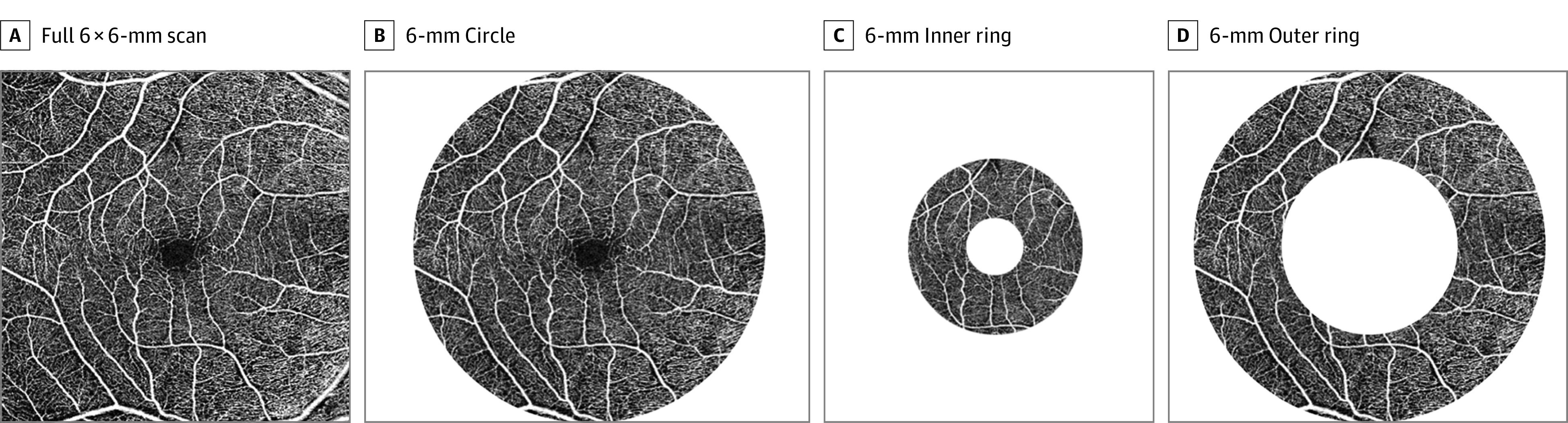
Main outcomes and measures: Generalized estimating equation analysis was used to characterize the association between imaging parameters and PD diagnosis. Superficial capillary plexus vessel density (VD) and perfusion density (PFD) were assessed within the ETDRS 6 × 6-mm circle, 6 × 6-mm inner ring, and 6 × 6-mm outer ring, as was the foveal avascular zone area. Peripapillary retinal nerve fiber layer thickness, macular ganglion cell-inner plexiform layer thickness, central subfield thickness, subfoveal choroidal thickness, total choroidal area, luminal area, and choroidal vascularity index (CVI) were measured.
Results: A total of 124 eyes of 69 participants with PD (39 men [56.5%]; mean [SD] age, 71.7 [7.0] years) and 248 eyes of 137 control participants (77 men [56.2%]; mean [SD] age, 70.9 [6.7] years) were analyzed. In the 6 × 6-mm ETDRS circle, VD (β coefficient = 0.37; 95% CI, 0.04-0.71; P = .03) and PFD (β coefficient = 0.009; 95% CI, 0.0003-0.018; P = .04) were lower in eyes of participants with PD. In the inner ring of the 6 × 6-mm ETDRS circle, VD (β coefficient = 0.61; 95% CI, 0.20-1.02; P = .003) and PFD (β coefficient = 0.015; 95% CI, 0.005-0.026; P = .004) were lower in eyes of participants with PD. Total choroidal area (β coefficient = -1.74 units2; 95% CI, -3.12 to -0.37 units2; P = .01) and luminal area (β coefficient = -1.02 units2; 95% CI, -1.86 to -0.18 units2; P = .02) were greater, but CVI was lower (β coefficient = 0.5%; 95% CI, 0.2%-0.8%; P < .001) in eyes of individuals with PD.
Conclusions and relevance: This study found that individuals with PD had decreased retinal VD and PFD as well as choroidal structural changes compared with age- and sex-matched control participants. Given the observed population differences in these noninvasive retinal biomarkers, further research into their clinical utility in PD is needed.
Conflict of interest statement
Figures
Comment in
-
Seeing Parkinson Disease in the Retina.JAMA Ophthalmol. 2021 Feb 1;139(2):189-190. doi: 10.1001/jamaophthalmol.2020.5719. JAMA Ophthalmol. 2021. PMID: 33355611 No abstract available.
-
Retinal Microvascular and Choroidal Changes in Parkinson Disease.JAMA Ophthalmol. 2021 Aug 1;139(8):921-922. doi: 10.1001/jamaophthalmol.2021.1728. JAMA Ophthalmol. 2021. PMID: 34110357 No abstract available.
-
Retinal Microvascular and Choroidal Changes in Parkinson Disease-Reply.JAMA Ophthalmol. 2021 Aug 1;139(8):922. doi: 10.1001/jamaophthalmol.2021.1731. JAMA Ophthalmol. 2021. PMID: 34110377 No abstract available.
References
MeSH terms
LinkOut - more resources
Full Text Sources
Other Literature Sources
Medical
