. 2021 Feb 25;29(24):3882-3891.
doi: 10.1093/hmg/ddaa276.

SPEG binds with desmin and its deficiency causes defects in triad and focal adhesion proteins

Affiliations

SPEG binds with desmin and its deficiency causes defects in triad and focal adhesion proteins

Shiyu Luo et al. Hum Mol Genet. .

Abstract

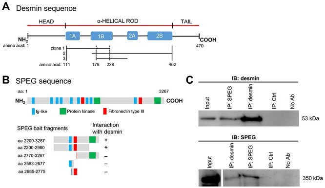
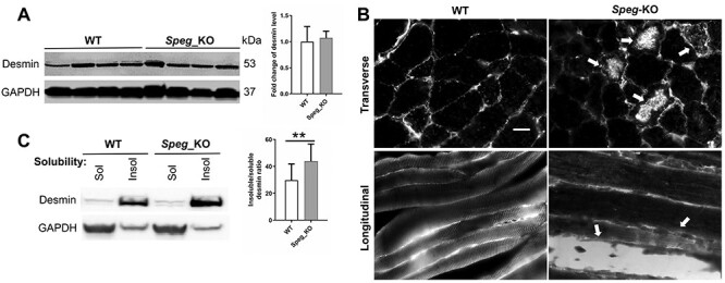
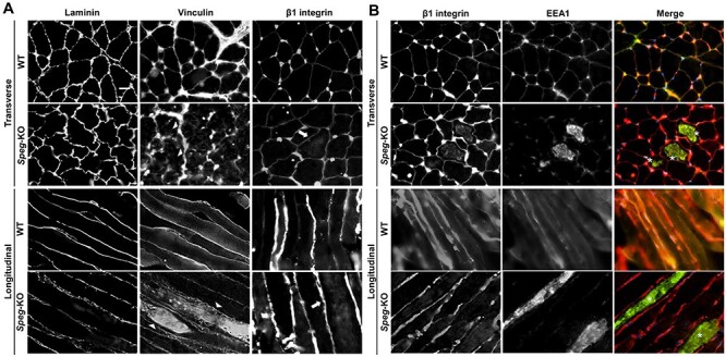
Striated preferentially expressed gene (SPEG), a member of the myosin light chain kinase family, is localized at the level of triad surrounding myofibrils in skeletal muscles. In humans, SPEG mutations are associated with centronuclear myopathy and cardiomyopathy. Using a striated muscle-specific Speg-knockout (KO) mouse model, we have previously shown that SPEG is critical for triad maintenance and calcium handling. Here, we further examined the molecular function of SPEG and characterized the effects of SPEG deficiency on triad and focal adhesion proteins. We used yeast two-hybrid assay, and identified desmin, an intermediate filament protein, to interact with SPEG and confirmed this interaction by co-immunoprecipitation. Using domain-mapping assay, we defined that Ig-like and fibronectin III domains of SPEG interact with rod domain of desmin. In skeletal muscles, SPEG depletion leads to desmin aggregates in vivo and a shift in desmin equilibrium from soluble to insoluble fraction. We also profiled the expression and localization of triadic proteins in Speg-KO mice using western blot and immunofluorescence. The amount of RyR1 and triadin were markedly reduced, whereas DHPRα1, SERCA1 and triadin were abnormally accumulated in discrete areas of Speg-KO myofibers. In addition, Speg-KO muscles exhibited internalized vinculin and β1 integrin, both of which are critical components of the focal adhesion complex. Further, β1 integrin was abnormally accumulated in early endosomes of Speg-KO myofibers. These results demonstrate that SPEG-deficient skeletal muscles exhibit several pathological features similar to those seen in MTM1 deficiency. Defects of shared cellular pathways may underlie these structural and functional abnormalities in both types of diseases.


Figures

Figure 1
Figure 1
SPEG interacts with desmin in skeletal muscles. (A) Schematic of the three different desmin Y2H prey clones that interact with SPEG fragment bait. These clones encode portions of the desmin (aa 111–402, as denoted between vertical solid lines) that occupy the central α-helical rod domain. The region of desmin (aa 179−228, as denoted between vertical dotted lines) that was overlapped in these clones was part of the helical segment 1B domain. (B) A schematic of SPEGβ isoform illustrates the location of three types of domains (Ig-like, protein kinase and fibronectin type III domains) above the map of fragments used for deletion mapping of the region responsible for interactions with desmin. Deletion analysis of SPEG showed that the Ig-like-9 and fibronectin III-2 domains together are necessary to mediate the interaction with desmin. (C) SPEG and desmin co-immunoprecipitated from C2C12 myotube lysates with the use of rabbit anti-SPEG generated against a FLAG-tagged APEG-1 fusion protein (34) and anti-desmin antibodies.
Figure 2
Figure 2
Effect of SPEG depletion on desmin expression and localization. (A) The protein expression level of desmin was not significantly changed in Speg-KO muscles compared with littermate controls. (B) Desmin aggregates in Speg-KO muscle biopsies. Arrows indicate the desmin aggregates. Scale bar, 20 μm. (C) Effect of SPEG deficiency on desmin expression and solubility (**P ˂ 0.01, n = 3).
Figure 3
Figure 3
SPEG colocalizes with DHPRα1, RyR1 and triadin in WT skeletal muscles. Longitudinal sections from frozen WT mouse TA muscles were co-immunostained with mouse anti-DHPRα1, mouse anti-RyR1, mouse anti-triadin and rabbit anti-SPEG, respectively. As seen in merged images, SPEG staining colocalizes with DHPRα1, RyR1 and triadin. Scale bar, 20 μm.
Figure 4
Figure 4
Effect of SPEG deficiency on the expression and localization of triadic proteins. (A) The protein expression levels of RyR1 and triadin were markedly reduced in the Speg-KO muscles compared to littermate controls (*P ˂ 0.05, n ≥ 3), whereas levels of DHPRα1, SERCA1 and JP1 were unchanged. (B) Transverse TA muscle sections stained for DHPRα1, RyR1, SERCA1, triadin and JP1. Abnormally accumulated DHPRα1 (denoted by arrowheads), SERCA1 (denoted by arrows) and triadin (denoted by asterisks) were observed in discrete areas of Speg-KO myofibers. Scale bar, 20 μm.
Figure 5
Figure 5
Abnormal localization of focal adhesion complex proteins associated with SPEG deficiency. (A) Transverse (upper panel) and longitudinal (lower panel) WT and Speg-KO TA muscles stained for the laminin, vinculin and β1 integrin. Arrowheads indicate internalized vinculin and arrows indicate internalized β1 integrin, respectively. Scale bar, 20 μm. (B) Transverse (upper panel) and longitudinal (lower panel) TA muscle sections stained for β1 integrin (red) and EEA1 (green). Asterisks indicate the colocalization between internalized β1 integrin and EEA1. Scale bar, 20 μm.

References

    1. Tasfaout, H., Cowling, B.S. and Laporte, J. (2018) Centronuclear myopathies under attack: a plethora of therapeutic targets. J. Neuromuscul. Dis., 5, 387–406. - PMC - PubMed
    1. Jungbluth, H., Wallgren-Pettersson, C. and Laporte, J. (2008) Centronuclear (myotubular) myopathy. Orphanet J. Rare Dis., 3, 26. - PMC - PubMed
    1. Ceyhan-Birsoy, O., Agrawal, P.B., Hidalgo, C., Schmitz-Abe, K., DeChene, E.T., Swanson, L.C., Soemedi, R., Vasli, N., Iannaccone, S.T., Shieh, P.B. et al. (2013) Recessive truncating titin gene, TTN, mutations presenting as centronuclear myopathy. Neurology, 81, 1205–1214. - PMC - PubMed
    1. Wilmshurst, J.M., Lillis, S., Zhou, H., Pillay, K., Henderson, H., Kress, W., Muller, C.R., Ndondo, A., Cloke, V., Cullup, T. et al. (2010) RYR1 mutations are a common cause of congenital myopathies with central nuclei. Ann. Neurol., 68, 717–726. - PubMed
    1. Nicot, A.S., Toussaint, A., Tosch, V., Kretz, C., Wallgren-Pettersson, C., Iwarsson, E., Kingston, H., Garnier, J.M., Biancalana, V., Oldfors, A. et al. (2007) Mutations in amphiphysin 2 (BIN1) disrupt interaction with dynamin 2 and cause autosomal recessive centronuclear myopathy. Nat. Genet., 39, 1134–1139. - PubMed

Publication types

MeSH terms