Review
. 2021 Sep 22;121(18):10908-10949.
doi: 10.1021/acs.chemrev.0c00923. Epub 2020 Dec 23.

Chemically Modified Biopolymers for the Formation of Biomedical Hydrogels

Affiliations
Review

Chemically Modified Biopolymers for the Formation of Biomedical Hydrogels

Victoria G Muir et al. Chem Rev. .

Abstract

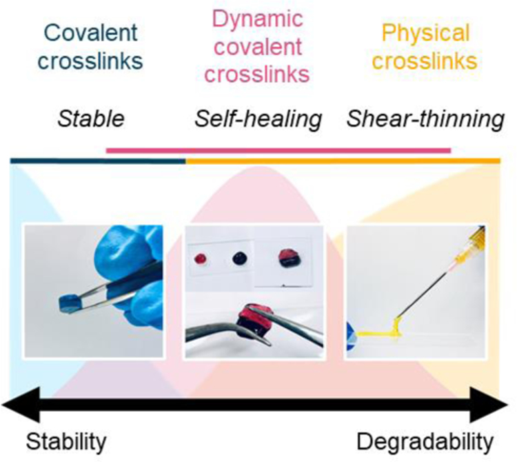
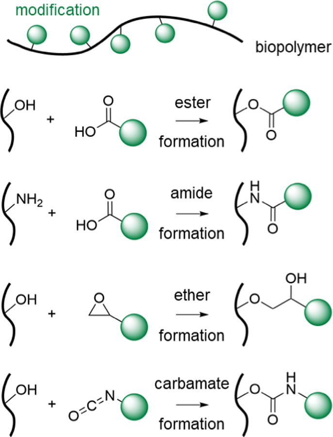
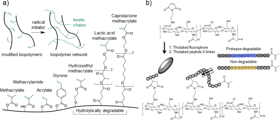
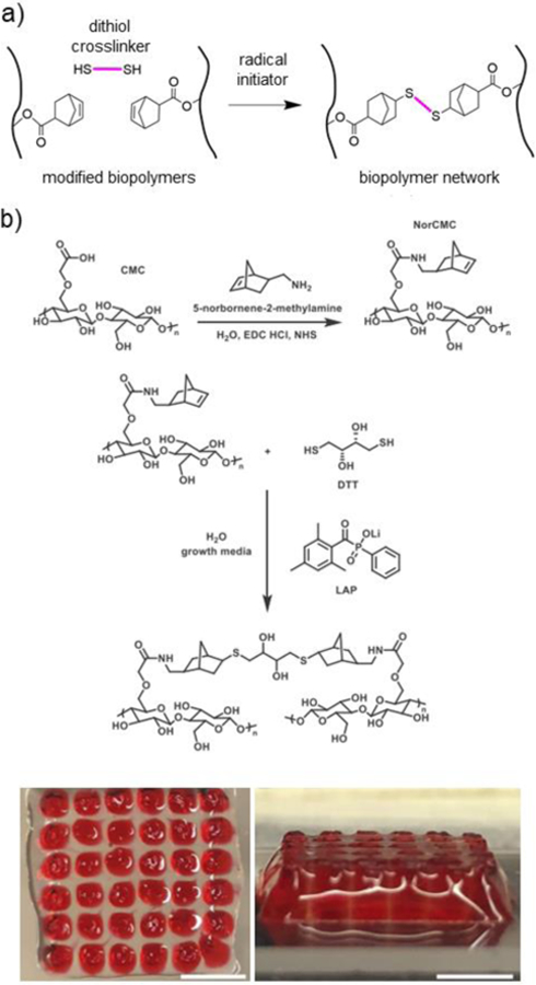
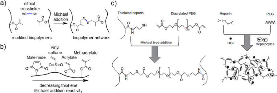
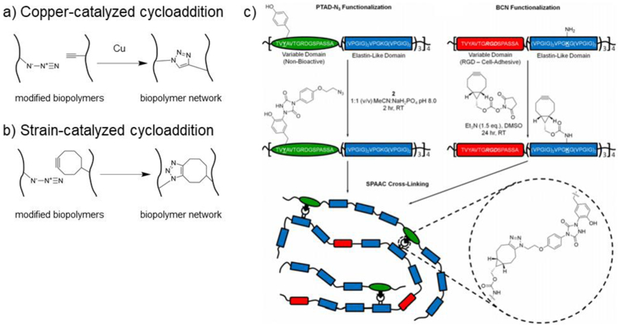
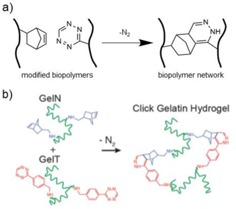
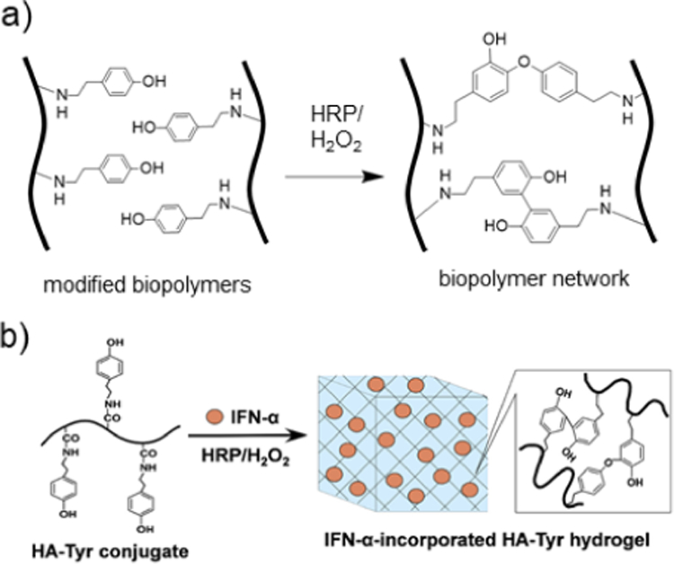
Biopolymers are natural polymers sourced from plants and animals, which include a variety of polysaccharides and polypeptides. The inclusion of biopolymers into biomedical hydrogels is of great interest because of their inherent biochemical and biophysical properties, such as cellular adhesion, degradation, and viscoelasticity. The objective of this Review is to provide a detailed overview of the design and development of biopolymer hydrogels for biomedical applications, with an emphasis on biopolymer chemical modifications and cross-linking methods. First, the fundamentals of biopolymers and chemical conjugation methods to introduce cross-linking groups are described. Cross-linking methods to form biopolymer networks are then discussed in detail, including (i) covalent cross-linking (e.g., free radical chain polymerization, click cross-linking, cross-linking due to oxidation of phenolic groups), (ii) dynamic covalent cross-linking (e.g., Schiff base formation, disulfide formation, reversible Diels-Alder reactions), and (iii) physical cross-linking (e.g., guest-host interactions, hydrogen bonding, metal-ligand coordination, grafted biopolymers). Finally, recent advances in the use of chemically modified biopolymer hydrogels for the biofabrication of tissue scaffolds, therapeutic delivery, tissue adhesives and sealants, as well as the formation of interpenetrating network biopolymer hydrogels, are highlighted.


Figures

Figure 1.
Figure 1.. General hydrogel properties as a function of crosslink type.
Schematic illustrating representative images of hydrogels formed from different crosslinking mechanisms (i.e., covalent [blue], dynamic covalent [pink], physical [yellow]).
Figure 2.
Figure 2.. Common chemical reactions for modification of biopolymers.
Schematic representation of chemically modifying biopolymers using common mechanisms. From top to bottom: esterification, amidation, etherification, and carbamate formation. The green circle denotes various chemical groups introduced onto biopolymers for potential use in hydrogel formation.
Figure 3.
Figure 3.. Crosslinking via free radical chain polymerization.
a) Schematic representation of the general crosslinking of modified biopolymers in the presence of an initiator to induce the formation of kinetic chains through the propagation of radical species (top), as well as common reactive groups used for biopolymer modification and hydrogel formation (bottom). b) Hyaluronic acid (HA) modified with maleimide groups to react with thiolated fluorophores and thiolated protease-degradable peptides capped with methacrylate groups for free radical chain polymerization. Peptide sequences are designed to be either protease degradable (blue) or non-degradable (yellow). Adapted with permission from Wade, et al. Copyright, 2015 Springer Nature.
Figure 4.
Figure 4.. Crosslinking via a thiol-ene radical addition.
a) Schematic representation of norbornene-modified biopolymers (black) crosslinked with a dithiol crosslinker (pink) in the presence of a radical initiator. b) Thiol-norbornene crosslinked CMC hydrogels for bioprinting, showing (top) schematic representation of amidation reaction to synthesize norbornene-modified CMC (NorCMC), (middle) schematic representation of photocrosslinking reaction, and (bottom) bioprinted NorCMC scaffolds (clear) filled with Pluronic (red). Scale bars represent 5mm. Adapted with permission from Ji, et al. Copyright, 2020 Elsevier.
Figure 5.
Figure 5.. Crosslinking via thiol-ene Michael addition.
a) Schematic representation of methacrylate-modified biopolymers (black) crosslinked with a dithiol crosslinker (blue) under Michael addition conditions. b) Schematic representation of a biopolymer modified with multiple ene groups that can undergo thiol-ene Michael addition. From left to right, in decreasing order of Michael addition reactivity: maleimide, vinyl sulfone, acrylate, and methacrylate. c) Thiolated heparin is crosslinked with diacrylated PEG (PEGDA) via a thiol-ene Michael addition reaction, which was used for the culture of primary rat hepatocytes and hepatocyte growth factor (HGF). Adapted with permission from Kim, et al. Copyright, 2010 Elsevier.
Figure 6.
Figure 6.. Crosslinking via azide-alkyne cycloaddition.
a) Schematic representation of copper-catalyzed azide-alkyne cycloaddition crosslinking of biopolymers. Biopolymers are modified with either azide or alkyne functional groups and upon combination in the presence of a copper catalyst, crosslinks form by azide-alkyne cycloaddition. b) Schematic representation of strain-catalyzed azide-alkyne cycloaddition crosslinking of biopolymers. Biopolymers are modified with either azide or strained alkyne (i.e., cyclooctyne) groups and upon combination, crosslinks form by azide-alkyne cycloaddition. c) Elastin-like polypeptides (ELPs) functionalized with either azide or bicyclononyne (BCN) groups for bio-orthogonal crosslinking due to the strain-promoted [3+2] azide-alkyne cycloaddition (SPAAC) reaction. Adapted with permission from Madl, et al. Copyright, 2017 American Chemical Society.
Figure 7.
Figure 7.. Crosslinking via tetrazine-norbornene reactions.
a) Schematic representation of biopolymers modified with either norbornene or tetrazine groups. Upon combination, crosslinks form by a tetrazine-norbornene reaction. b) Gelatin modified with either norbornene (GelN) or tetrazine (GelT) are mixed to form a tetrazine-norbornene click biopolymer network, which was cell-adhesive and degradable for use in cell encapsulation. Adapted with permission from Koshy, et al. Copyright 2016, Wiley.
Figure 8.
Figure 8.. Crosslinking via tyramine enzymatic reactions.
a) Schematic representation of biopolymers modified with tyramine that crosslink in the presence of HRP and H2O2 to form dityramine adducts. b) HA is modified with tyramine and subsequently exposed to horse radish peroxidase (HRP) and H2O2 to undergo enzymatic crosslinking by oxidation of tyramine groups, forming covalent dityramine adducts. Interferon-α (IFN-α) is encapsulated in the hydrogel for use as a prolonged-release delivery vehicle for renal carcinoma treatment. Adapted with permission from Ueda, et al. Copyright, 2016 Elsevier.
Figure 9.
Figure 9.. Crosslinking via catechol reactions.
a) Schematic representation of biopolymers modified with catechol and crosslinking in the presence of NaIO4 to form dicatechol adducts. b) A mussel-inspired, HA hydrogel is formed by modifying HA with catechol moieties (HA-CA). HA-CA is covalently crosslinked in the presence of sodium periodate (NaIO4). Image shows HA-CA hydrogel before (clear) and after (red) gelation. Adapted with permission from Shin, et al. Copyright, 2015 Wiley.
Figure 10.
Figure 10.. Crosslinking via Schiff base formation.
a) Schematic representation of imine dynamic covalent crosslinking by combining biopolymers modified with either primary amine or aldehyde groups. b) Schematic representation of hydrazone dynamic covalent crosslinking by combining biopolymers modified with either hydrazide or aldehyde groups. c) N-carboxyethyl chitosan (CEC) is combined with dialdehyde PEG (PEGDA, blue), where imine dynamic covalent crosslinks are formed between the amine groups on CEC and the aldehyde groups on PEGDA. Images show the self-healing (a-d) and shear-thinning (e-h) properties of the hydrogel. Adapted from Qu, et al. Copyright, 2017 Elsevier.
Figure 11.
Figure 11.. Crosslinking via disulfide bond formation.
a) Schematic representation of thiolated biopolymers forming dynamic covalent crosslinks by disulfide bond formation (blue) under oxidative conditions. Upon the addition of a mono-thiolated component, disulfide exchange can result in disassembly of the hydrogel. b) Various thiolated HA biopolymers are synthesized for disulfide dynamic covalent crosslinking, including: HA-thiol (HA-SH, green), HA-acetyl cysteine (HA-ActCys, red), and HA-cysteine (HA-Cys, blue). Among the thiol groups, HA-Cys has the strongest electron-withdrawing group in the β-position, resulting in the most disulfide bond formation under neutral conditions. Adapted with permission from Bermejo-Velasco, et al. Copyright, 2019 American Chemical Society.
Figure 12.
Figure 12.. Crosslinking via reversible Diels-Alder reactions.
a) Schematic representation of dynamic covalent crosslinks formed by combining biopolymers modified with either furan or maleimide groups. b) Hydroxypropyl chitin (HPC, black) modified with furan groups and combined with PEG-bismaleimide crosslinks for hydrogel formation. Immediately, a thermo-responsive physical hydrogel forms due to interactions between HPC molecules, and over time, reversible Diels-Alder crosslinks form to stabilize the hydrogel structure. Adapted from Bi, et al. Copyright, 2019 Elsevier.
Figure 13.
Figure 13.. Crosslinking via guest-host complexation.
a) Schematic representation of biopolymers (black) modified with either host β-cyclodextrin (β-CD, pink) or guest (blue) functional groups to undergo reversible crosslinking due to guest-host complexation. Common guest groups for β-CD include adamantane (Ad) and azobenzene (Az). b) Schematic representation of biopolymers (black) modified with guest groups (yellow) and combined with host cucurbit[8]uril (CB[8]) to undergo reversible crosslinking due to guest-host complexation. CB[8] has a large host cavity to accommodate two guest groups, which commonly include naphthalene and phenylalanine. c) Dextran (green) is modified with either β-CD (blue) or trans Az (red). Upon mixing, hydrogel formation occurs due to guest-host complexation between β-CD and trans Az. Upon exposure to UV light, Az groups convert from trans to cis state, resulting in hydrogel disassembly and photoresponsive release of encapsulated proteins. Adapted from Peng, et al. Copyright, 2010, Royal Society of Chemistry.
Figure 14.
Figure 14.. Crosslinking via hydrogen bonding.
a) Schematic representation of biopolymers modified with ureidopyrimidone (UPy) and crosslinking due to hydrogen bonding. b) Schematic representation of biopolymers modified with gallol groups and crosslinking due to hydrogen bonding. c) Dextran is modified with UPy (Dex-UPy) to undergo hydrogel formation due to hydrogen bonding. The resulting hydrogel is shear-thinning and self-healing. Images show self-healing behavior of Dex-UPy hydrogels. Adapted with permission form Hou, et al. Copyright, 2015 Wiley.
Figure 15.
Figure 15.. Crosslinking via metal-ligand complexation.
a) Schematic representation of biopolymers modified with catechol groups forming metal-ligand complexes with Fe(III). As pH increases, bis- and tris-complexation occurs, resulting in crosslink formation. b) Chitosan (green) is modified with catechol groups and forms a hydrogel metal-ligand complexation with Fe(III). The injectable hydrogel was used as a localized delivery vehicle for multiple chemotherapeutics. An increase in median survival rate was observed in murine lung and breast cancer models upon localized delivery of anticancer drugs from the hydrogel. Adapted with permission from Yavvari, et al. Copyright, 2017 American Chemical Society.
Figure 16.
Figure 16.. Crosslinking via interactions between synthetic polymers grafted to biopolymers.
a) Schematic representation of poly(N-isopropylacrylamide) (PNIPAAm, pink) grafted to a biopolymer (black) (left). The grafted biopolymer can undergo reversible physical crosslinking above the lower critical solution temperature (LCST) (~30°C) of PNIPAAm due to hydrophobic interactions between PNIPAAm groups (right). b) Schematic representation of Pluronic grafted to a biopolymer (black). Pluronic is an A-B-A triblock copolymer consisting of poly(ethylene glycol) (PEG) blocks (blue) and poly(propylene glycol) (PPG) blocks (red) (left). The grafted biopolymer can undergo reversible physical crosslinking above the critical micelle temperature (CMT) (~25–40°C) of Pluronic due to hydrophobic interactions between PPG blocks. (right) c) A biopolymer of PNIPAAm grafted to keratin (keratin-g-PNIPAAm) exhibited an LCST around 28–32°C, resulting in thermo-responsive hydrogel formation due to hydrophobic interactions between PNIPAAm groups. Keratin-g-PNIPAAm was explored for applications in brain injury repair. Adapted with permission from Zhu, et al. Copyright 2019, Elsevier.
Figure 17:
Figure 17:. Representative examples of biofabricated hydrogel scaffolds made from chemically-modified biopolymers.
a) Silk modified with GMA (Sil-MA) is photocrosslinked using LAP as a photoinitiator (left). Scaffolds that mimic the shape of trachea, heart, lung, and vessel are printed using dynamic light processing (DLP) (right). Adapted with permission from Kim et al. Copyright, 2018 Springer Nature. b) GelMA (red) and ChitoMA (gray) microgels are fabricated using a microfluidic device and then mixed to form a self-healing granular hydrogel scaffold with ionic interparticle interactions. The scaffold is combined with human adipose-derived stem cells (hADSCs) to form a cell-laden network. Adapted with permission from Hsu, et al. Copyright 2019, Wiley. c) HA modified with norbornene (NorHA) and either hydrazides (NorHA-Hyd, red) or aldehydes (NorHA-Ald, green) are electrospun to create a multifiber network with dynamic covalent inter-fiber crosslinks (left). Luminal scaffolds are created by wrapping the multifiber network around a needle and visualized i) while removing the scaffold from the needle and ii) while extruding rhodamine-labeled dextran dye through the lumen. Adapted with permission from Davidson et al. Copyright, 2020 Wiley.
Figure 18.
Figure 18.. Interpenetrating network (IPN) biopolymer hydrogels.
a) IPN hydrogels are formed through various synthesis techniques, including the sequential (swelling of first network in a secondary monomer/macromer) or simultaneous (orthogonal crosslinking of both first and second networks) introduction of networks. Adapted with permission from Dhand, et al. Copyright 2020, Elsevier. b) An IPN is formed by combining bis-maleimide-PEG, furan-modified gelatin (Gel-Furan), and chitosan grafted with Pluronic F127 (Chitosan-g-Pluronic). Initially, Chitosan-g-Pluronic formed a physically crosslinked, thermosensitive hydrogel network. After 2 h, Diels-Alder crosslinks between bis-maleimide-PEG and Gel-Furan covalently stabilize the hydrogel. Adapted with permission from Abandansari, et al. Copyright 2018, Elsevier.

Similar articles

Cited by

References

    1. Caló E; Khutoryanskiy VV Biomedical Applications of Hydrogels: A Review of Patents and Commercial Products. Eur. Polym. J 2015, 65, 252–267.
    1. Janouskova O Synthetic Polymer Scaffolds for Soft Tissue Engineering. Physiol. Res 2018, 67. - PubMed
    1. Van Vlierberghe S; Dubruel P; Schacht E Biopolymer-Based Hydrogels as Scaffolds for Tissue Engineering Applications: A Review. Biomacromolecules 2011, 12, 1387–1408. - PubMed
    1. Burdick JA; Prestwich GD Hyaluronic Acid Hydrogels for Biomedical Applications. Adv. Mater 2011, 23, 41–56. - PMC - PubMed
    1. Necas J; Bartosikova L; Brauner P; Kolar J Hyaluronic Acid (Hyaluronan): A Review. Vet. Med. (Praha) 2008, 53, 397–411.

Publication types