. 2021 Feb;41(2):e82-e96.
doi: 10.1161/ATVBAHA.120.315485. Epub 2020 Dec 24.

Intracellular AIBP (Apolipoprotein A-I Binding Protein) Regulates Oxidized LDL (Low-Density Lipoprotein)-Induced Mitophagy in Macrophages

Affiliations

Intracellular AIBP (Apolipoprotein A-I Binding Protein) Regulates Oxidized LDL (Low-Density Lipoprotein)-Induced Mitophagy in Macrophages

Soo-Ho Choi et al. Arterioscler Thromb Vasc Biol. 2021 Feb.

Abstract

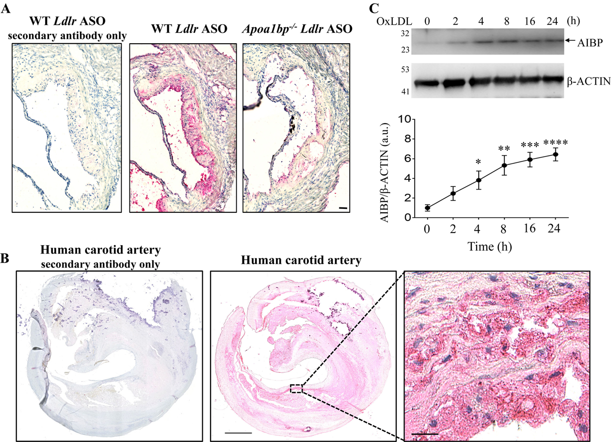
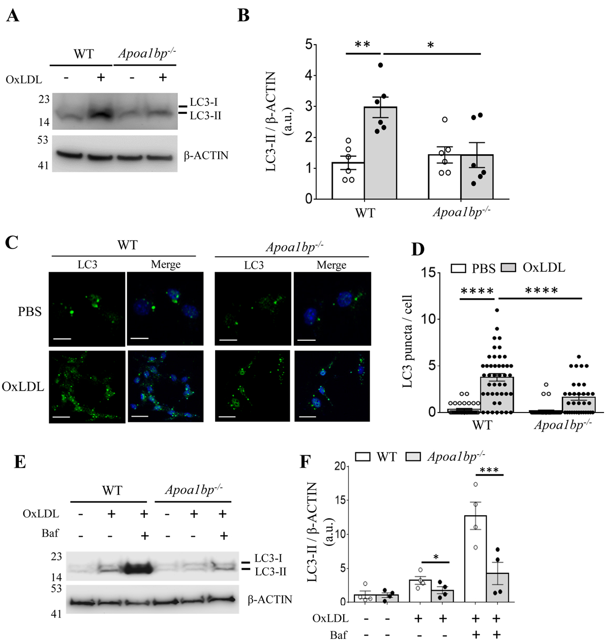
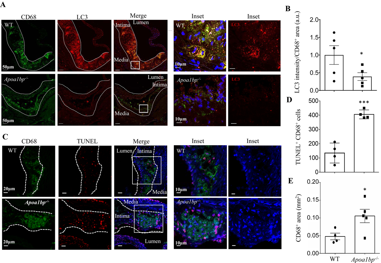
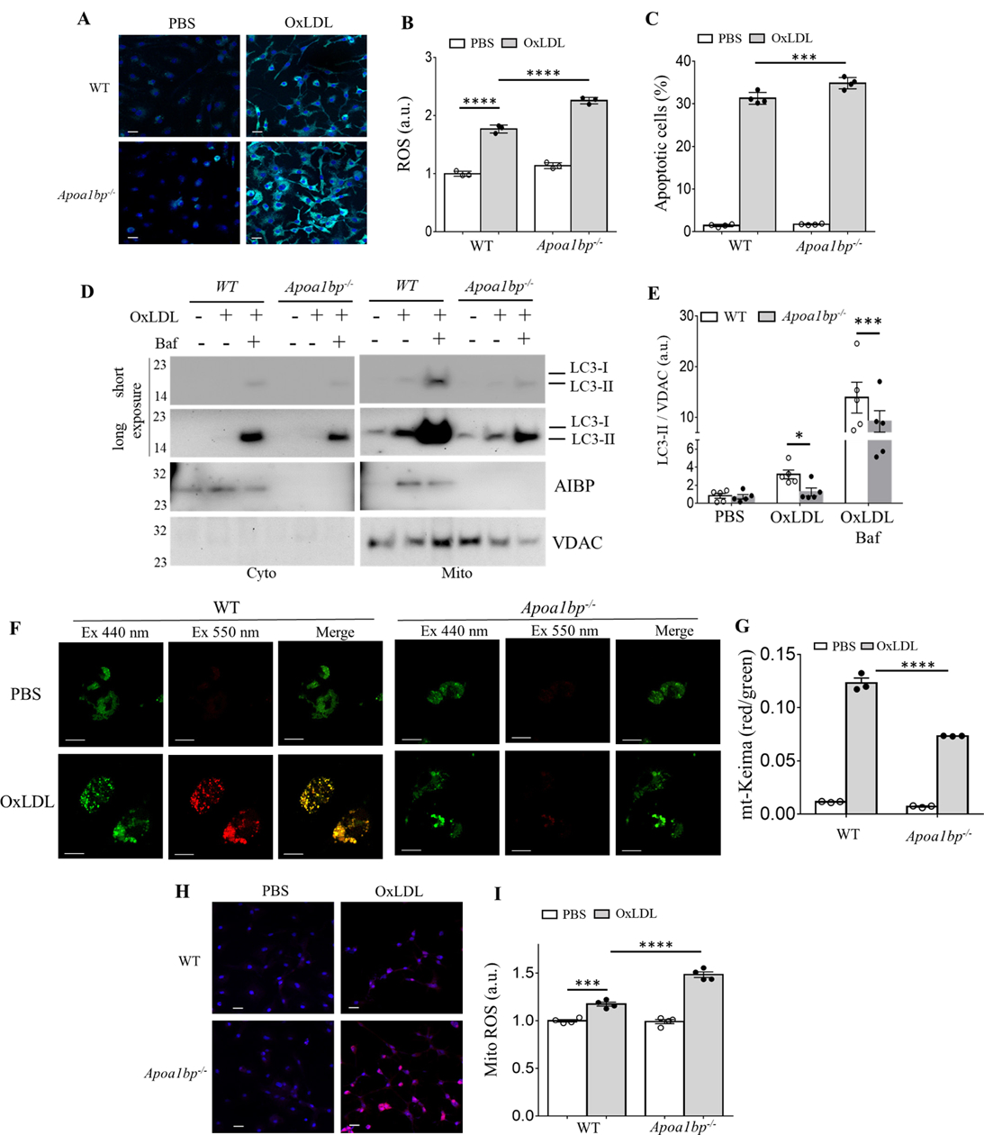
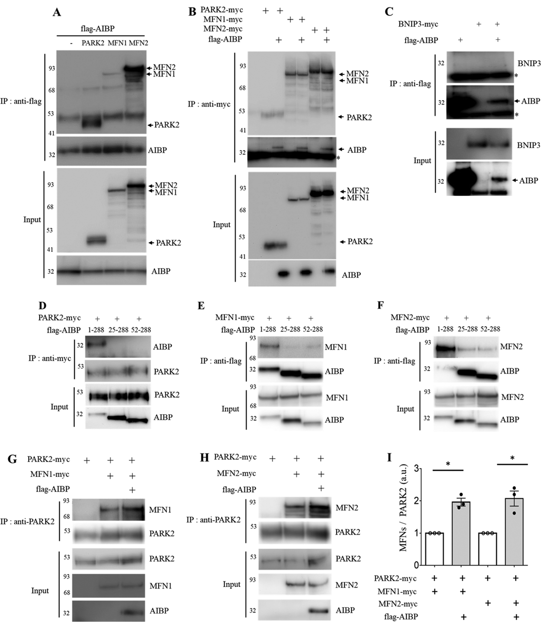
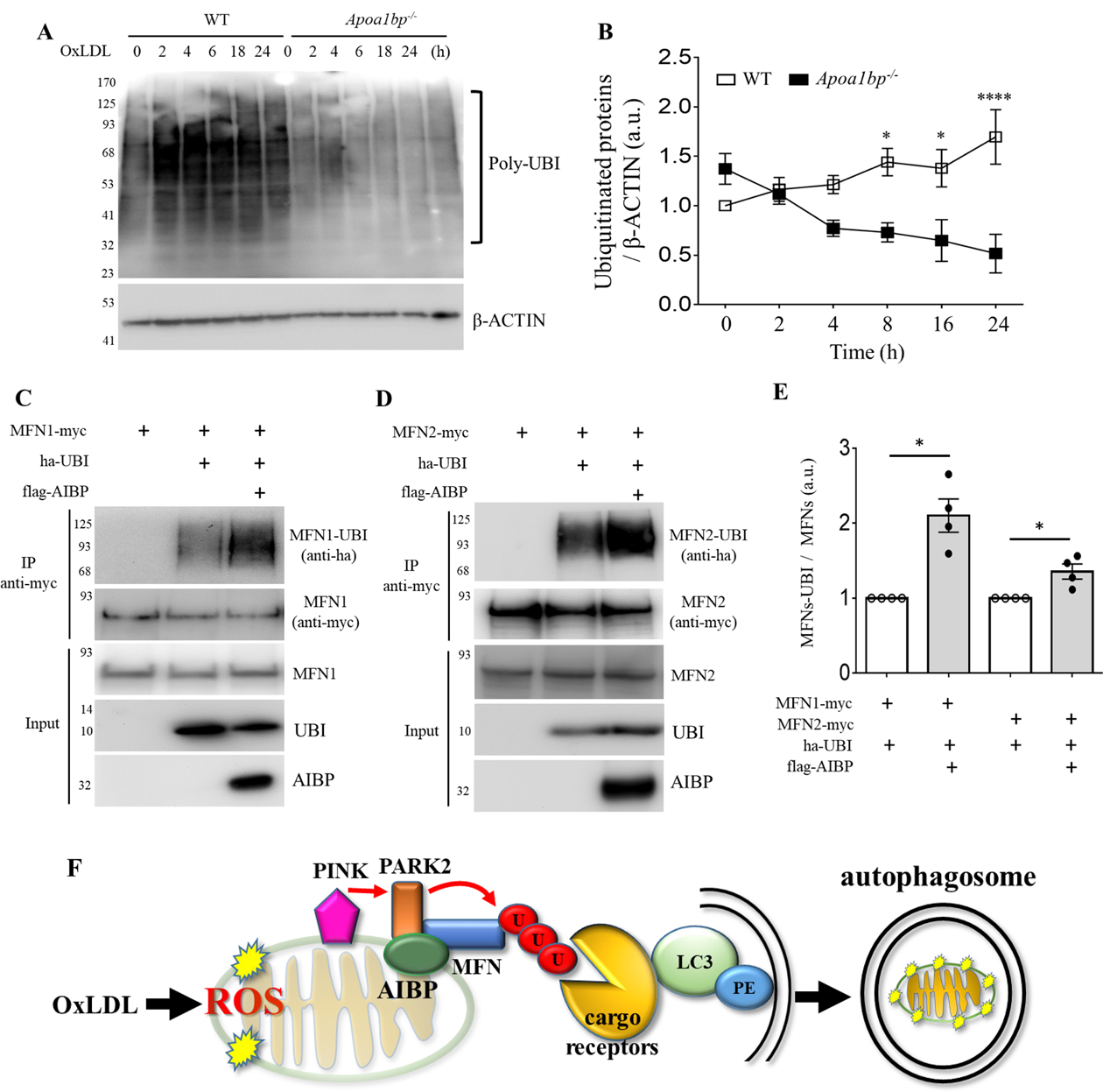
Objective: Atherosclerotic lesions are often characterized by accumulation of OxLDL (oxidized low-density lipoprotein), which is associated with vascular inflammation and lesion vulnerability to rupture. Extracellular AIBP (apolipoprotein A-I binding protein; encoded by APOA1BP gene), when secreted, promotes cholesterol efflux and regulates lipid rafts dynamics, but its role as an intracellular protein in mammalian cells remains unknown. The aim of this work was to determine the function of intracellular AIBP in macrophages exposed to OxLDL and in atherosclerotic lesions. Approach and Results: Using a novel monoclonal antibody against human and mouse AIBP, which are highly homologous, we demonstrated robust AIBP expression in human and mouse atherosclerotic lesions. We observed significantly reduced autophagy in bone marrow-derived macrophages, isolated from Apoa1bp-/- compared with wild-type mice, which were exposed to OxLDL. In atherosclerotic lesions from Apoa1bp-/- mice subjected to Ldlr knockdown and fed a Western diet, autophagy was reduced, whereas apoptosis was increased, when compared with that in wild-type mice. AIBP expression was necessary for efficient control of reactive oxygen species and cell death and for mitochondria quality control in macrophages exposed to OxLDL. Mitochondria-localized AIBP, via its N-terminal domain, associated with E3 ubiquitin-protein ligase PARK2 (Parkin), MFN (mitofusin)1, and MFN2, but not BNIP3 (Bcl2/adenovirus E1B 19-kDa-interacting protein-3), and regulated ubiquitination of MFN1 and MFN2, key components of mitophagy.

Conclusions: These data suggest that intracellular AIBP is a new regulator of autophagy in macrophages. Mitochondria-localized AIBP augments mitophagy and participates in mitochondria quality control, protecting macrophages against cell death in the context of atherosclerosis.

Keywords: atherosclerosis; autophagy; inflammation; macrophages; mitophagy.


Conflict of interest statement

Disclosures

YIM is a scientific co-founder of Raft Pharmaceuticals LLC. ST is a consultant to Boston Heart Diagnostics and is a co-inventor and receives royalties from patents owned by UCSD on oxidation-specific antibodies and of biomarkers related to oxidized lipoproteins, has a dual appointment at UCSD and Ionis Pharmaceuticals, and is a co-founder and has an equity interest in Oxitope, Inc and Kleanthi Diagnostics, LLC. Although these relationships have been identified for conflict of interest management based on the overall scope of the project and its potential benefit to Oxitope and Kleanthi Diagnostics LLC, the research findings included in this particular publication may not necessarily relate to the interests of Oxitope, Inc and Kleanthi Diagnostics, LLC. The terms of this arrangement have been reviewed and approved by the University of California, San Diego in accordance with its conflict of interest policies. Other authors declare that they have no competing interests.

Figures

Figure 1.
Figure 1.
AIBP expression in atherosclerotic lesions and in OxLDL-stimulated macrophages. Sections of the aortic root of hypercholesterolemic WT and Apoa1bp−/− mice (A) and human carotid artery (B) were stained with the monoclonal anti-AIBP antibody BE-1, followed by a secondary antibody or by the secondary antibody only, and counterstained with H&E. Representative images of three specimens tested. (C) BMDM isolated from WT mice were stimulated with 25 μg/ml OxLDL for indicated time. Cells lysates were immunoblotted with anti-AIBP and β-ACTIN antibodies. Band intensities were quantified. Mean±SEM; N=4–5. *, p<0.05; **, p<0.005; ***, p<0.0005; ****, p<0.0001 vs. time zero. Scale bars: 50 μm in A, 1 mm in B, and 25 μm in the zoomed-in region in B.
Figure 2.
Figure 2.
AIBP regulates OxLDL-induced autophagy. (A to D) BMDM isolated from WT and Apoa1bp−/− mice were incubated with PBS or 25 μg/ml OxLDL for 24 h. Cell lysates were immunoblotted with antibodies against LC3 and β-ACTIN, and band intensities were quantified. For imaging, cells were stained with an anti-LC3 antibody and DAPI. The numbers of LC3 puncta per cell were counted from three independent experiments. (E and F) Cells were pretreated with or without 100 nM Baf for 1h and then incubated with 25 μg/ml OxLDL for 24 h. Cell lysates were immunoblotted with indicated antibodies, and band intensities were quantified. Mean±SEM; N=3–5. *, p<0.05; **, p<0.005; ***, p<0.0005; ****; p<0.0001. Scale bar: 20 μm.
Figure 3.
Figure 3.
AIBP expression regulates autophagy and cell survival in atherosclerotic lesions of hypercholesterolemic mice. (A and B) Aortic root sections from hypercholesterolemic WT and Apoa1bp−/− mice were stained with anti-LC3 (red) and anti-CD68 (green) antibodies to assay for autophagy in macrophage-rich areas. The LC3 fluorescent intensities were measured in CD68+ areas and specific LC3 intensities per mm2 of CD68+ areas were calculated. Data are from immunohistochemical staining conducted on 5 separate days and because fluorescence intensity varies from day-to-day, repeated measures t-test was conducted to calculate the p-value. (C and D) TUNEL staining showing apoptotic cells in the aortic root sections. The numbers of TUNEL and CD68 double-positive cells were counted. (E) CD68-positive area in each lesion. Mean±SEM; N=4–5. ***, p<0.0005.
Figure 4.
Figure 4.
Macrophage AIBP regulates OxLDL-induced ROS, apoptosis and mitophagy. BMDM isolated from WT and Apoa1bp−/− mice were incubated with PBS or 25 μg/ml OxLDL for 24 h and ROS generation was measured using (A) a confocal microscope or (B) flow cytometer. Geometric means of FACS histograms were measured and presented as scattered bar graphs. (C) Apoptotic cells were detected by Annexin V staining using a flow cytometry analysis. (D-E) LC3-II and AIBP expression in isolated mitochondria were measured by immunoblot. (F-G) BMDM were infected with retrovirus encoding mt-Keima for 24 h and incubated with PBS or 25 μg/ml OxLDL for additional 24 h. Mitophagy flux was analyzed by confocal microscopy, capturing images at both 440nm (green, mitochondria) and 550nm (red, lysosome) (F), and flow cytometry (G). Mitochondrial ROS were measured using (H) confocal microscopy and (I) flow cytometry. Mean±SEM; N=3–5. *, p<0.05; **, p<0.005; ***, p<0.0005; ****, p<0.0001. Scale bars, 20μm in A and H, 10μm in F.
Figure 5.
Figure 5.
AIBP associates with PARK2, MFN1 and MFN2 and enhances PARK2-MFN1/2 interactions. (A-B) HEK 293 cells were co-transfected with flag-tagged AIBP (1–289 aa) and PARK2-myc, MFN1-myc, or MFN2-myc. (A) AIBP-driven immunoprecipitation with an anti-flag antibody. (B) Immunoprecipitation driven by PARK2, MFN1, or MFN2 with an anti-myc antibody. (C) AIBP does not bind to BNIP3. HEK 293 cells were co-transfected with flag-tagged AIBP and BNIP3-myc. Cell lysates were immunoprecipitated with an anti-flag antibody. *, IgG light chain. (D-F) HEK293 cells were co-transfected with flag-tagged AIBPs (1–289 aa, 25–289 aa, or 52–289 aa) together with (D) PARK2-myc, (E) MFN1-myc, or (F) MFN2-myc. (D) PARK2 and (E and F) AIBPs were immunoprecipitated with an anti-myc or anti-flag antibody, respectively. (G-I) HEK 293 cells were co-transfected with flag-tagged AIBP, PARK2-myc, and MFN1-myc or MFN2-myc. Cell lysates were immunoprecipitated with an anti-PARK2 antibody, and the bound (G) MFN1 and (H) MFN2 were detected by immunoblotting with an anti-myc antibody. (I) Band intensities were measured and MFN/PARK2 ratios calculated. Because of variation in independent transient transfection/pull-down/blot experiments conducted on separate days, experimental samples (with AIBP) were normalized to the control samples in which AIBP was not added. With control samples (no AIBP) set as 1, one-sample t-test was conducted to test the hypothesis whether the experimental group (with AIBP) was different from 1, i.e., whether AIBP significantly increased MFN1 and MFN2 binding to PARK2 compared to no-AIBP controls. Mean±SEM; N=3–4. *, p<0.05; **, p<0.005; ***, p<0.0005; ****, p<0.0001.
Figure 6.
Figure 6.
AIBP regulates the ubiquitination pathway. (A and B) BMDM isolated from WT and Apoa1bp−/− mice were incubated with PBS or 25 μg/ml OxLDL for indicated time. Cells were harvested and immunoblotted with anti-UBI and β-ACTIN antibodies. The band intensities were quantified. (C and D) HEK 293 cells were co-transfected with flag-tagged AIBP, ha-ubiquitin, and MFN1-myc or MFN2-myc. Cell lysates were immunoprecipitated with an anti-myc antibody, and ubiquitination was detected by immunoblotting with an anti-ha antibody. (E) Band intensities were measured and MFN-UBI/MFN ratios calculated. Because of variation in independent transient transfection/pull-down/blot experiments conducted on separate days, experimental samples (with AIBP) were normalized to the control samples in which AIBP was not added. With control samples (no AIBP) set as 1, one-sample t-test was conducted to test the hypothesis whether the experimental group (with AIBP) was different from 1, i.e., whether AIBP significantly increased MFN1 and MFN2 ubiquitination compared to no-AIBP controls. Mean±SEM; N=3–4. *, p<0.05; **, p<0.005; ***, p<0.0005; ****, p<0.0001. (F) Schematic overview of the regulation of mitophagy by AIBP. Intracellular AIBP associates with mitochondrial proteins Parkin and MFN1/MFN2 and upregulates ubiquitination of MFN1 and MFN2; in turn ubiquitinated MFN are recognized by cargo receptors interacting with lipidated LC3 anchored in the membrane to form an autophagosome. In the context of atherosclerosis, this regulation is particularly important in removal of damaged mitochondria in macrophages exposed to atherogenic OxLDL.

References

    1. Libby P Inflammation in atherosclerosis. Arterioscler Thromb Vasc Biol. 2012;32:2045–2051 - PMC - PubMed
    1. Hansson GK, Libby P The immune response in atherosclerosis: A double-edged sword. Nat Rev Immunol. 2006;6:508–519 - PubMed
    1. Miller YI, Choi SH, Wiesner P, Fang L, Harkewicz R, Hartvigsen K, Boullier A, Gonen A, Diehl CJ, Que X, Montano E, Shaw PX, Tsimikas S, Binder CJ, Witztum JL. Oxidation-specific epitopes are danger-associated molecular patterns recognized by pattern recognition receptors of innate immunity. Circ Res. 2011;108:235–248 - PMC - PubMed
    1. Peng N, Meng N, Wang S, Zhao F, Zhao J, Su L, Zhang S, Zhang Y, Zhao B, Miao J. An activator of mtor inhibits oxldl-induced autophagy and apoptosis in vascular endothelial cells and restricts atherosclerosis in apolipoprotein e(-)/(-) mice. Sci Rep. 2014;4:5519. - PMC - PubMed
    1. Choi SH, Gonen A, Diehl CJ, Kim J, Almazan F, Witztum JL, Miller YI. Syk regulates macrophage MHC-II expression via activation of autophagy in response to oxidized ldl. Autophagy. 2015;11:785–795 - PMC - PubMed

Publication types

MeSH terms