. 2021 Jan;13(2):129-144.
doi: 10.2217/epi-2020-0189. Epub 2020 Dec 24.

Epigenetic targeting of Waldenström macroglobulinemia cells with BET inhibitors synergizes with BCL2 or histone deacetylase inhibition

Affiliations

Epigenetic targeting of Waldenström macroglobulinemia cells with BET inhibitors synergizes with BCL2 or histone deacetylase inhibition

Stephan J Matissek et al. Epigenomics. 2021 Jan.

Erratum in

  • Corrigendum.
    [No authors listed] [No authors listed] Epigenomics. 2021 Jun;13(11):909-911. doi: 10.2217/epi-2020-0189c1. Epub 2021 Apr 26. Epigenomics. 2021. PMID: 33899492 Free PMC article. No abstract available.

Abstract

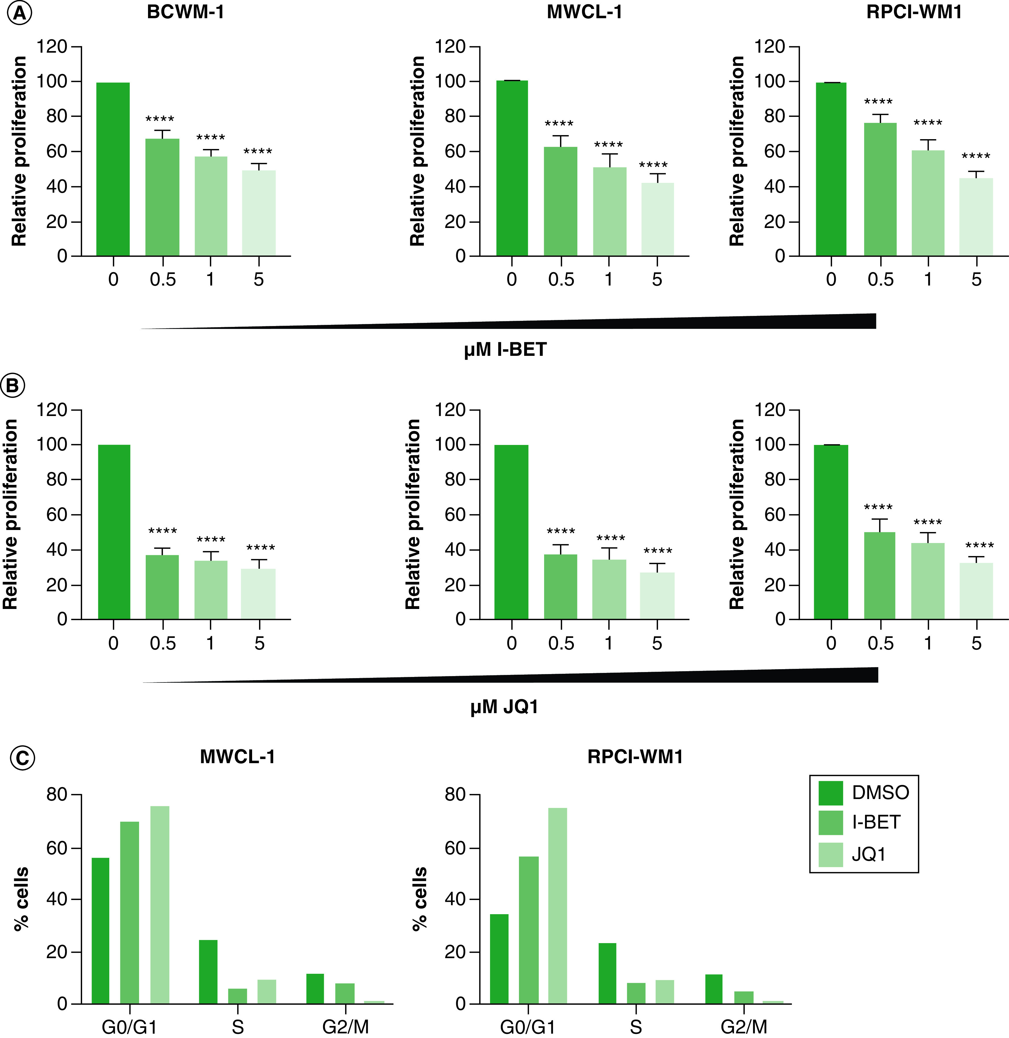
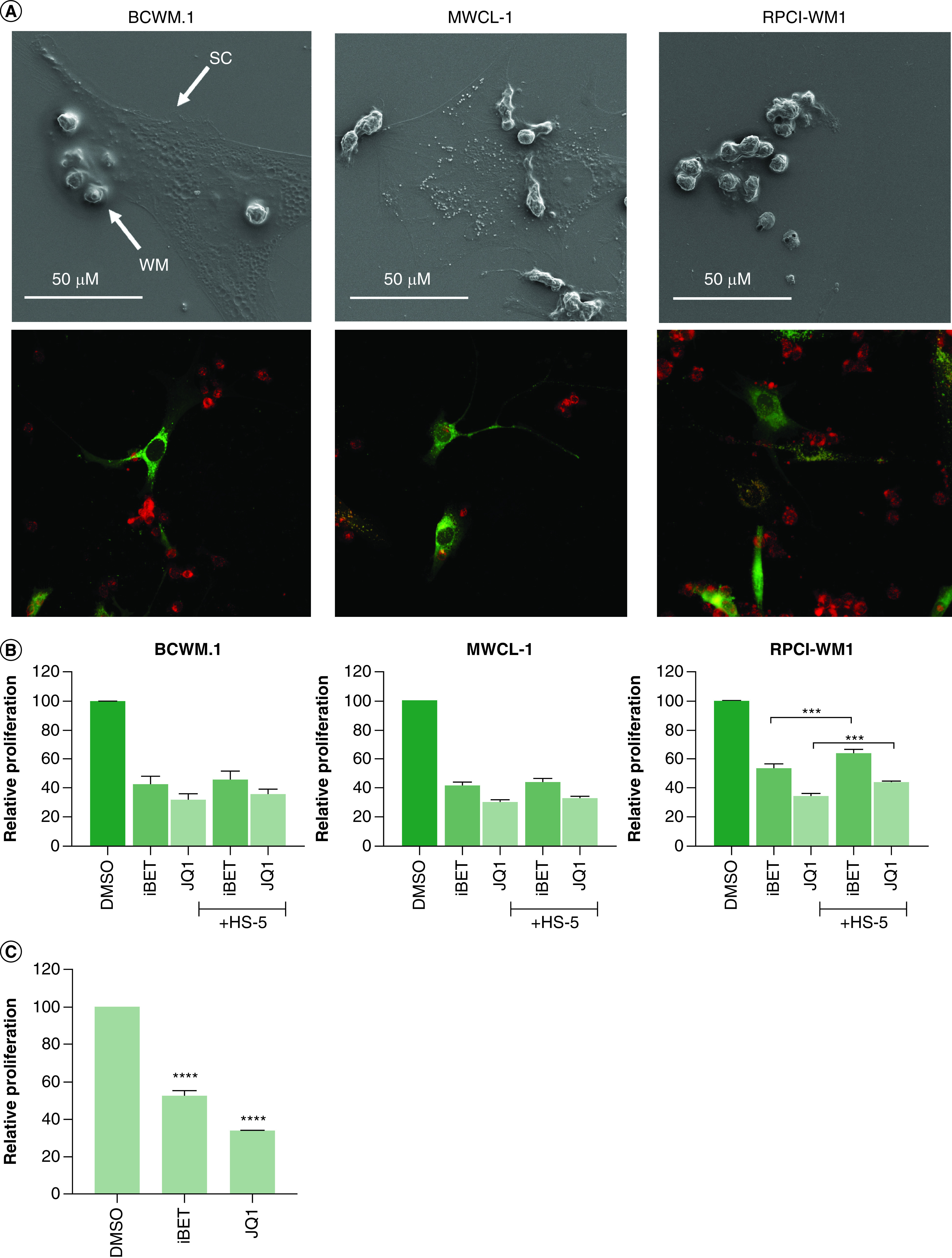
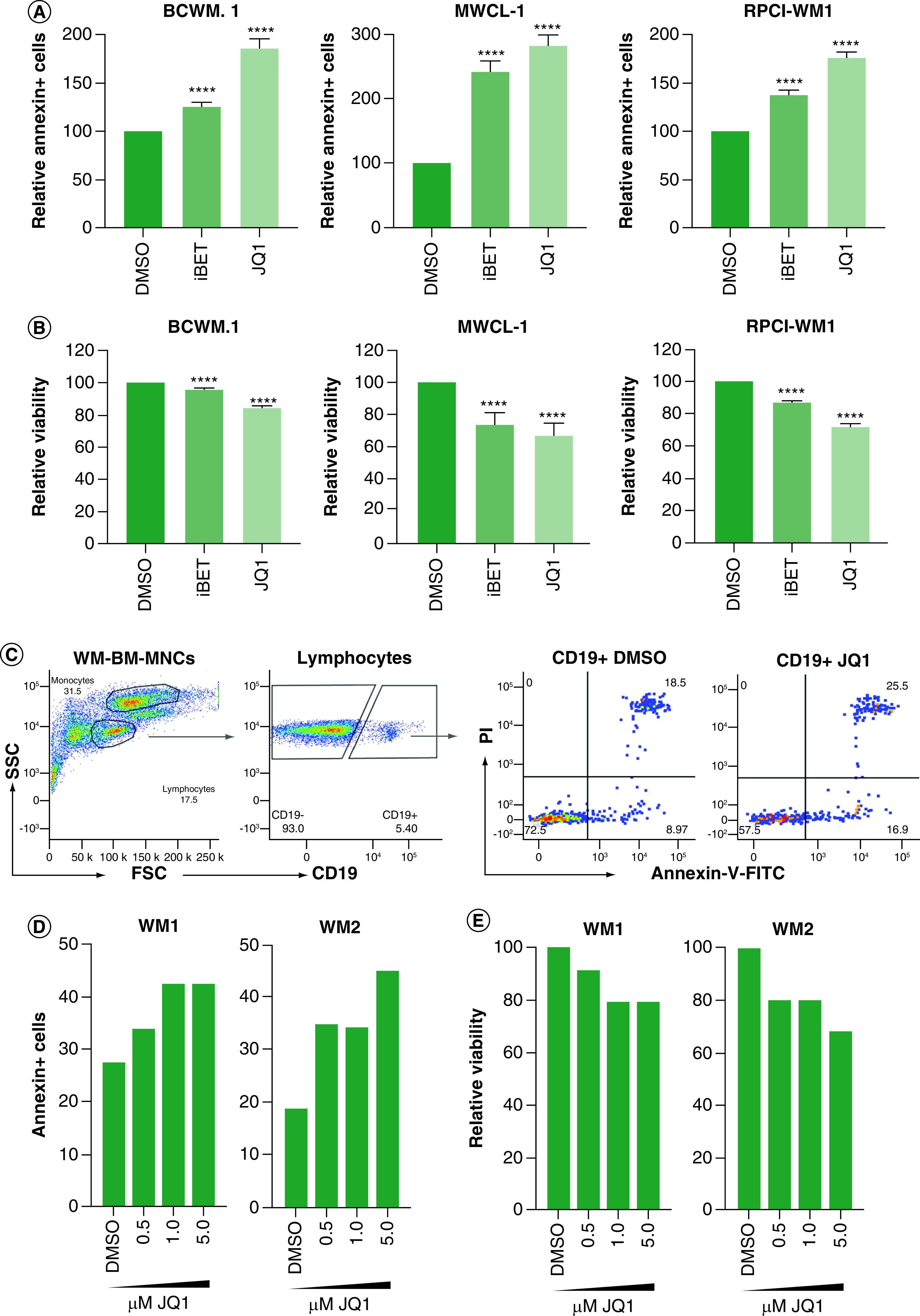
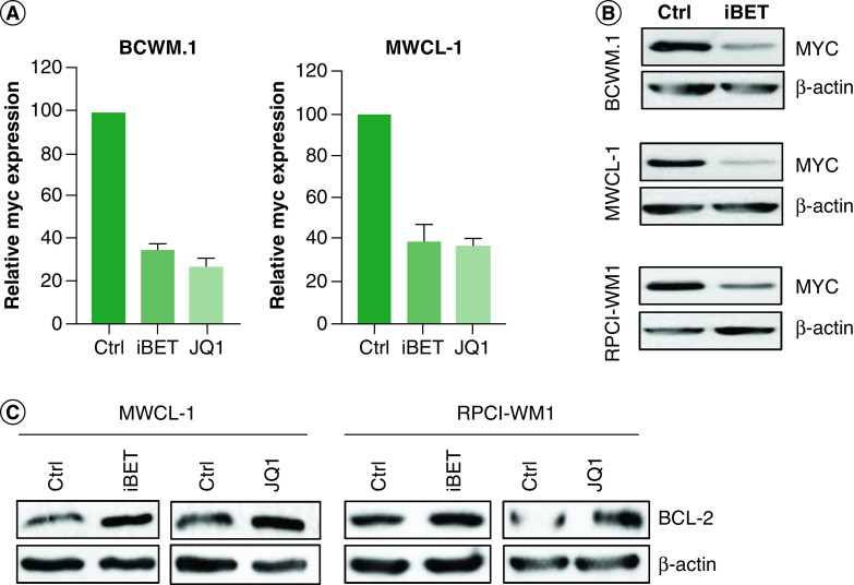
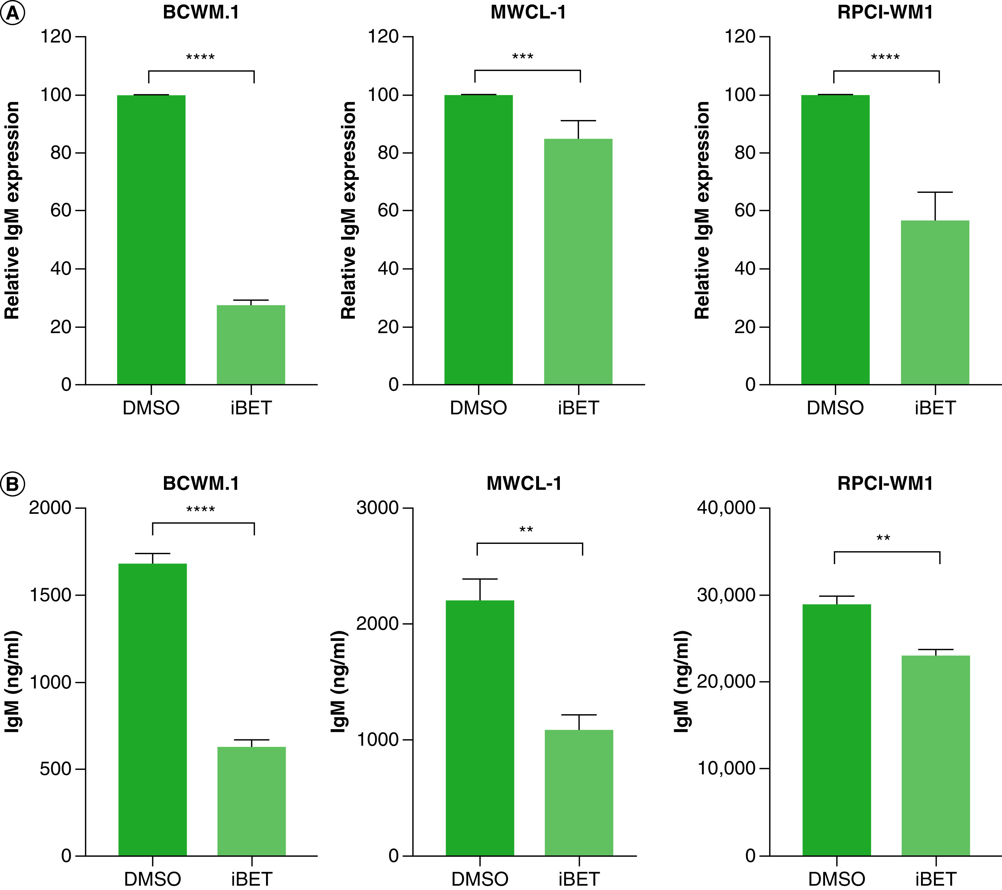
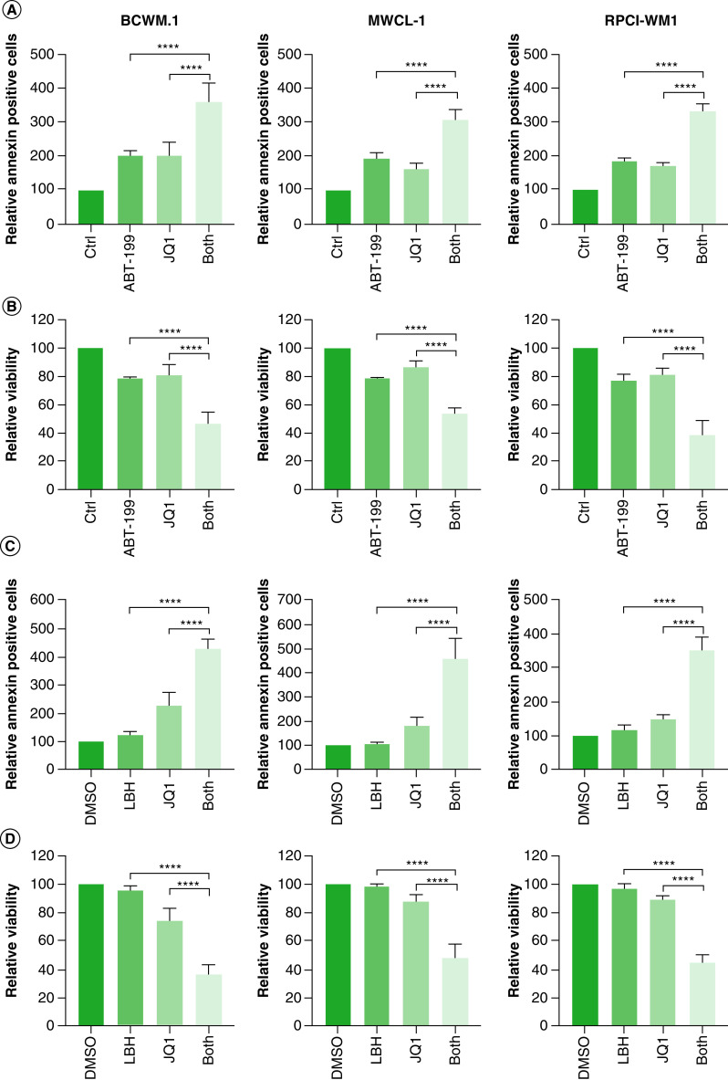
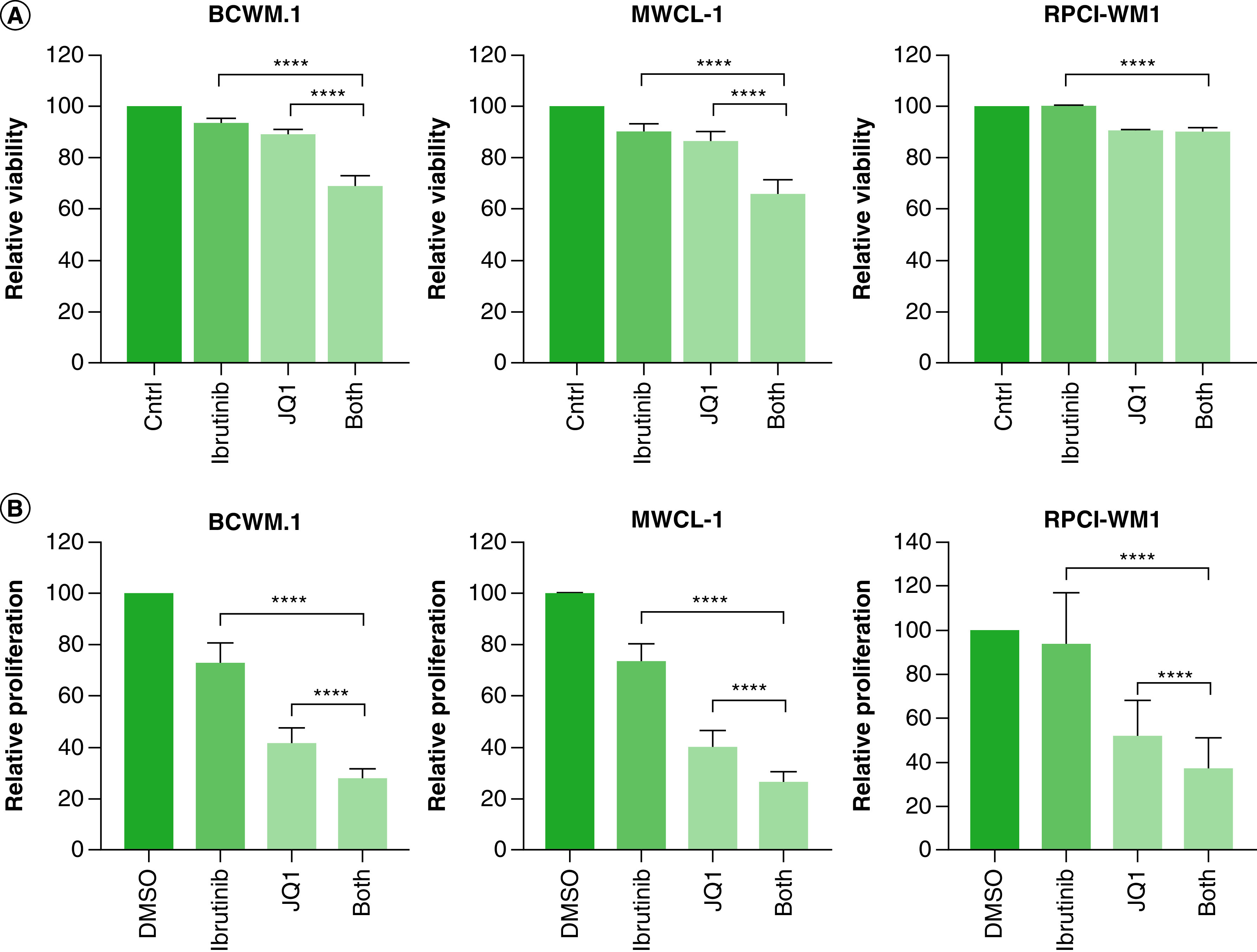
Aim: Waldenström macroglobulinemia (WM) is a low-grade B-cell lymphoma characterized by overproduction of monoclonal IgM. To date, there are no therapies that provide a cure for WM patients, and therefore, it is important to explore new therapies. Little is known about the efficiency of epigenetic targeting in WM. Materials & methods: WM cells were treated with BET inhibitors (JQ1 and I-BET-762) and venetoclax, panobinostat or ibrutinib. Results: BET inhibition reduces growth of WM cells, with little effect on survival. This finding was enhanced by combination therapy, with panobinostat (LBH589) showing the highest synergy. Conclusion: Our studies identify BET inhibitors as effective therapy for WM, and these inhibitors can be enhanced in combination with BCL2 or histone deacetylase inhibition.

Keywords: BET inhibitors; Waldenström macroglobulinemia; epigenetics; panobinostat; venetoclax.


Conflict of interest statement

Financial & competing interests disclosure

This research was supported by an NIH COBRE Center of Integrated Biomedical and Bioengineering Research (CIBBR, P20 GM113131) through an Institutional Development Award (IDeA) from the National Institute of General Medical Sciences. This work was supported in part by a grant from the International Waldenström Macroglobulinemia Foundation and the Leukemia & Lymphoma Society (IWMF-LLS). The authors have no other relevant affiliations or financial involvement with any organization or entity with a financial interest in or financial conflict with the subject matter or materials discussed in the manuscript apart from those disclosed.

No writing assistance was utilized in the production of this manuscript.

Figures

Figure 1.
Figure 1.. BET bromodomain inhibitors reduce Waldenström macroglobulinemia cell growth.
(A) WM cells (0.25 × 10³) were treated with the indicated doses of iBET or DMSO control (0). (B) WM cells (0.25 × 10³) were treated with the indicated doses of JQ1 or DMSO control. Cells were cultured at 37°C for 72 h, followed by the determination of cell proliferation using an XTT assay. Each experiment was repeated at least five-times. Bars represent the mean ± SE of at least three biological replicates of each experiment. (C) WM cells (0.5 × 10⁶) were treated with 5 mM iBET, JQ1 or DMSO control for 72 h followed by cell-cycle analysis as described in the Materials & methods section. This experiment was repeated twice with similar results and shown is a representative experiment. ****p < 0.0001. DMSO: Dimethyl sulfoxide; iBET: I-BET-762; SM: Standard Error; WM: Waldenström macroglobulinemia.
Figure 2.
Figure 2.. The tumor microenvironment does not protect against BET inhibitors.
(A) HS-5 stromal cells (0.1 × 10⁶) and WM cells (0.5 × 10⁶) were co-cultured on coverslips as described in the Materials & methods section for 2 days. Coverslips were carefully removed and used for imaging using SEM (top panel) or confocal microscopy (lower panel). (B) Bone marrow stromal cells (HS-5; 0.1 × 10³) were plated in 96-well plates and allowed to adhere overnight. Cells were fixed using 10 µg/ml of mitomycin C for 3 h, followed by washing with DPBS. WM cells (0.5 × 10³) were added to each well and treated with either 5 mM of iBET, JQ1 or DMSO control. Cells were incubated at 37°C for 72 h, and cell proliferation was determined by XTT assay, as described in Materials & methods section. (C) HS-5 cells (0.25 × 10³) were treated with 5 mM iBET, 5 mM JQ1 or DMSO control for 72 h, followed by the determination of cell proliferation using an XTT assay. These experiments were repeated at least three-times. Bars represent the mean ± SE of at least three biological replicates of each experiment. ***p < 0.001; ****p < 0.0001. DMSO: Dimethyl sulfoxide; DPBS: Dulbecco’s phosphate-buffered saline; iBET: I-BET-762; SC: Stromal cells; SE: Standard error; WM: Waldenström macroglobulinemia.
Figure 3.
Figure 3.. BET inhibitors induce modest cell death.
WM cells (0.5 × 10⁶) were treated with 5 mM of iBET or JQ1 or DMSO control for 72 h. Cells were harvested and viability was determined by annexin-V/PI staining. (A) Percent annexin-V-positive cells were determined by flow cytometry. (B) Percent viable cells as determined by flow cytometry. Each experiment was repeated at least three-times. Bars represent the mean ± SE of all experiments. (C) MNCs (2 × 10⁶ cells) from BM biopsy specimens from WM patients were treated with the indicated doses of JQ1 or DMSO control as described in Materials & methods section. Cells were harvested and viability was determined by annexin-V/PI and CD19 staining. Gating was done on lymphocytes from total MNCs followed by gating on CD19+ cells. Annexin-V/PI was analyzed on CD19+ cells. (D) Annexin-V-positive cells were determined by flow cytometry in two WM patient samples (WM1 and WM2). (E) Percent viable cells were determined for primary WM patient samples. ****p < 0.0001. BM: Bone marrow; DMSO: Dimethyl sulfoxide; iBET: I-BET-762; MNC: Mononuclear cell; PI: Propidium iodide; SE: standard error; WM: Waldenström macroglobulinemia.
Figure 4.
Figure 4.. BET inhibitors reduce MYC but induce BCL-2 expression.
(A) WM cells (2 × 10⁶) were treated with 5 mM of either iBET or JQ1 or DMSO control for 24 h. Cells were harvested and RNA was extracted and used to determine MYC expression by qRT-PCR. This experiment was repeated at least three-times and data were presented as mean ± SE of all experiments. (B) WM cells (5 × 10⁶) were treated with either 5 mM of iBET, JQ1 or DMSO control for 24 h. Cells were harvested and lysed and lysates were used to determine protein levels by western blot. (C) WM cells (5 × 10⁶) were treated with either 5 mM of iBET, JQ1 or DMSO control for 24 h. Cells were lysed and lysates were used to determine BCL-2 protein levels by Western blot. Western blot experiments were repeated at least three-times with similar results. DMSO: Dimethyl sulfoxide; iBET: I-BET-762; SE: Standard error; WM: Waldenström macroglobulinemia.
Figure 5.
Figure 5.. BET inhibitor reduces IgM levels.
(A) WM cells (2 × 10⁶) were treated with 5 mM of iBET or DMSO control for 24 h. Cells were harvested and RNA was extracted and used in qRT-PCR to determine IgM expression. (B) WM cells (2 × 10⁶) were treated with either 5 mM iBET or DMSO control for 72 h. Supernatant was used to quantify IgM secretion by ELISA. Each experiment was repeated at least three-times. Bars represent the mean ± SE of all experiments. **p < 0.01; ***p < 0.001; ****p < 0.0001. DMSO: Dimethyl sulfoxide; iBET: I-BET-762; qRT: quantitative reverse-transcription; SE: Standard error; WM: Waldenström macroglobulinemia.
Figure 6.
Figure 6.. Combined targeting of BET proteins and BCL-2/histone deacetylase enhances cell death in Waldenström macroglobulinemia cells.
(A & B) WM cells (0.5 × 10⁶) were treated with either 500 nM of ABT-199, 5 mM of JQ1, a combination of venetoclax (ABT-199) and JQ1, or DMSO control for 72 h. Cells were harvested and stained with annexin-V/PI followed by data acquisition using flow cytometry. Annexin-V-FITC positive cells are shown in (A) and viable cells are shown in (B). (C & D) WM cells (0.5 × 10⁶) were treated with either 25 nM of panobinostat (LBH589), 5 mM of JQ1, a combination of LBH589 and JQ1, or DMSO control for 72 h. Cells were harvested and stained with annexin-V/PI followed by data acquisition using flow cytometry. Annexin-V-FITC positive cells are shown in (C) and viable cells are shown in (D). Each experiment was repeated at least three-times. Bars represent the mean ± SE of all experiments. ****p < 0.0001. DMSO: Dimethyl sulfoxide; PI: Propidium iodide; SE: Standard error; WM: Waldenström macroglobulinemia.
Figure 7.
Figure 7.. Combined targeting of BET proteins and Bruton’s tyrosine kinase has a modest effect on cell viability in Waldenström macroglobulinemia cells.
(A) WM cells (0.5 × 10⁶) were treated with either 5 mM of ibrutinib, 5 mM of JQ1, combination of ibrutinib and JQ1 or DMSO control for 72 h, followed by determination of cell viability by annexin-V/PI double staining. (B) WM cells (0.25 × 10³) were treated with either 5 mM of ibrutinib, 5 mM of JQ1, combination of ibrutinib and JQ1 or DMSO control for 72 h, followed by the determination of cell proliferation using an XTT assay as described in Materials & methods section. Each experiment was repeated at least three-times. Bars represent the mean ± SE of all experimental. ****p < 0.0001. DMSO: Dimethyl sulfoxide; PI: Propidium iodide; SE: Standard error; WM: Waldenström macroglobulinemia.

Similar articles

Cited by

References

    1. Kapoor P, Paludo J, Vallumsetla N, Greipp PR. Waldenström macroglobulinemia: what a hematologist needs to know. Blood Rev. 29(5), 301–319 (2015). - PubMed
    2. •• Discusses aspects of Waldenström macroglobulinemia (WM) disease.

    1. Ansell SM, Kyle RA, Reeder CB et al. Diagnosis and management of Waldenström macroglobulinemia: mayo stratification of macroglobulinemia and risk-adapted therapy (mSMART) guidelines. Mayo Clin. Proc. 85(9), 824–833 (2010). - PMC - PubMed
    1. Treon SP, Gustine J, Meid K et al. Ibrutinib monotherapy in symptomatic, treatment-naïve patients with Waldenström macroglobulinemia. J. Clin. Oncol. 36(27), 2755–2761 (2018). - PubMed
    2. • Discusses the role of ibrutinib in WM.

    1. Han W, Matissek SJ, Jackson DA, Sklavanitis B, Elsawa SF. Targeting IL-6 receptor reduces IgM levels and tumor growth in Waldenström macroglobulinemia. Oncotarget 10(36), 3400–3407 (2019). - PMC - PubMed
    2. • Highlights the efficacy of targeting the tumor microenvironment.

    1. Sun JY, Xu L, Tseng H et al. Histone deacetylase inhibitors demonstrate significant preclinical activity as single agents, and in combination with bortezomib in Waldenström’s macroglobulinemia. Clin. Lymphoma Myeloma Leuk. 11, 152-156 (2011). - PubMed
    2. • Efficacy of epigenetic targeting in WM using histone deacetylase inhibitors.

Publication types

MeSH terms

LinkOut - more resources