

Optimizing In Vitro Osteogenesis in Canine Autologous and Induced Pluripotent Stem Cell-Derived Mesenchymal Stromal Cells with Dexamethasone and BMP-2
- PMID: 33356875
- PMCID: PMC7891305
- DOI: 10.1089/scd.2020.0144
Optimizing In Vitro Osteogenesis in Canine Autologous and Induced Pluripotent Stem Cell-Derived Mesenchymal Stromal Cells with Dexamethasone and BMP-2
Abstract
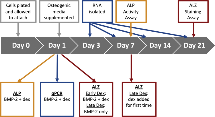
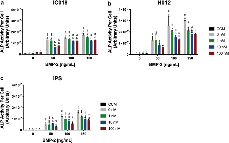
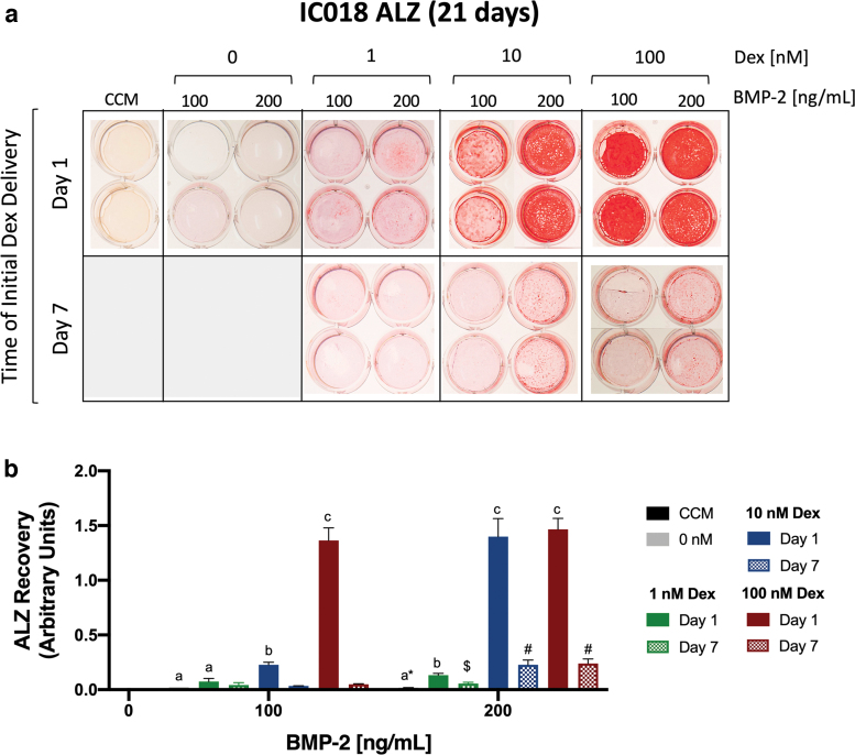
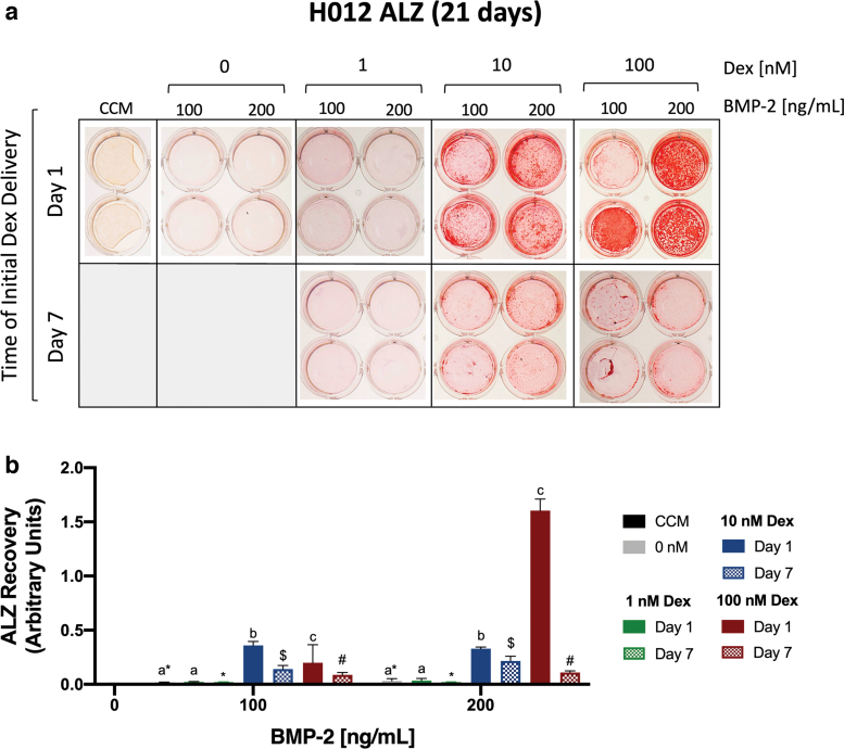
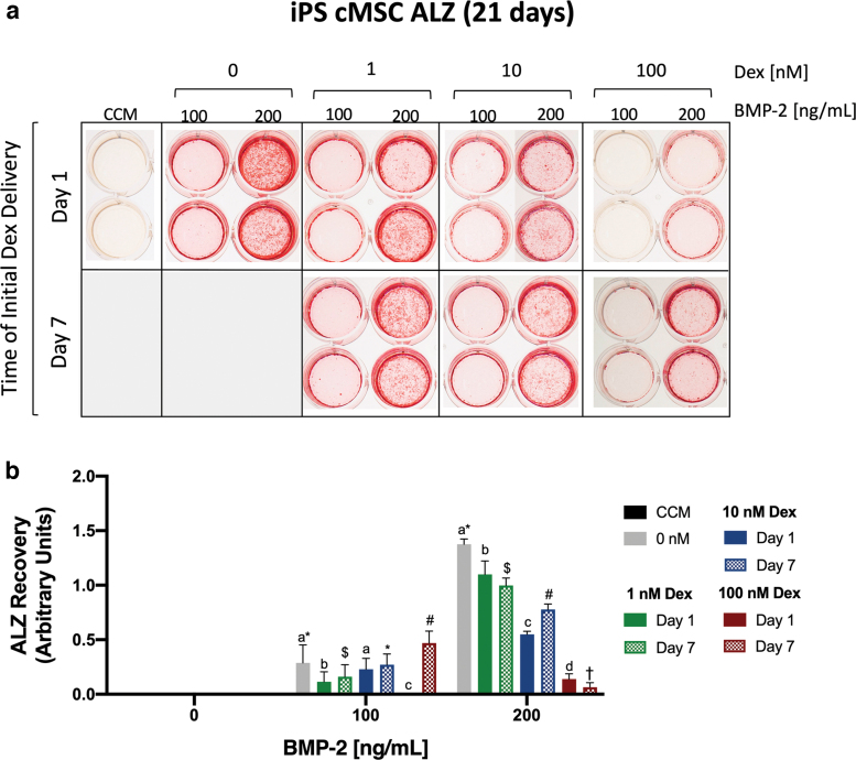
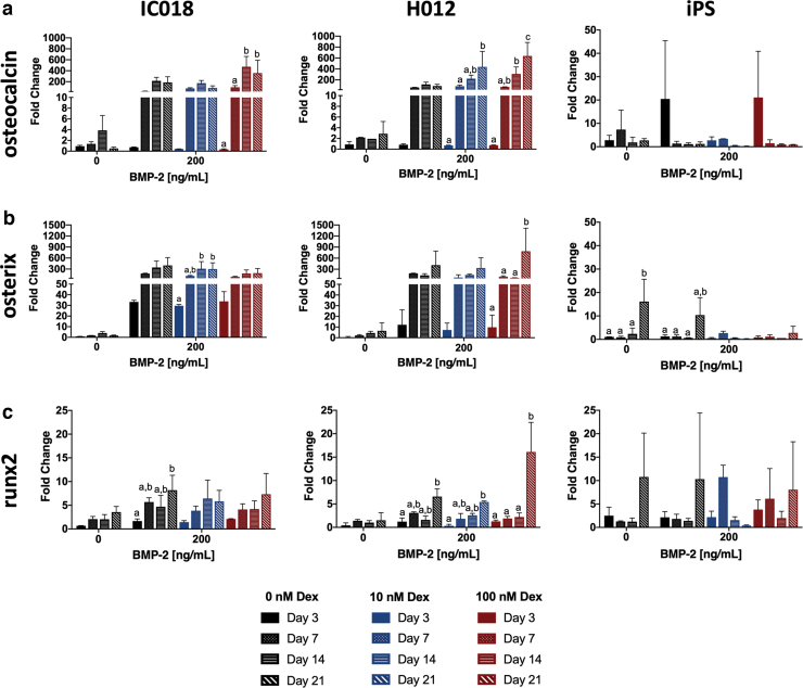
A growing body of work suggests that canine mesenchymal stromal cells (cMSCs) require additional agonists such as bone morphogenic protein-2 (BMP-2) for consistent in vitro osteogenic differentiation. BMP-2 is costly and may challenge the translational relevance of the canine model. Dexamethasone enhances osteogenic differentiation of human MSCs (hMSCs) and is widely utilized in osteogenic protocols. The aim of this study was to determine the effect of BMP-2 and dexamethasone on early- and late-stage osteogenesis of autologous and induced pluripotent stem cell (iPS)-derived cMSCs. Two preparations of marrow-derived cMSCs were selected to represent exceptionally or marginally osteogenic autologous cMSCs. iPS-derived cMSCs were generated from canine fibroblasts. All preparations were evaluated using alkaline phosphatase (ALP) activity, Alizarin Red staining of osteogenic monolayers, and quantitative polymerase chain reaction. Data were reported as mean ± standard deviation and compared using one- or two-way analysis of variance and Tukey or Sidak post hoc tests. Significance was established at P < 0.05. In early-stage assays, dexamethasone decreased ALP activity for all cMSCs in the presence of BMP-2. In late-stage assays, inclusion of dexamethasone and BMP-2 at Day 1 of culture produced robust monolayer mineralization for autologous cMSCs. Delivering 100 nM dexamethasone at Day 1 improved mineralization and reduced the BMP-2 concentrations required to achieve mineralization of the marginal cMSCs. For iPS-cMSCs, dexamethasone was inhibitory to both ALP activity and monolayer mineralization. There was increased expression of osteocalcin and osterix with BMP-2 in autologous cMSCs but a more modest expression occurred in iPS cMSCs. While autologous and iPS-derived cMSCs respond similarly in early-stage osteogenic assays, they exhibit unique responses to dexamethasone and BMP-2 in late-stage mineralization assays. This study demonstrates that dexamethasone and BMP-2 can be titrated in a time- and concentration-dependent manner to enhance osteogenesis of autologous cMSC preparations. These results will prove useful for investigators performing translational studies with cMSCs while providing insight into iPS-derived cMSC osteogenesis.
Keywords: MSCs; bone morphogenic protein-2; canine; induced pluripotent cells; mesenchymal stromal cells; osteogenic differentiation.
Conflict of interest statement
No competing financial interests exist.
Figures
References
-
- Marcacci M, Kon E, Moukhachev V, Lavroukov A, Kutepov S, Quarto R, Mastrogiacomo M and Cancedda R (2007). Stem cells associated with macroporous bioceramics for long bone repair: 6- to 7-year outcome of a pilot clinical study. Tissue Eng 13:947–955 - PubMed
-
- Viateau V, Logeart-Avramoglou D, Guillemin G and Petite H (2008). Animal models for bone tissue engineering purposes. In: Sourcebook of Models for Biomedical Research. Conn PM, ed. Humana Press, Totowa, NJ, pp 725–736
-
- Reichert JC, Saifzadeh S, Wullschleger ME, Epari DR, Schütz MA, Duda GN, Schell H, van Griensven M, Redl H and Hutmacher DW (2009). The challenge of establishing preclinical models for segmental bone defect research. Biomaterials 30:2149–2163 - PubMed
Publication types
MeSH terms
Substances
Grants and funding
LinkOut - more resources
Full Text Sources
Other Literature Sources
