Review
. 2020 Dec 24;24(1):702.
doi: 10.1186/s13054-020-03369-5.

Multi-organ point-of-care ultrasound for COVID-19 (PoCUS4COVID): international expert consensus

Affiliations
Review

Multi-organ point-of-care ultrasound for COVID-19 (PoCUS4COVID): international expert consensus

Arif Hussain et al. Crit Care. .

Abstract

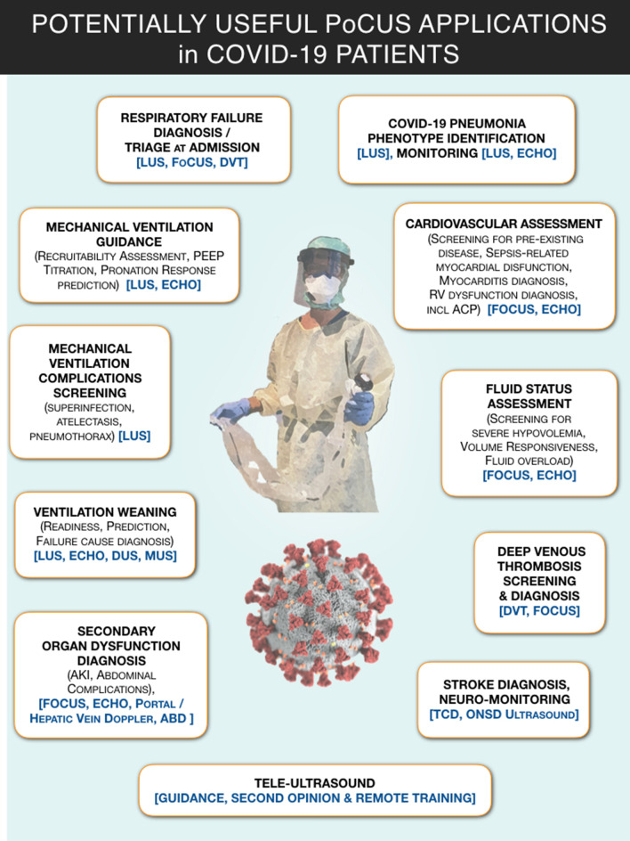
COVID-19 has caused great devastation in the past year. Multi-organ point-of-care ultrasound (PoCUS) including lung ultrasound (LUS) and focused cardiac ultrasound (FoCUS) as a clinical adjunct has played a significant role in triaging, diagnosis and medical management of COVID-19 patients. The expert panel from 27 countries and 6 continents with considerable experience of direct application of PoCUS on COVID-19 patients presents evidence-based consensus using GRADE methodology for the quality of evidence and an expedited, modified-Delphi process for the strength of expert consensus. The use of ultrasound is suggested in many clinical situations related to respiratory, cardiovascular and thromboembolic aspects of COVID-19, comparing well with other imaging modalities. The limitations due to insufficient data are highlighted as opportunities for future research.

Keywords: COVID-19; Echocardiography; Focused cardiac ultrasound (FoCUS); Lung ultrasound (LUS); Point-of-care ultrasound (PoCUS); SARS-CoV-2.


Conflict of interest statement

Except for the following authors, none have declared any disclosures with respect to the present work: A Hussain: Currently serving President of WINFOCUS—G Tavazzi: Received fees for lectures from GE Healthcare unrelated to this work—R Hoppmann: EchoNous Advisory Board—F Mojoli: Received fees for lectures from Hamilton Medical, GE Healthcare, Seda Spa and institutional relationship between University of Pavia and Hamilton Medical—G Via: eMedical Academy co-founder—B Nelson: Echonous and DiA Advisory Board.

Figures

Fig. 1
Fig. 1
Graphical synopsis of potentially useful applications of point-of-care ultrasound (PoCUS) in COVID-19 patients. ABD, abdominal ultrasound; ACP, acute cor pulmonale; AKI, acute kidney injury; DUS, diaphragmatic ultrasound; DVT, ultrasound for deep venous thrombosis screening; ECHO, echocardiography; FoCUS, focused cardiac ultrasound; LUS, lung ultrasound; MUS, parasternal intercostal muscles ultrasound; ONSD, optic nerve sheath diameter; PEEP, positive end expiratory pressure; PoCUS, point-of-care ultrasound; TCD, transcranial Doppler; VASC, ultrasound for venous and arterial access
Fig. 2
Fig. 2
Literature search strategy. A literature search of Pubmed, Pubmed Central, Embase, Scopus and Cochrane library databases was conducted by 2 independent researchers from 01/01/2020–01/08/2020 to identify all publications on point-of-care ultrasound in COVID-19 adult patients, using English language restriction, and the following MeSH query: ((“lung” AND “ultrasound”) OR “echocardiography” OR “Focused cardiac ultrasound” OR “point-of-care ultrasound” OR “venous ultrasound”) AND (“COVID-19” OR “SARS-CoV2”). Non-pertinent findings were discarded. The references of relevant papers were hand-searched for missed papers. Duplicates were removed. An additional search of pre-print publications was made through ResearchGate, preprint online repositories and social medias
Fig. 3
Fig. 3
Examples of lung ultrasound cumulative patterns of patients presenting with a similar degree of hypoxemia, but very different degree of aeration and respiratory mechanics characteristics, and recalling the recently proposed COVID-19 pneumonia phenotypes [89]. Patient on upper panel presents a nearly normal respiratory system compliance and LUS evidence of a milder lung involvement, reflected in a total LUS score of 11. This suggests a lung condition matching which has been recently described as “Phenotype L,” based on CT findings, and characterized by low lung elastance and low ventilation/perfusion ratio (explaining the severe hypoxia). Based on this imaging and on respiratory mechanics findings, final PEEP was set at 10 cm H20. Upper panel shows LUS evidence of a more diffuse and severe diffuse sonographic interstitial syndrome (cause of the shunt and the severe hypoxia), yielding a total LUS score of 27. Respiratory mechanics characteristics recall what has been described as “Phenotype H” (COVID-19 pneumonia: high lung elastance, high right-to-left shunt). Based on this imaging and on respiratory mechanics findings, PEEP was set at 14 cm H20 after a stepwise recruiting maneuver. LUS, lung ultrasound
Fig. 4
Fig. 4
Use of lung ultrasound to monitor lung aeration and guide ventilatory management in 2 COVID-19 patients. a COVID-19 patient on day 2 after intubation and ICU admission, initially with PEEP 12 cmH2O: diffuse bilateral B-pattern with crowded, coalescent B-lines (“white lung appearance”) is visible, consistent with a sonographic interstitial syndrome and severe loss of aeration/increase of extravascular lung water. Based on these findings and on respiratory mechanics, a stepwise recruitment maneuver with a final PEEP set at 15 cmH20 was performed, with improvement in gas exchange. b A different COVID-19 patient on day 4; PEEP set at 14 cmH2O: in comparison with previous patient, less B-lines are visible in ventral scans, with asymmetric distribution (more on the left scan); dorsal areas show lung consolidations, larger on the right side, with air bronchograms (dynamic at live scan). A pronation trial was successful, yielding immediate improvement in gas exchange and subsequent re-aeration of dorsal areas. (Ventral scans are taken with a linear, high frequency probe, dorsal ones with a phased array low-frequency one)
Fig. 5
Fig. 5
Lung ultrasound to monitor adequacy of re-aeration of dorsal areas upon pronation and recruitment maneuvers in a COVID-19 patient. Same patient of Fig. 2B, before (upper panels) and after (lower panels) pronation and a series of stepwise recruitment maneuvers up to PEEP 26 cmH2O, and final PEEP setting at 16 cmH20
Fig. 6
Fig. 6
Example of highly portable ultrasound device covered for use on COVID-19 patients. The iPAD, with which the device works, is tightly enveloped in plastic film wrap, while the probe is covered with a dedicated sheath (normally used for sterile ultrasound-guided procedures). Donning and doffing the device requires assistance and involves stepwise uncovering, with multiple steps of disinfection before and after removing the covering. Use of the device is restricted to the COVID-19 unit

References

    1. Coronavirus disease 2019 (COVID-19) Situation Report – 92 [https://www.who.int/docs/default-source/coronaviruse/situation-reports/2...]
    1. Wu Z, McGoogan JM. Characteristics of and Important Lessons From the Coronavirus Disease 2019 (COVID-19) Outbreak in China: Summary of a Report of 72314 Cases From the Chinese Center for Disease Control and Prevention. JAMA 2020. - PubMed
    1. Mojoli F, Mongodi S, Orlando A, Arisi E, Pozzi M, Civardi L, Tavazzi G, Baldanti F, Bruno R, Iotti GA, et al. Our recommendations for acute management of COVID-19. Crit Care. 2020;24(1):207. doi: 10.1186/s13054-020-02930-6. - DOI - PMC - PubMed
    1. Alhazzani W, Moller MH, Arabi YM, Loeb M, Gong MN, Fan E, Oczkowski S, Levy MM, Derde L, Dzierba A et al. Surviving Sepsis Campaign: Guidelines on the Management of Critically Ill Adults with Coronavirus Disease 2019 (COVID-19). Crit Care Med 2020. - PMC - PubMed
    1. Clinical management of severe acute respiratory infection when COVID-19 is suspected - Interim guidance. https://www.who.int/publications-detail/clinical-management-of-severe-ac...