. 2021 Feb 10;29(2):236-249.e6.
doi: 10.1016/j.chom.2020.12.010. Epub 2020 Dec 16.

Intranasal vaccination with a lentiviral vector protects against SARS-CoV-2 in preclinical animal models

Affiliations

Intranasal vaccination with a lentiviral vector protects against SARS-CoV-2 in preclinical animal models

Min-Wen Ku et al. Cell Host Microbe. .

Abstract

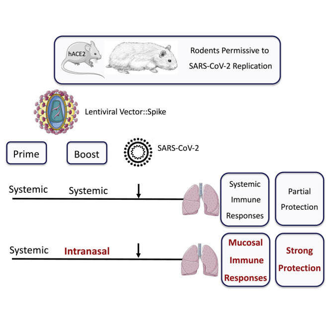
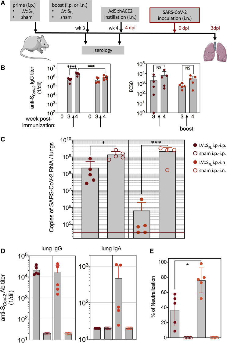
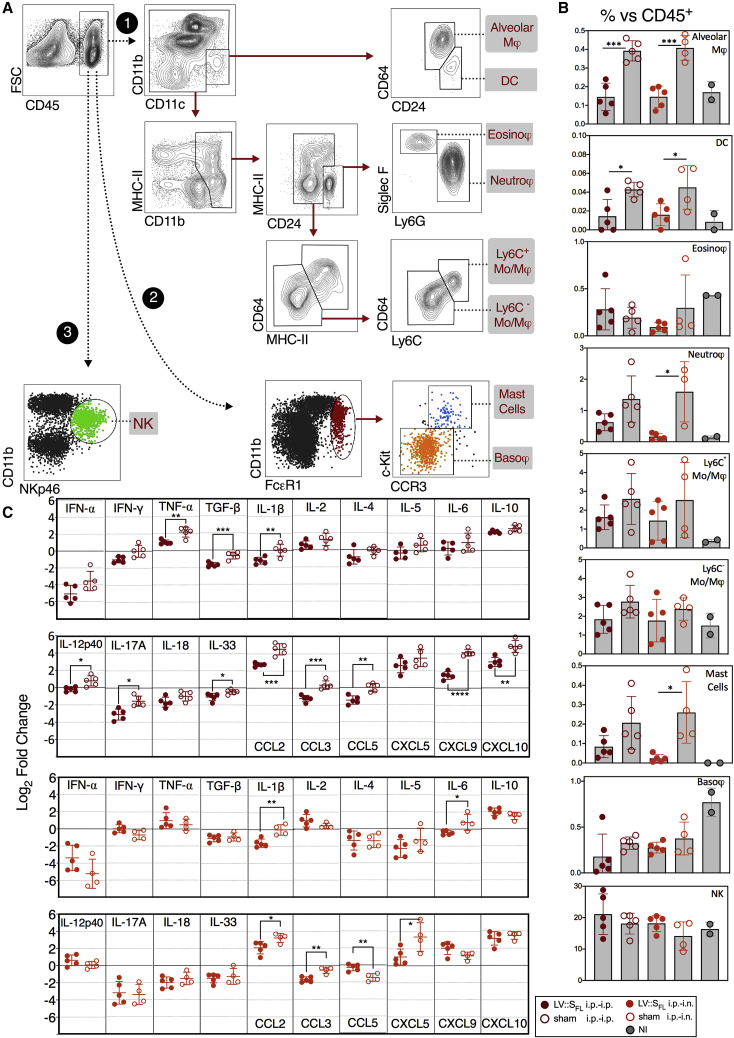
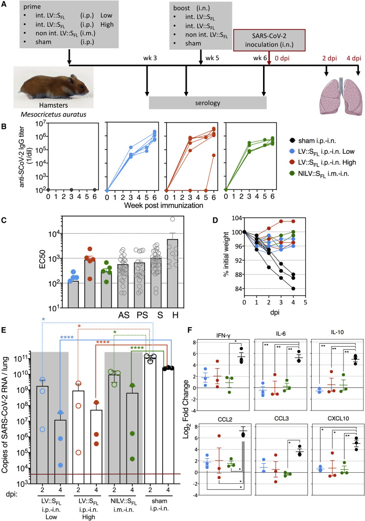
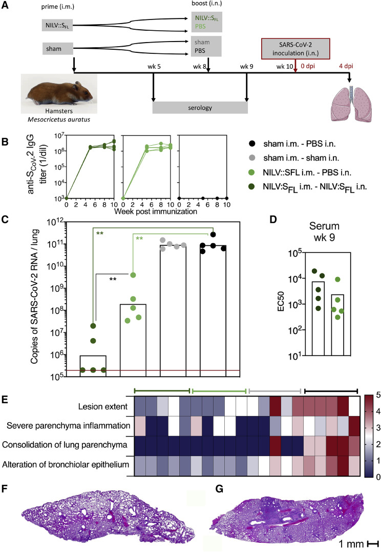
To develop a vaccine candidate against coronavirus disease 2019 (COVID-19), we generated a lentiviral vector (LV) eliciting neutralizing antibodies against the Spike glycoprotein of SARS-CoV-2. Systemic vaccination by this vector in mice, in which the expression of the SARS-CoV-2 receptor hACE2 has been induced by transduction of respiratory tract cells by an adenoviral vector, confers only partial protection despite high levels of serum neutralizing activity. However, eliciting an immune response in the respiratory tract through an intranasal boost results in a >3 log10 decrease in the lung viral loads and reduces local inflammation. Moreover, both integrative and non-integrative LV platforms display strong vaccine efficacy and inhibit lung deleterious injury in golden hamsters, which are naturally permissive to SARS-CoV-2 replication and closely mirror human COVID-19 physiopathology. Our results provide evidence of marked prophylactic effects of LV-based vaccination against SARS-CoV-2 and designate intranasal immunization as a powerful approach against COVID-19.

Keywords: beta-coronavirus; boost-target; golden hamsters; immunoglobulin A; in vivo Ad5 transduction; intranasal vaccination; lentiviral vectors; lung inflammation; mucosal immunity; neutralizing antibodies; respiratory tracts.


Conflict of interest statement

Declaration of interests P.C. is the founder and CSO of TheraVectys. M.-W.K., P.A., J.L., K.N., F.M., A.N., B.V., F.N., and F.A. are employees of TheraVectys. M.-W.K., M.B., P.A., N.E., L.M., and P.C. are inventors of a pending patent directed to a vaccine candidate against SARS-CoV2.

Figures

None
Graphical abstract
Figure 1
Figure 1
Induction of anti-SCoV-2 Ab responses by LV (A) Schematic representation of three forms of SCoV-2 protein (SFL, S1-S2, and S1) encoded by LV injected into mice. RBD, S1/S2 and S2′ cleavage sites, fusion peptide (FP), transmembrane domain (TM), and short internal tail (T) are indicated. (B) Dynamic of anti-SCoV-2 Ab response after LV immunization. C57BL/6 mice (n = 4 per group) received an i.p. injection of 1 × 107 TU of LV::GFP as a negative control, LV::S1, LV::S1-S2, or LV::SFL. Sera were collected 2, 3, 4, and 6 weeks after immunization. Anti-SCoV-2 IgG responses were evaluated by ELISA and expressed as mean endpoint dilution titers. (C) Neutralization capacity of anti-SCoV-2 Abs induced by LV::SFL immunization. Mouse sera were evaluated in a sero-neutralization assay for determination of EC50 neutralizing titers. (D) Correlation between the Ab titers and neutralization activity in various experimental groups. Statistical significance was determined by a two-sided Spearman rank-correlation test. NS, not significant. (E) Head-to-head comparison at a 1:40 dilution between mouse sera taken 3 or 4 weeks after immunization and a cohort of mildly symptomatic individuals living in Crépy-en-Valois, Ile de France. These patients did not seek medical attention and recovered from COVID-19. Results are expressed as mean ± SEM percentages of inhibition of luciferase activity. See also Figure S1.
Figure 2
Figure 2
Induction of T cell responses by LV::SFL C57BL/6 mice (n = 3) were immunized with 1 × 107 TU of LV::SFL or a negative control LV via i.p. injection. (A) Splenocytes collected 2 weeks after immunization were subjected to an IFN-γ ELISPOT with 16 distinct pools of 15-mer peptides spanning the entire SCoV-2 (1–1273 aa) and overlapping each other by 10 aa residues. SFU, spot-forming units. (B) Deconvolution of the positive peptide pools by ELISPOT applied to splenocytes pooled from three LV::SFL-or control-LV-immunized mice. (C) Intracellular IFN-γ versus IL-2 staining of CD4+ or CD8+ T splenocytes after stimulation with individual peptides encompassing the immuodominant epitopes.
Figure 3
Figure 3
Setup of a murine model expressing hACE2 in the respiratory tracts (A) Detection of hACE2 expression by RT-PCR in HEK293 T cells transduced with Ad5::hACE2 2 days after transduction. NT, not transduced. (B) hACE2 protein detection by western blot in lung cell extracts recovered 4 days after i.n. instillation of Ad5::hACE2 or empty Ad5 into C57BL/6 mice (n = 2 per group). (C) GFP expression in lung cells prepared 4 days after i.n. instillation of Ad5::GFP or PBS into C57BL/6 mice, as assessed by flow cytometry in CD45+ hematopoietic or EpCam+ epithelial cells. (D) Lung viral loads in mice pretreated with 2.5 × 109 IGU of Ad5::hACE2, control empty Ad5, or PBS and then inoculated with 1 × 105 TCID50 of SARS-CoV-2 via i.n. administration 4 days later. In one group, we inoculated the Ad5::hACE2-pretreated mice with an equivalent amount of heat-killed virus to measure the input viral RNA in the absence of viral replication. Viral load was quantitated by qRT-PCR in the lung homogenates at 2, 4, or 7 dpi. The red line indicates the detection limit. (E) Percentages of CD45+ cells in the lungs as determined 4 days after pretreatment with various doses of Ad5::hACE2. (F) Lung viral loads in mice pretreated with various doses of Ad5::hACE2 and then inoculated with 1 × 105 TCID50 of SARS-CoV-2 via i.n. administration 4 days later. Viral loads were determined at 3 dpi. See also Figure S2.
Figure 4
Figure 4
Intranasal boost with LV::SFL strongly protects against SARS-CoV-2 in mice (A) Timeline of the LV-based prime-boost strategy, followed by Ad5::hACE2 pretreatment and SARS-CoV-2 challenge. (B) Titers of anti-SCoV-2 IgG as quantitated by ELISA in the sera of C57BL/6 mice primed at week 0 via the i.p. route and boosted at week 3 via the i.p. or i.n. route (left). Titers were determined as a mean endpoint dilution before the boost (week 3) and challenge (week 4). ∗∗∗p < 0.001, ∗∗∗∗p < 0.0001; two-way ANOVA followed by Sidak’s multiple-comparison test. NS, not significant. The neutralization capacity of these sera is indicated as EC50 (right). See also Figure S4A. (C). Lung viral loads at 3 dpi in mice primed (i.p. route) and boosted (i.p. or i.n. route) with LV::SFL. Sham-vaccinated mice received an empty LV. The red line indicates the detection limit. Statistical significance of the differences in the viral loads was evaluated by a two-tailed unpaired t test; p < 0.0139, ∗∗∗p < 0.0088. (D) Titers of anti-SCoV-2 IgG and IgA Abs determined in the clarified lung homogenates by ELISA with foldon-trimerized SCoV-2 for coating. See also Figure S4B. (E) Neutralizing activity of the clarified lung homogenates as determined for 1/5 dilution. Statistical significance of the difference was evaluated by a Mann-Whitney U test (p < 0.0159). See also Figure S3.
Figure 5
Figure 5
LV::SFL vaccination reduces SARS-Co-2-mediated lung inflammation in mice (A) Cytometric strategy to identify and quantify distinct lung innate immune cell subsets. Lung hematopoietic CD45+ cells were analyzed with the use of Abs specific to surface markers or a combination of surface markers, allowing characterization of innate immune cell populations, via three distinct paths and by sequential gating. Cell populations are highlighted in gray. (B) Percentages of each cell subset versus total lung CD45+ cells at 3 dpi in mice sham vaccinated or vaccinated with LV::SFL after various prime-boost regimens versus non-infected (NI) controls. All mice were pretreated with Ad5::hACE-2 4 days prior to SARS-CoV-2 inoculation. (C) Relative log2 fold change in mRNA expression of cytokines and chemokines in mice sham vaccinated or vaccinated with LV::SFL after various prime-boost regimens at 3 dpi. Data were normalized to those of PBS-treated, unchallenged controls. Statistical significance was evaluated by a two-tailed unpaired t test; p < 0.05, ∗∗p < 0.01, ∗∗∗p < 0.001, ∗∗∗∗p < 0.0001.
Figure 6
Figure 6
Intranasal vaccination with LV::SFL strongly protects against SARS-CoV-2 in golden hamsters (A) Timeline of prime-boost or prime-target immunization regimen and challenge in hamsters. Sham-vaccinated mice received an empty LV. (B) Dynamic of anti-SCoV-2 Ab response following immunization. Sera were collected 3, 5 (before the boost), and 6 (after the boost) weeks after priming. Anti-SCoV-2 IgG responses were evaluated by ELISA. (C) EC50 serum neutralizing titers after the boost or target regimen versus sera from a cohort of asymptomatic (AS), pauci-symptomatic (PS), or symptomatic (S) COVID-19 cases or of hospitalized (H) humans. (D) Weight follow-up in hamsters either sham- or LV::SFL-vaccinated with diverse regimens. For further clarity, only the individuals reaching 4 dpi are shown. Those sacrificed at 2 dpi had the same mean weight as their counterparts between 0 and 2 dpi. (E) Lung viral loads at 2 or 4 dpi in LV::SFL-vaccinated hamsters. Statistical significance was evaluated by a two-tailed unpaired t test; p < 0.0402, ∗∗∗∗p < 0.0001. See also Figure S4C. (F) Relative log2 fold changes in cytokine and chemokine expression in LV::SFL-vaccinated and protected hamsters versus sham-vaccinated individuals, as determined at 4 dpi by qRT-PCR in the total lung homogenates and normalized to untreated controls. Statistical significance was evaluated by one-way ANOVA; p < 0.05, ∗∗p < 0.01. See also Figures S5 and S6.
Figure 7
Figure 7
Protective efficacy of NILV::SFL in a systemic prime and i.n. boost regimen in hamsters (A) Timeline of the NILV::SFL prime-boost or prime-target immunization regimen and challenge. (B) Profile of serum anti-SCoV-2 IgG response following a single (i.m.) injection or a prime (i.m.)-boost (i.n.) immunization with NILV::SFL. (C) Lung viral loads at 4 dpi with SARS-CoV-2 in controls or NILV::SFL-vaccinated hamsters. Statistical significance was evaluated by a two-tailed unpaired t test; ∗∗p < 0.01. (D) EC50 serum neutralizing titers after the boost-target regimen. (E) Lung histological H&E analysis shown in a heatmap recapitulating the histological scores for each parameter defined in Figure S7C and determined for individuals of various groups at 4 dpi. (F and G) Representative whole-lung section from NILV::SFL i.m.-NILV::SFL i.n. (F) or sham i.m.-sham i.n. (G) hamsters. See also Figure S7.

References

    1. Amanat F., Krammer F. SARS-CoV-2 vaccines: status report. Immunity. 2020;52:583–589. - PMC - PubMed
    1. Anna F., Goyard S., Lalanne A.I., Nevo F., Gransagne M., Souque P., Louis D., Gillon V., Turbiez I., Bidard F.-C., et al. High seroprevalence but short-lived immune response to SARS-CoV-2 infection in Paris. Eur. J. Immunol. 2020 doi: 10.1002/eji.202049058. - DOI - PMC - PubMed
    1. Beignon A.S., Mollier K., Liard C., Coutant F., Munier S., Rivière J., Souque P., Charneau P. Lentiviral vector-based prime/boost vaccination against AIDS: pilot study shows protection against Simian immunodeficiency virus SIVmac251 challenge in macaques. J. Virol. 2009;83:10963–10974. - PMC - PubMed
    1. Belouzard S., Chu V.C., Whittaker G.R. Activation of the SARS coronavirus Spike protein via sequential proteolytic cleavage at two distinct sites. Proc. Natl. Acad. Sci. USA. 2009;106:5871–5876. - PMC - PubMed
    1. Bourgine M., Crabe S., Lobaina Y., Guillen G., Aguilar J.C., Michel M.L. Nasal route favors the induction of CD4+ T cell responses in the liver of HBV-carrier mice immunized with a recombinant hepatitis B surface- and core-based therapeutic vaccine. Antiviral Res. 2018;153:23–32. - PubMed

Publication types

MeSH terms