. 2020 Dec 22;33(12):108546.
doi: 10.1016/j.celrep.2020.108546.

RTEL1 Regulates G4/R-Loops to Avert Replication-Transcription Collisions

Affiliations

RTEL1 Regulates G4/R-Loops to Avert Replication-Transcription Collisions

Panagiotis Kotsantis et al. Cell Rep. .

Abstract

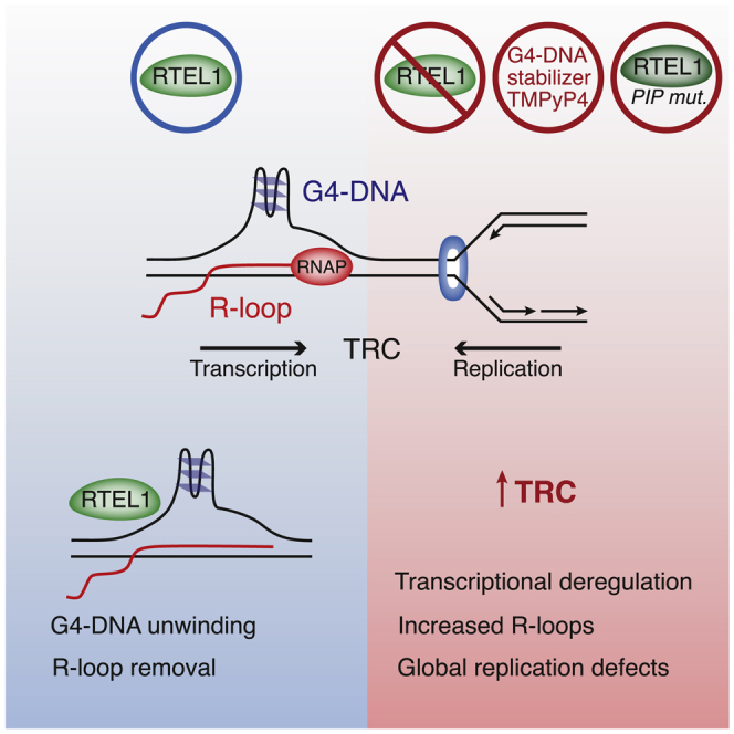
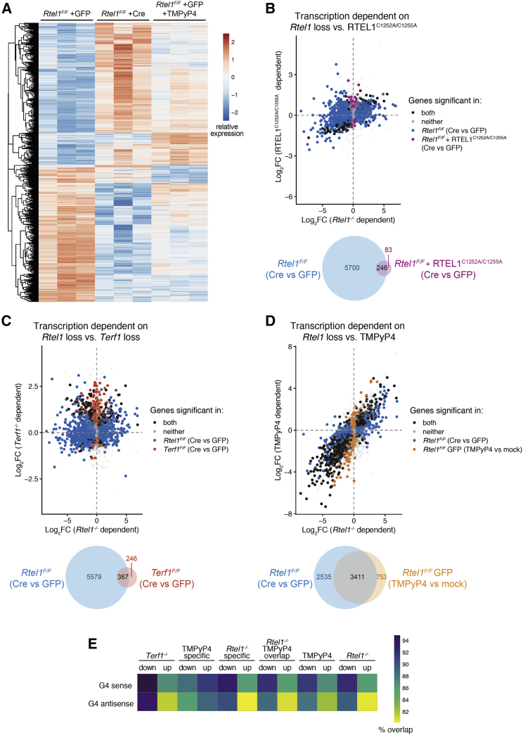
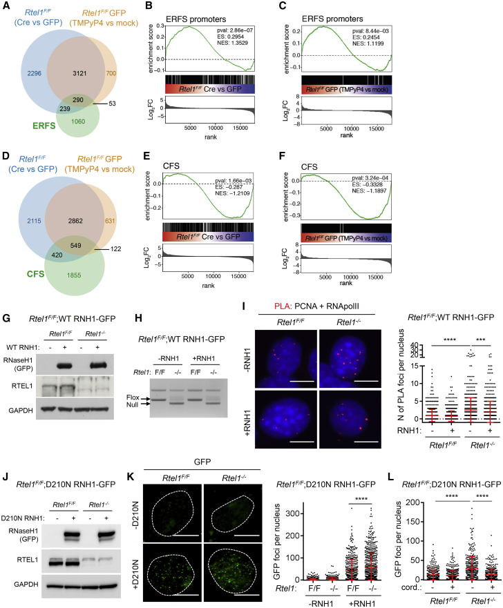
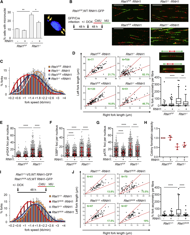
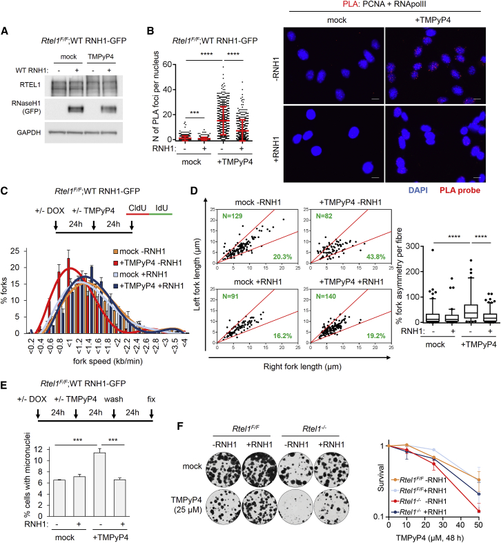
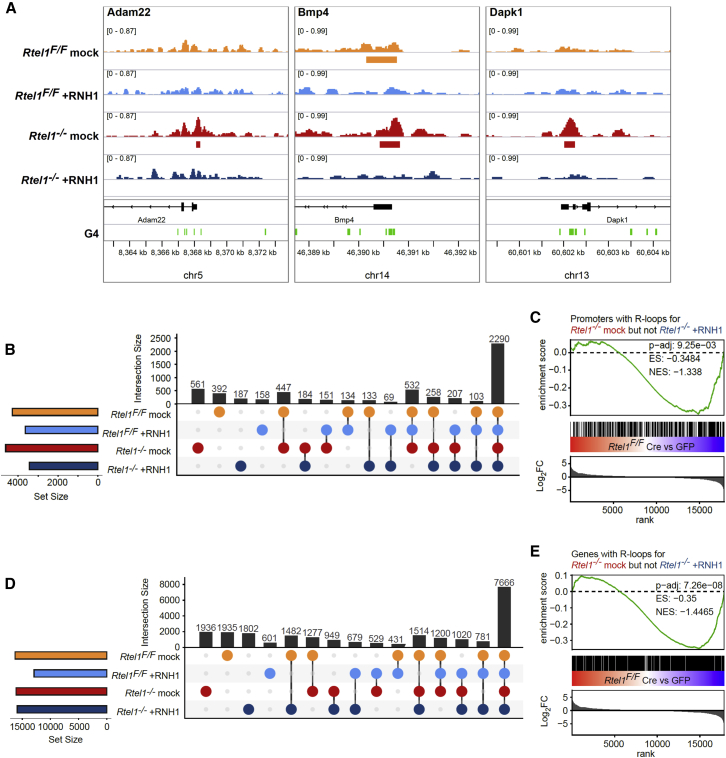
Regulator of telomere length 1 (RTEL1) is an essential helicase that maintains telomere integrity and facilitates DNA replication. The source of replication stress in Rtel1-deficient cells remains unclear. Here, we report that loss of RTEL1 confers extensive transcriptional changes independent of its roles at telomeres. The majority of affected genes in Rtel1-/- cells possess G-quadruplex (G4)-DNA-forming sequences in their promoters and are similarly altered at a transcriptional level in wild-type cells treated with the G4-DNA stabilizer TMPyP4 (5,10,15,20-Tetrakis-(N-methyl-4-pyridyl)porphine). Failure to resolve G4-DNAs formed in the displaced strand of RNA-DNA hybrids in Rtel1-/- cells is suggested by increased R-loops and elevated transcription-replication collisions (TRCs). Moreover, removal of R-loops by RNaseH1 overexpression suppresses TRCs and alleviates the global replication defects observed in Rtel1-/- and Rtel1PIP_box knockin cells and in wild-type cells treated with TMPyP4. We propose that RTEL1 unwinds G4-DNA/R-loops to avert TRCs, which is important to prevent global deregulation in both transcription and DNA replication.

Keywords: G-quadruplexes; G4-DNA structures; R-loops; RTEL1; genome instability; replication stress; transcription.


Conflict of interest statement

Declaration of Interests S.J.B. is also scientific co-founder and VP Science Strategy at Artios Pharma, Babraham Research Campus, Cambridge, UK.

Figures

None
Graphical abstract
Figure 1
Figure 1
Rtel1 Deletion Induces Transcriptional Changes That Are Independent of Its Role in Telomere Maintenance and That Overlap with Those Caused by G4 Stabilization (A) RTEL1F/F MEFs were infected with GFP or Cre-GFP adenovirus and collected after 96 h or infected with GFP adenovirus for 48 h, treated with TMPyP4, and collected after 48 h. RNA was isolated, and gene expression levels were analyzed. Heatmap of norm transformed counts per significantly deregulated gene (p < 0.01 in any comparison between groups). Data are scaled by row and clustered with hierarchical clustering. (B) Top: comparative differential gene expression between samples when Rtel1 was deleted (Rtel1F/F, Cre versus GFP) or contained a t-loop unwinding defect (Rtel1F/F + RTEL1C1252A/C1255A, Cre versus GFP). Differentially expressed genes are differentiated by their significance (p < 0.01) in the respective comparisons. Bottom: Venn diagram of differentially expressed genes. (C) Top: comparative differential gene expression between samples when Rtel1 was deleted (Rtel1F/F, Cre versus GFP) or are shelterin defective (Terf1F/F, Cre versus GFP). Bottom: Venn diagram of differentially expressed genes. (D) Top: comparative differential gene expression between samples when Rtel1 was deleted (Rtel1F/F, Cre versus GFP) or treated with TMPyP4 (Rtel1F/F GFP, TMPyP4 versus mock). Bottom: Venn diagram of differentially expressed genes. (E) Heatmap of comparative proportions of G4-containing promoters in sense and antisense of differentially regulated genes following various treatments as indicated.
Figure 2
Figure 2
RTEL1 Counteracts Transcription-Replication Conflicts by Regulating R-Loops (A) Venn diagram of differentially regulated genes of samples with deleted Rtel1 (Rtel1F/F, Cre versus GFP) or samples treated with TMPyP4 (Rtel1F/F GFP, TMPyP4 versus mock), and their overlap with ERFSs. (B) GSEA that shows enrichment of genes with ERFSs in their promoters. Genes are ranked dependent of logarithm of fold change (Log2FC) of differential expression with deleted Rtel1 (Rtel1F/F, Cre versus GFP), and the overall enrichment score (ES) and normalized enrichment score (NES) with the respective p value (pval) of the enrichment were determined. (C) GSEA that shows enrichment of genes with ERFSs in their promoters. Genes are ranked dependent of Log2FC of differential expression upon TMPyP4 treatment, and the overall ES and NES with the respective pval of the enrichment were determined. (D) Venn diagram of differentially regulated genes of samples with deleted Rtel1 (Rtel1F/F, Cre versus GFP) or treated with TMPyP4 (Rtel1F/F GFP, TMPyP4 versus mock), and their overlap with CFSs. (E) GSEA that shows enrichment of genes overlapping with CFSs. Genes are ranked dependent of Log2FC of differential expression with deleted Rtel1 (Rtel1F/F, Cre versus GFP), and the overall ES and NES with the respective pval of the enrichment were determined. (F) GSEA that shows enrichment of genes with CFSs in their promoters. Genes are ranked dependent of Log2FC of differential expression upon TMPyP4 treatment, and the overall ES and NES with the respective pval of the enrichment were determined. (G) Rtel1F/F;WT RNH1-GFP MEFs were infected with GFP or Cre-GFP adenovirus. After 48 h, doxycycline was added, and cells were collected after 48 h. The cells were then lysed, and whole-cell extracts were analyzed by SDS-PAGE and immunoblotted for GFP, RTEL1, and glyceraldehyde 3-phosphate dehydrogenase (GAPDH). (H) Cells were treated as in (G), and genomic DNA was isolated and loss of Rtel1 was verified by PCR. (I) Cells were treated as in (G), and the interaction between PCNA and RNA polymerase II (RNApolII) was assessed by PLA. Left: representative images of PLA. Right: quantification of PLA. Data are represented as mean ± SD (n = 4). (J) Rtel1F/F;D210N RNH1-GFP MEFs were infected with red fluorescent protein (RFP) or improved Cre-RFP (iCre-RFP) adenovirus. After 48 h, doxycycline was added, and cells were collected after 48 h. The cells were then lysed, and whole-cell extracts were analyzed by SDS-PAGE and immunoblotted for GFP, RTEL1, and GAPDH. (K) Cells were treated as in (J) and immunostained for GFP. Left: representative images of GFP immunofluorescence. Right: quantification of RNaseH1D210N-GFP foci per nucleus of cells. Data are represented as mean ± SD (n = 3). (L) Rtel1F/F;D210N RNH1-GFP MEFs were infected with RFP or iCre-RFP adenovirus. After 96 h, cells were treated with cordycepin for 3.5 h and immunostained for GFP. Quantification of RNaseH1D210N-GFP foci per nucleus of cells. Data are represented as mean ± SD (n = 3). The pvals were determined by unpaired t test, with ∗∗∗p < 0.001 and ∗∗∗∗p < 0.0001. Scale bars, 10 μm.
Figure 3
Figure 3
Rtel1 Deletion Causes Genome-wide R-Loop-Dependent Replication Stress (A) Rtel1F/F;WT RNH1-GFP MEFs were infected with GFP or Cre-GFP adenovirus. After 48 h, doxycycline was added, and cells were collected after 48 h. The cells were then fixed, and the percentage of cells with >1 micronucleus was quantified. Left: quantification. Right: representative images of micronuclei. Data are represented as mean ± SE (n = 3). (B) Rtel1F/F;WT RNH1-GFP MEFs were treated as in (A), and a DNA fiber assay was performed. Left: experimental setup. Right: representative images of DNA fibers. (C) Distribution of replication fork speeds of DNA fibers as prepared in (B). Data are represented as mean ± SE (n = 3). (D) Left: scatterplot of fork asymmetry of DNA fibers as prepared in (B). Right, top: representative images of symmetric and asymmetric DNA fibers. Right, bottom: quantification of fork asymmetry of DNA fibers as prepared in (B) (n = 4). (E) Rtel1F/F;WT RNH1-GFP MEFs were treated as in (A) and immunostained for γH2AX. Quantification of γH2AX foci per nucleus. Data are represented as mean ± SD (n = 3). (F) Rtel1F/F;WT RNH1-GFP MEFs were treated as in (A) and immunostained for 53BP1. Quantification of 53BP1 foci per nucleus. Data are represented as mean ± SD (n = 3). (G) Rtel1F/F;WT RNH1-GFP MEFs were treated as in (A) and immunostained for pATR-S428. Quantification of pATR-S428 foci per nucleus. Data are represented as mean ± SD (n = 3). (H) Colony formation capacity in Rtel1F/F;WT RNH1-GFP MEFs infected with GFP or Cre-GFP adenovirus with or without doxycycline. Data are mean ± SD normalized to GFP and 0 μg/mL doxycycline condition (n = 3). (I) Rtel1+/+-V5;WT RNH1-GFP and Rtel1IA/IA-V5;WT RNH1-GFP were treated with doxycycline for 48 h, and a DNA fiber assay was performed. Top: experimental setup. Bottom: distribution of replication fork speeds of DNA fibers. Data are represented as mean ± SE (n = 3). (J) Left: scatterplot of fork asymmetry of DNA fibers prepared as in (I). Right, bottom: quantification of fork asymmetry of DNA fibers prepared as in (I) (n = 3). In boxplots, horizontal line denotes the mean; whiskers denote the 5th and 95th percentiles. The pvals were determined by unpaired t test, with p < 0.05, ∗∗p < 0.01, ∗∗∗p < 0.001, and ∗∗∗∗p < 0.0001. Scale bars, 10 μm.
Figure 4
Figure 4
G4-DNA Stabilization Causes Genome-wide R-Loop-Dependent Replication Stress (A) Rtel1F/F;WT RNH1-GFP MEFs were treated with doxycycline. After 24 h, TMPyP4 was added for 24 h. Cells were then collected and lysed, and whole-cell extracts were analyzed by SDS-PAGE and immunoblotted for GFP, RTEL1, and GAPDH. (B) Rtel1F/F;WT RNH1-GFP MEFs were treated as in (A), and the interaction between PCNA and RNApolII was assessed by PLA. Right: representative images of PLA. Left: quantification of PLA (n = 3). (C) Rtel1F/F;WT RNH1-GFP MEFs were treated as in (A), and DNA fiber assay was performed. Top: experimental setup. Bottom: distribution of replication fork speeds of DNA fibers. Data are represented as mean ± SE (n = 3). (D) Left: scatterplot of fork asymmetry of DNA fibers as prepared in (C). Right, bottom: quantification of fork asymmetry of DNA fibers as prepared in (C). In boxplots, horizontal line denotes the mean; whiskers denote the 5th and 95th percentiles (n = 3). (E) Rtel1F/F;WT RNH1-GFP MEFs were treated with doxycycline. After 24 h, TMPyP4 was added for 24 h and then removed. Cells were incubated for another 24 h and then fixed. The percentage of cells with micronuclei was quantified. (F) Right: TMPyP4 colony survival assay in Rtel1F/F;WT RNH1-GFP MEFs. Data are mean ± SEM normalized to untreated cells (n = 3). Left: representative images of colonies. The pvals were determined by unpaired t test, with ∗∗∗p < 0.001 and ∗∗∗∗p < 0.0001. Scale bars, 10 μm.
Figure 5
Figure 5
Rtel1−/−-Induced R-Loops Are Related to Rtel1−/−-Induced Transcriptional Changes (A) Rtel1F/F;WT RNH1-GFP MEFs were infected with GFP or Cre-GFP adenovirus. After 48 h, doxycycline was added, and cells were collected after 48 h and used for R-loop detection with DRIP-seq. Genome browser plots of normalized read coverage, called peaks of DRIP-seq, and associated predicted G4 structures in three different genomic locations. (B) Overlap analysis of DRIP-seq peaks in promoters. Upset plot that depicts the numbers of promoters with R-loops shared between Rtel1F/F and Rtel1−/−, dependent on RNaseH1. (C) GSEA that shows transcriptional enrichment of genes with Rtel1−/−-specific and RNH1-sensitive promoter R-loops. Genes are ranked dependent of Log2FC of differential expression with deleted Rtel1 (Rtel1F/F, Cre versus GFP). The overall ES and NES with the respective pvals were determined of the enrichment with R-loop peaks that are present with Rtel1 deletion, yet not present with RNaseH1 overexpression. (D) Overlap analysis of DRIP-seq peaks in genes. Upset plot that depicts the numbers of genes with R-loops shared between Rtel1F/F and Rtel1−/−, dependent on RNaseH1. (E) GSEA that shows transcriptional enrichment of genes with Rtel1−/−-specific and RNaseH1-sensitive R-loops. Genes are ranked dependent of Log2FC of differential expression with deleted Rtel1 (Rtel1F/F, Cre versus GFP). The overall ES and NES with the respective pvals were determined of the enrichment with R-loop peaks that are present with Rtel1 deletion, yet not present with RNaseH1 overexpression.

References

    1. Arora R., Lee Y., Wischnewski H., Brun C.M., Schwarz T., Azzalin C.M. RNaseH1 regulates TERRA-telomeric DNA hybrids and telomere maintenance in ALT tumour cells. Nat. Commun. 2014;5:5220. - PMC - PubMed
    1. Barber L.J., Youds J.L., Ward J.D., McIlwraith M.J., O’Neil N.J., Petalcorin M.I., Martin J.S., Collis S.J., Cantor S.B., Auclair M. RTEL1 maintains genomic stability by suppressing homologous recombination. Cell. 2008;135:261–271. - PMC - PubMed
    1. Barlow J.H., Faryabi R.B., Callén E., Wong N., Malhowski A., Chen H.T., Gutierrez-Cruz G., Sun H.W., McKinnon P., Wright G. Identification of early replicating fragile sites that contribute to genome instability. Cell. 2013;152:620–632. - PMC - PubMed
    1. Barnett D.W., Garrison E.K., Quinlan A.R., Strömberg M.P., Marth G.T. BamTools: a C++ API and toolkit for analyzing and managing BAM files. Bioinformatics. 2011;27:1691–1692. - PMC - PubMed
    1. Bedrat A., Lacroix L., Mergny J.L. Re-evaluation of G-quadruplex propensity with G4Hunter. Nucleic Acids Res. 2016;44:1746–1759. - PMC - PubMed

Publication types