

Paper-based electrochemical biosensor for diagnosing COVID-19: Detection of SARS-CoV-2 antibodies and antigen
- PMID: 33358057
- PMCID: PMC7746088
- DOI: 10.1016/j.bios.2020.112912
Paper-based electrochemical biosensor for diagnosing COVID-19: Detection of SARS-CoV-2 antibodies and antigen
Abstract
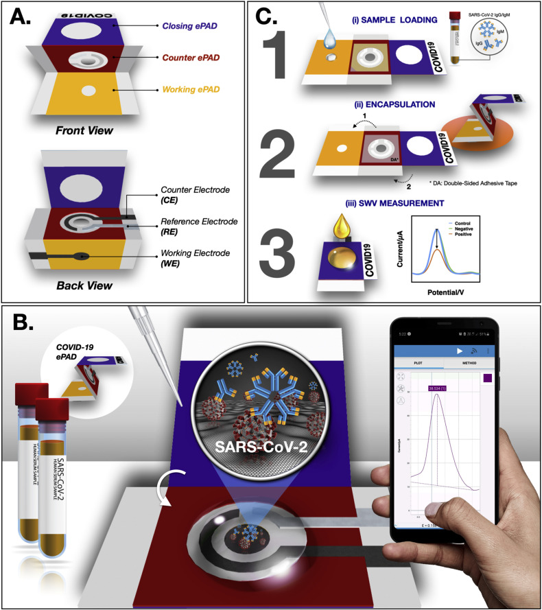
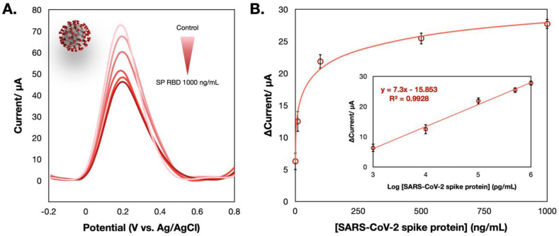
Coronavirus disease 2019 (COVID-19) caused by severe acute respiratory syndrome coronavirus 2 (SARS-CoV-2) is emerging as a global pandemic outbreak. To date, approximately one million deaths and over 32 million cases have been reported. This ongoing pandemic urgently requires an accurate testing device that can be used in the field in a fast manner. Serological assays to detect antibodies have been proven to be a great complement to the standard method of reverse transcription-polymerase chain reaction (RT-PCR), particularly after the second week of infection. We have developed a specific and sensitive immunosensor for immunoglobulin detection produced against SARS-CoV-2. Unlike other lateral flow-based assays (LFAs) involving the utilization of multiple antibodies, we have reported a label-free paper-based electrochemical platform targeting SARS-CoV-2 antibodies without the specific requirement of an antibody. The presence of SARS-CoV-2 antibodies will interrupt the redox conversion of the redox indicator, resulting in a decreased current response. This electrochemical sensor was proven effective in real clinical sera from patients with satisfactory results. In addition, the proposed format was also extended to antigen detection (the spike protein of SARS-CoV-2), which presents new possibilities for diagnosing COVID-19.
Keywords: COVID-19; Electrochemical detection; Paper-based sensors; SARS-CoV-2.
Copyright © 2020 Elsevier B.V. All rights reserved.
Conflict of interest statement
The authors declare that they have no known competing financial interests or personal relationships that could have appeared to influence the work reported in this paper.
Figures
References
-
- Boonkaew S., Teengam P., Jampasa S., Rengpipat S., Siangproh W., Chailapakul O. Analyst. 2020;145(14):5019–5026. - PubMed
-
- Burrell C.J., Howard C.R., Murphy F.A. In: Fenner and White's Medical Virology. fifth ed. Burrell C.J., Howard C.R., Murphy F.A., editors. Academic Press; London: 2017. Chapter 6 - adaptive immune responses to infection; pp. 65–76.
-
- Carrilho E., Martinez A.W., Whitesides G.M. Anal. Chem. 2009;81(16):7091–7095. - PubMed
-
- Carvajal D.N., Rowe P.C. Research and statistics. Pediatr. Rev. 2010;31(12):511. - PubMed
-
- Casali P. In: Encyclopedia of Immunology. second ed. Delves P.J., editor. Elsevier; Oxford: 1998. IgM; pp. 1212–1217.
MeSH terms
Substances
LinkOut - more resources
Full Text Sources
Other Literature Sources
Medical
Miscellaneous
