Review
. 2021 Feb;32(2):118-129.
doi: 10.1016/j.tem.2020.11.008. Epub 2020 Dec 23.

Key Metabolic Functions of β-Arrestins: Studies with Novel Mouse Models

Affiliations
Review

Key Metabolic Functions of β-Arrestins: Studies with Novel Mouse Models

Sai P Pydi et al. Trends Endocrinol Metab. 2021 Feb.

Abstract

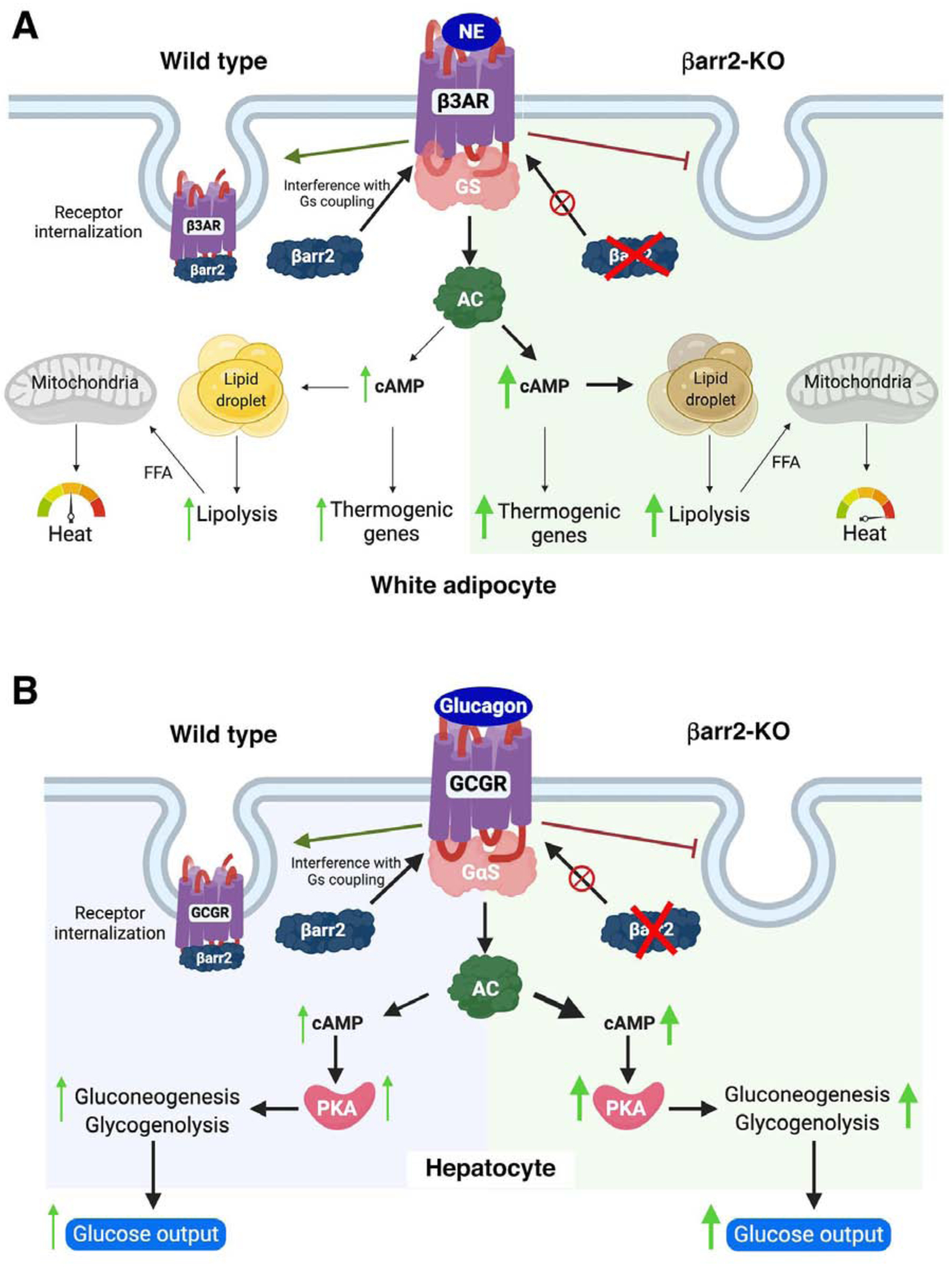
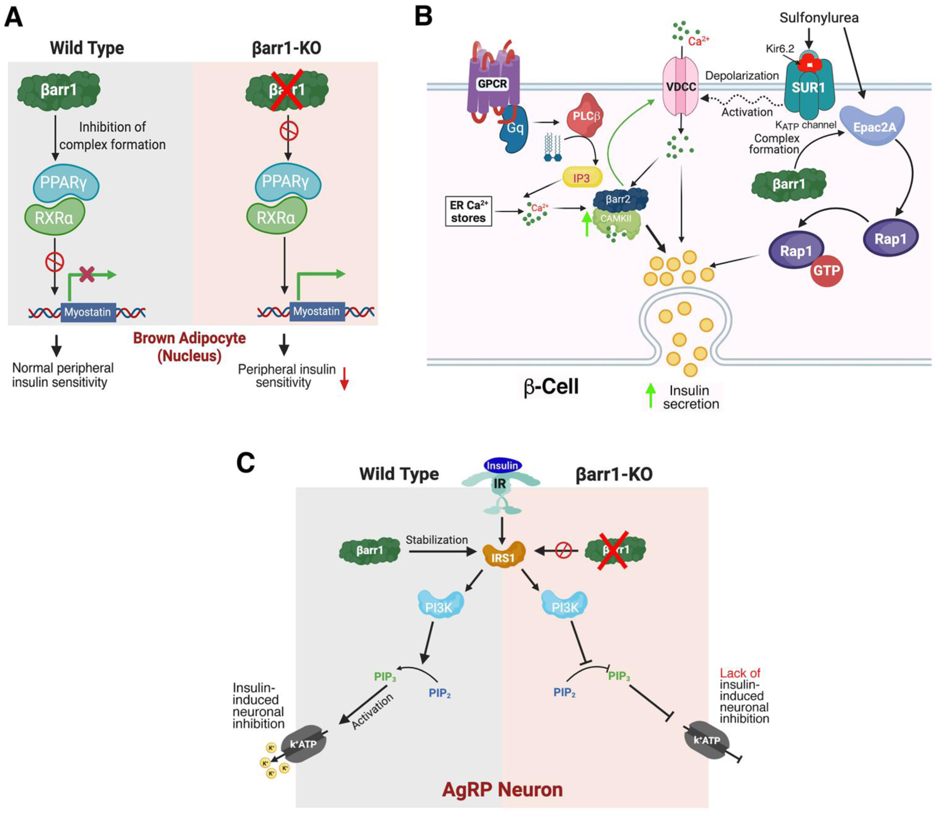
β-Arrestin-1 and -2 are intracellular proteins that are able to inhibit signaling via G protein-coupled receptors (GPCRs). However, both proteins can also modulate cellular functions in a G protein-independent fashion. During the past few years, studies with mutant mice selectivity lacking β-arrestin-1 and/or -2 in metabolically important cell types have led to novel insights into the mechanisms through which β-arrestins regulate key metabolic processes in vivo, including whole-body glucose and energy homeostasis. The novel information gained from these studies should inform the development of novel drugs, including β-arrestin- or G protein-biased GPCR ligands, that could prove useful for the therapy of several important pathophysiological conditions, including type 2 diabetes and obesity.

Keywords: G protein-coupled receptors; diabetes; metabolism; mutant mice; obesity; β-arrestins.


Conflict of interest statement

Disclosure Statement

The authors have no financial conflicts of interest to disclose.

Figures

Figure 1.
Figure 1.
Canonical and non-canonical functions of β-arrestins.
Figure 2.
Figure 2.
Canonical functions of βarr2 in mouse adipocytes and hepatocytes. A) Adipocytes. In white adipocytes from wild type mice, βarr2 is involved in terminating β3-AR signaling by interacting with activated β3-ARs and promoting receptor internalization. In white adipocytes lacking βarr2, this inhibitory regulation of β3-AR function is absent, resulting in enhanced β3-AR-mediated cellular effects [34]. B) Hepatocytes. In hepatocytes from wild type mice, βarr2 acts as a negative regulator of glucagon receptor (GCGR) signaling. In βarr2-deficient hepatocytes, GCGR signaling is no longer subject to inhibition by βarr2, leading to enhanced glucose output [49]. AC, adenyl cyclase; FFA, free fatty acids; NE, norepinephrine; PKA; protein kinase A.
Figure 3.
Figure 3.
Non-canonical functions of β-arrestins in mouse adipocytes, pancreatic β-cells, and AgRP neurons. A) Adipocytes. In brown adipocytes from wild type mice, βarr1 binding to the PPARγ/RXRα complex is predicted to prevent activation of the myostatin (Mstn) promoter [51] In the absence of βarr1, the PPARγ/RXRα complex can activate the Mstn promoter, leading to increased plasma Mstn levels which cause peripheral insulin resistance [51]. B) β-Cells. The left side of the cartoon shows that βarr2 is required for the proper function of CAMKII in β-cells. βarr2 is predicted to form a complex with CAMKII that stimulates CAMKII activity in β-cells [21]. Activated CAMKII stimulates insulin secretion via phosphorylation of various signaling proteins involved in insulin exocytosis and has been shown to facilitate glucose-dependent calcium influx by acting on VDCCs [89]. Impaired CAMKII activity can fully account for the metabolic impairments observed with beta-βarr2-KO mice [21]. The right side of the cartoon shows that β-cell βarr1 is required for the proper function of most sulphonylurea drugs (SUs). By binding to the SUR1 subunit of the β-cell ATP-gated K+ channel, SU drugs cause channel closure, triggering membrane depolarization, Ca2+ influx through VDCCs, and eventually insulin secretion. Most SUs also stimulate the formation of a βarr1/Epac2a complex, which promotes Rap1-mediated insulin secretion [22]. C) AgRP neurons. In AgRP neurons from wild type mice, insulin induces hyperpolarization and reduced firing frequency by activating a pathway that leads to the opening of KATP channels. In the absence of βarr1, insulin-mediated inhibition of AgRP neurons is abolished, most likely due to reduced stability of IRS1 (IRS1 is stabilized by βarr1 in wild type neurons) [83]. AC, adenylyl cyclase; DAG, diacylglycerol; ER, endoplasmic reticulum; IP3, inositol trisphosphate; IR, insulin receptor; IRS1, insulin receptor substrate 1; PIP2, phosphatidylinositol 4,5-bisphosphate; PIP3, phosphatidylinositol 3,4,5-trisphosphate; PI3K, phosphoinositide 3-kinase; PKA, protein kinase A; PKC, protein kinase C; PPARγ, peroxisome proliferator-activated receptor γ; RXRα, retinoid X receptor α, VDCC, voltage-dependent calcium channel.

References

    1. Sriram K and Insel PA (2018) G Protein-Coupled Receptors as Targets for Approved Drugs: How Many Targets and How Many Drugs? Mol Pharmacol 93 (4), 251–258. - PMC - PubMed
    1. Pierce KL and Lefkowitz RJ (2001) Classical and new roles of beta-arrestins in the regulation of G-protein-coupled receptors. Nat Rev Neurosci 2 (10), 727–33. - PubMed
    1. Tian X et al. (2014) β-arrestins and G protein-coupled receptor trafficking. Handb Exp Pharmacol 219, 173–86. - PMC - PubMed
    1. Gurevich VV and Gurevich EV (2019) Plethora of functions packed into 45 kDa arrestins: biological implications and possible therapeutic strategies. Cell Mol Life Sci 76 (22), 4413–4421. - PMC - PubMed
    1. Peterson YK and Luttrell LM (2017) The Diverse Roles of Arrestin Scaffolds in G Protein-Coupled Receptor Signaling. Pharmacol Rev 69 (3), 256–297. - PMC - PubMed

Publication types

Substances