Review
. 2021 Mar:145:111200.
doi: 10.1016/j.exger.2020.111200. Epub 2020 Dec 24.

Aging of the immune system and impaired muscle regeneration: A failure of immunomodulation of adult myogenesis

Affiliations
Review

Aging of the immune system and impaired muscle regeneration: A failure of immunomodulation of adult myogenesis

James G Tidball et al. Exp Gerontol. 2021 Mar.

Abstract

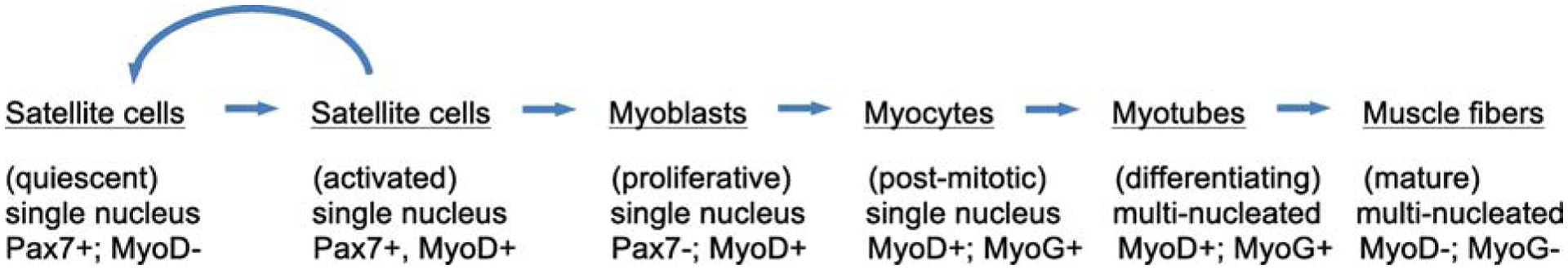
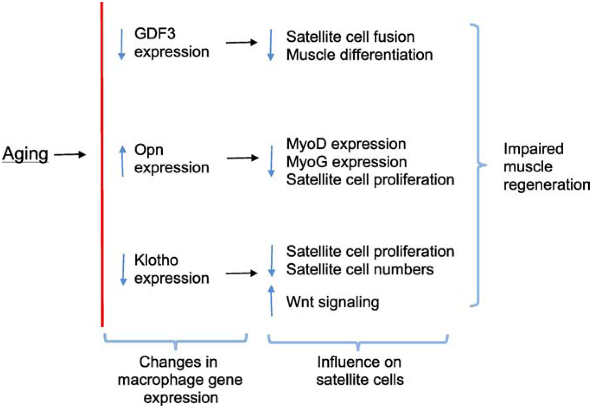
Skeletal muscle regeneration that follows acute injury is strongly influenced by interactions with immune cells that invade and proliferate in the damaged tissue. Discoveries over the past 20 years have identified many of the key mechanisms through which myeloid cells, especially macrophages, regulate muscle regeneration. In addition, lymphoid cells that include CD8+ T-cells and regulatory T-cells also significantly affect the course of muscle regeneration. During aging, the regenerative capacity of skeletal muscle declines, which can contribute to progressive loss of muscle mass and function. Those age-related reductions in muscle regeneration are accompanied by systemic, age-related changes in the immune system, that affect many of the myeloid and lymphoid cell populations that can influence muscle regeneration. In this review, we present recent discoveries that indicate that aging of the immune system contributes to the diminished regenerative capacity of aging muscle. Intrinsic, age-related changes in immune cells modify their expression of factors that affect the function of a population of muscle stem cells, called satellite cells, that are necessary for normal muscle regeneration. For example, age-related reductions in the expression of growth differentiation factor-3 (GDF3) or CXCL10 by macrophages negatively affect adult myogenesis, by disrupting regulatory interactions between macrophages and satellite cells. Those changes contribute to a reduction in the numbers and myogenic capacity of satellite cells in old muscle, which reduces their ability to restore damaged muscle. In addition, aging produces changes in the expression of molecules that regulate the inflammatory response to injured muscle, which also contributes to age-related defects in muscle regeneration. For example, age-related increases in the production of osteopontin by macrophages disrupts the normal inflammatory response to muscle injury, resulting in regenerative defects. These nascent findings represent the beginning of a newly-developing field of investigation into mechanisms through which aging of the immune system affects muscle regeneration.

Keywords: Aging; Muscle inflammation; Muscle injury; Muscle regeneration; Skeletal muscle.


Figures

Figure 1.
Figure 1.
Approximate, relative timelines for mice, rats and humans.
Figure 2.
Figure 2.
Time course of muscle regeneration after intramuscular BaCl2 injection. Hematoxylin-stained cross-sections of TA muscles from 3-month-old (A-C) and 24-month-old (D-F) mice, either non-injured (A, D), or 3-dpi (B, E) or 7-dpi (C, F). Representative fibers with central nuclei (white arrows) and areas of inflammation or edema between damaged fibers (*) are indicated. Note that 3-month-old muscle at 7-dpi shows numerous, relatively-large, regenerating fibers that contain multiple, central nuclei and few inflammatory cells between fibers (C). Muscle from 24-month-old mice at 7-dpi shows fewer and smaller central-nucleated fibers and more numerous inflammatory cells between fibers. Bars = 120 μm.
Figure 3.
Figure 3.
Pax7-expressing cells in non-injured (A) and BaCl2-injured TA muscles at 7-dpi (B, C) from 6-month-old mice. A. In non-injured muscle, Pax7+ cells satellite cells appear at relatively-low numbers in tissue sections, flattened on the surface of healthy muscle fibers. An anti-Pax7-labeled cell is stained red (arrow). B: At 7-dpi, areas of relatively little muscle damage show elevated numbers of satellite cells located on the surface of muscle fibers. Some fibers are associated with more than one satellite cell at the fiber surface (*), indicating satellite cell proliferation at the fiber surface. C: At 7-dpi, areas of more extensive muscle damage show elevated numbers of satellite cells, many of which have left the surface of muscle fibers, to occupy the extracellular space (arrows), where they may undergo further differentiation and fusion to form myotubes. Bars = 50 μm.
Figure 4.
Figure 4.
General overview of the stages of satellite cell activation and muscle cell differentiation and maturation following acute injury. Activated satellite cells can either return to quiescence or proceed along a course of differentiation to contribute to mature muscle fibers. A few of the key transcription factors that are expressed at different stages of adult myogenesis are indicated.
Figure 5.
Figure 5.
General overview of the stages of hematopoiesis that give rise to myeloid and lymphoid cells that can influence muscle regeneration. Hematopoietic stem cells that reside in the bone marrow enter either the myeloid or lymphoid cell lineage by differentiating to become either common myeloid progenitors (CMPs) or common lymphoid progenitors (CLPs). CLPs then further differentiate to enter either the B-cell or T-cell lineage. B-cells are not known to have any direct influences on adult myogenesis. T-cells then differentiate to become CD8+ or CD4+ T-cells, which have direct effects on muscle regeneration. CMPs differentiate to become granulocyte progenitors (GPs) or monocyte progenitors (MPs). MPs then further differentiate to monocytes and then become macrophages, which have the most prominent influence on muscle regeneration of all immune cell populations.
Figure 6.
Figure 6.
Non-injured and BaCl2-injured (3 dpi) TA muscles, immunolabeled for F/80+ macrophages (A, B) and CD206+ macrophages (C, D). Note that macrophages in non-injured muscle (A, C) are small, indicating lack of activation, versus macrophages in areas of extensive injury and inflammation where they are greatly enlarged, reflecting their activation (B, D). Representative macrophages in each sample are indicated by arrows. Bars = 50 μm.
Figure 7.
Figure 7.
Summary diagram of changes in gene expression in intramuscular macrophages that occur during aging and the consequence of those changes on satellite cell function and numbers.

References

    1. Ahrens HE, Huettemeister J, Schmidt M, Kaether C, von Maltzahn J, 2018. Klotho expression is a prerequisite for proper muscle stem cell function and regeneration of skeletal muscle. Skeletal Muscle 8, 20 10.1186/s13395-018-0166-x. - DOI - PMC - PubMed
    1. Allbrook DB, Han MF, Hellmuth AE, 1971. Population of muscle satellite cells in relation to age and mitotic activity. Pathology 3, 223–243. 10.3109/00313027109073739. - DOI - PubMed
    1. Allen RE, Boxhorn LK, 1989. Regulation of skeletal muscle satellite cell proliferation and differentiation by transforming growth factor-beta, insulin-like growth factor I, and fibroblast growth factor. J. Cell. Physiol 138, 311–315. 10.1002/jcp.1041380213. - DOI - PubMed
    1. Arnold L, Henry A, Poron F, Baba-Amer Y, van Rooijen N, Plonquet A, Gherardi RK, Chazaud B, 2007. Inflammatory monocytes recruited after skeletal muscle injury switch into 38nti-inflammatory macrophages to support myogenesis. J. Exp. Med 204, 1057–1069. 10.1084/jem.20070075. - DOI - PMC - PubMed
    1. Ashkar S, Weber GF, Panoutsakopoulou V, Sanchirico ME, Jansson M, Zawaideh S, Rittling SR, Denhardt DT, Glimcher MJ, Cantor H, 2000. Eta-1 (osteopontin): an early component of type-1 (cell-mediated) immunity. Science 287, 860–864. 10.1126/science.287.5454.860. - DOI - PubMed

Publication types