

Unusual collision tumor with infiltrating ductal carcinoma and breast skin squamous cell carcinoma: A case report and literature review
- PMID: 33360037
- PMCID: PMC7758454
- DOI: 10.1016/j.ijscr.2020.12.010
Unusual collision tumor with infiltrating ductal carcinoma and breast skin squamous cell carcinoma: A case report and literature review
Abstract
Introduction: Breast cancer is the most common diagnosed cancer among women worldwide. Invasive ductal carcinoma (IDC) is the most common type, on the other hand, squamous cell carcinoma of the skin (SCC) overlying the breast is a rare tumor. The co-presence of two tumor types in one organ is even a rarer entity, termed as collision tumor. Only 3 known cases of collision tumor with breast invasive ductal and skin squamous carcinoma were reported in the literature.
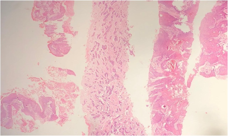
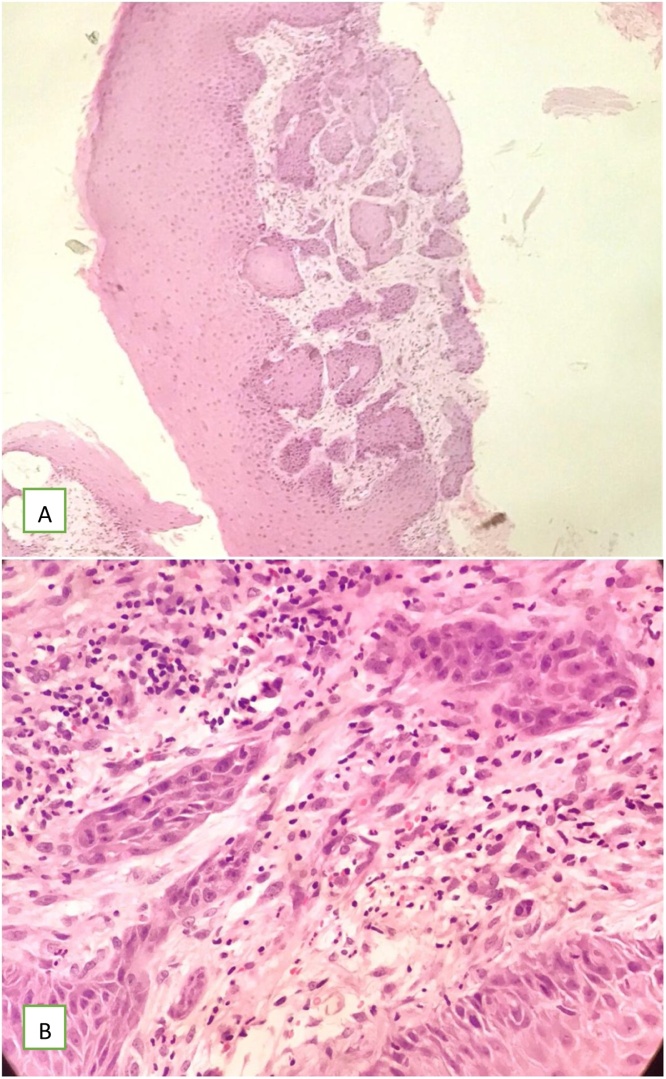
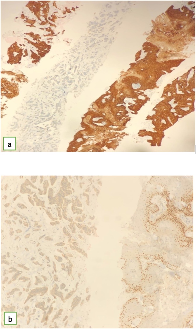
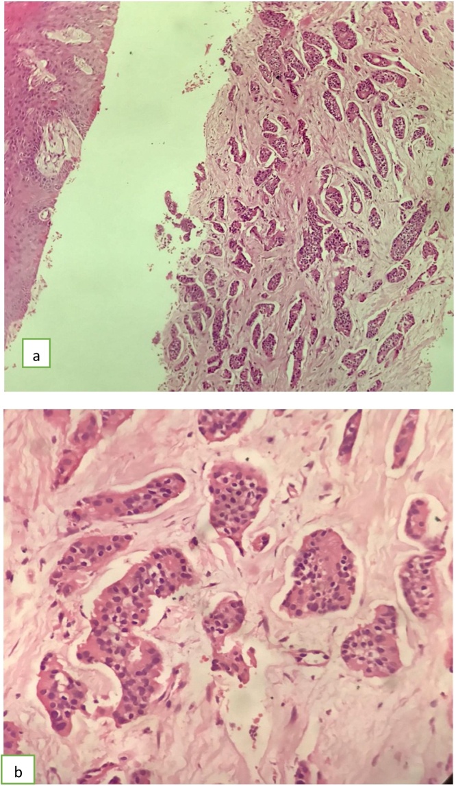
Case presentation: An otherwise medically free 91-year-old, postmenopausal, female presented with left breast fungating mass for four months. Pre-operative core tissue biopsy and incisional skin biopsy revealed two distinct tumor subtypes of invasive ductal carcinoma, positive for progesterone, estrogen receptors and negative for human epidermal growth factor receptor 2, as well as skin squamous cell carcinoma, and axillary lymph node metastasis. Patient underwent left breast modified radical mastectomy and split skin grafting for wound closure. The final histopathology was consistent with grade 2 IDC. The nipple and areola complex were involved by moderately differentiated squamous cell carcinoma. Currently patient on adjuvant hormonal treatment. Follow up showed no local recurrence or distal metastasis.
Conclusion: Collision tumors of the breast with IDC and SCC of the overlying skin is very rare. The surgeon has to be aware of of such entity as the proper peri-operative management should be tailored to target the most aggressive histologic subtype.
Keywords: Breast; Cancer; Case report; Collision; Invasice ductal cancer; Squamous cell cancer.
Copyright © 2020 The Authors. Published by Elsevier Ltd.. All rights reserved.
Figures


References
-
- Agha R.A., Borrelli M.R., Farwana R., Koshy K., Fowler A., Orgill D.P., For the SCARE Group The SCARE 2018 Statement: Updating Consensus Surgical CAse REport (SCARE) guidelines. Int. J. Surg. 2018;60:132–136. - PubMed
-
- Siegel R.L., Miller K.D., Jemal A. Cancer statistics, 2020. CA Cancer J. Clin. 2020;70:7. - PubMed
-
- Rosen P.R. Lippincott Williams & Wilkins, 2001; Philadelphia, PA: 1997. Rosen’s Breast Pathology; pp. 455–461.
-
- Tayeb K., Saadi I., Kharmash M. Primary squamous cell carcinoma of the breast: report of three cases [French] Cancer Radiother. 2002;6:366–368. - PubMed
-
- Behranwala K.A., Nasiri N., Abdullah N. Squamous cell carcinoma of the breast:clinicopathologic implications and outcome. Eur. J. Surg. Oncol. 2003;29(386) - PubMed
Publication types
LinkOut - more resources
Full Text Sources
Other Literature Sources
Research Materials
