. 2021 Jan-Jun:296:100247.
doi: 10.1074/jbc.RA120.016350. Epub 2021 Jan 14.

Functional metagenomics of the thioredoxin superfamily

Affiliations

Functional metagenomics of the thioredoxin superfamily

Sebastian Nilewski et al. J Biol Chem. 2021 Jan-Jun.

Abstract

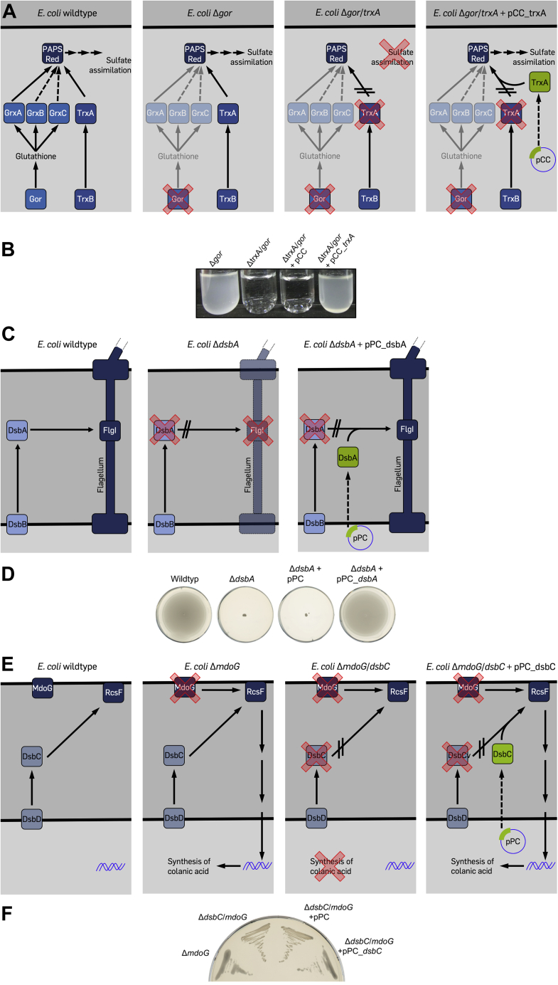
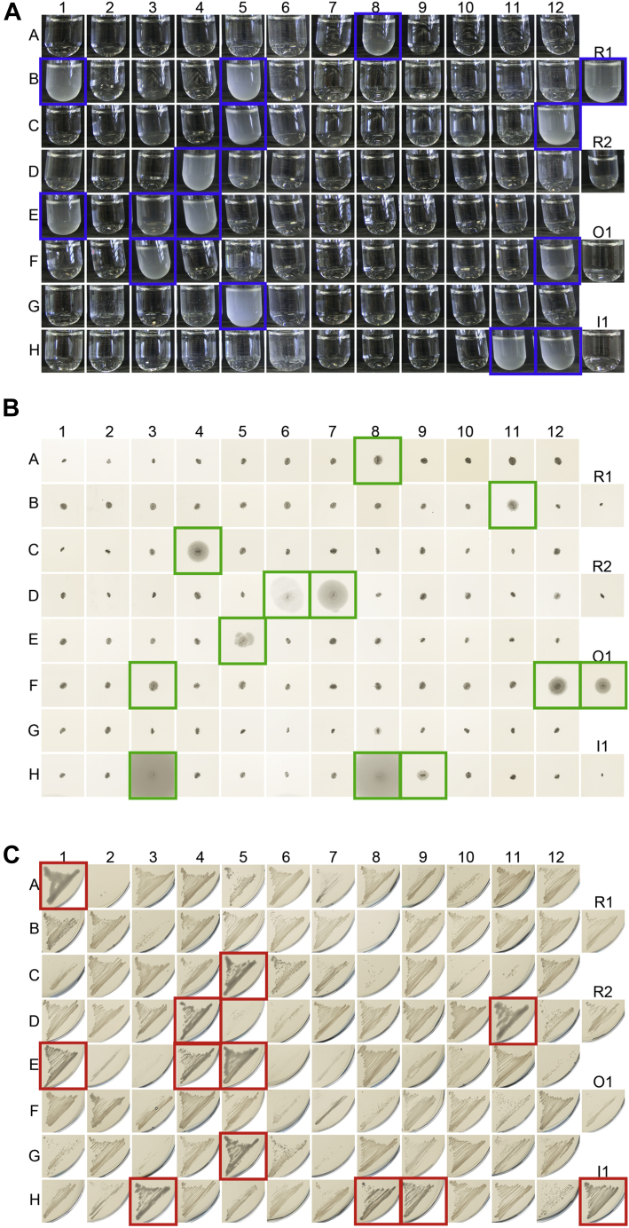
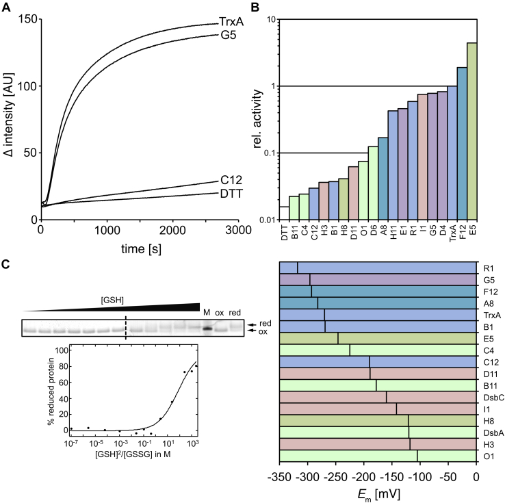
Environmental sequence data of microbial communities now makes up the majority of public genomic information. The assignment of a function to sequences from these metagenomic sources is challenging because organisms associated with the data are often uncharacterized and not cultivable. To overcome these challenges, we created a rationally designed expression library of metagenomic proteins covering the sequence space of the thioredoxin superfamily. This library of 100 individual proteins represents more than 22,000 thioredoxins found in the Global Ocean Sampling data set. We screened this library for the functional rescue of Escherichia coli mutants lacking the thioredoxin-type reductase (ΔtrxA), isomerase (ΔdsbC), or oxidase (ΔdsbA). We were able to assign functions to more than a quarter of our representative proteins. The in vivo function of a given representative could not be predicted by phylogenetic relation but did correlate with the predicted isoelectric surface potential of the protein. Selected proteins were then purified, and we determined their activity using a standard insulin reduction assay and measured their redox potential. An unexpected gel shift of protein E5 during the redox potential determination revealed a redox cycle distinct from that of typical thioredoxin-superfamily oxidoreductases. Instead of the intramolecular disulfide bond formation typical for thioredoxins, this protein forms an intermolecular disulfide between the attacking cysteines of two separate subunits during its catalytic cycle. Our functional metagenomic approach proved not only useful to assign in vivo functions to representatives of thousands of proteins but also uncovered a novel reaction mechanism in a seemingly well-known protein superfamily.

Keywords: DsbA; DsbC; Escherichia coli (E. coli); TrxA; oxidase; protein disulfide isomerase; reductase; thiol; thiol-disulfide oxidoreductase; thioredoxin.


Conflict of interest statement

Conflict of interest The authors declare that they have no conflicts of interest with the contents of this article.

Figures

Figure 1
Figure 1
Phylogenetic tree of all 4678 complete thioredoxin superfamily members found in the Global Ocean Sampling data set. Different colors represent phylogenetic clades identified by Markov clustering. Representative sequences chosen for this study are indicated by arrows. See also Table S1 and Supporting Information S1.
Figure 2
Figure 2
Plasmids used in this study. pCC (plasmid for Cytoplasmic Complementation) and pPC (plasmid for Periplasmic Complementation) are derivatives of pTAC-MAT-Tag-2 (Sigma–Aldrich). pOE (plasmid for OverExpression) is a derivative of pET-11a (Agilent). Thioredoxin superfamily members were inserted between NdeI (containing the original gene's start codon ATG) and EcoRI. Inserted genes contained a stop codon before EcoRI. For vector sequences, see also Supporting Information S2.
Figure 3
Figure 3
Rescue of phenotypes of thioredoxin superfamily member knockouts in Escherichia coli.A, schematic of the trxA complementation assay. E. coli wildtype is able to grow on sulfate as the only sulfur source using 3'-phosphoadenosine-5'-phosphosulfate-reductase (PAPS red)-dependent sulfate assimilation. PAPS red itself is reduced by thioredoxin (TrxA) or one of the several glutaredoxins (GrxA, GrxB, and GrxC). The knockout mutant of glutathione oxidoreductase (E. coli Δgor) effectively inactivates the glutaredoxins, but the strain is still able to grow on sulfate as the sole sulfur source because TrxA is sufficient to reduce PAPS red. A Δgor ΔtrxA double mutant (E. coli Δgor/trxA) is no longer able to reduce PAPS red. The introduction of trxA from a plasmid (pCC_trxA) restores PAPS red reduction. B, a Δgor/trxA double mutant is not viable in minimal medium containing sulfate as the sole sulfur source. Expression of E. coli TrxA from plasmid pCC rescues the phenotype. C, E. coli wildtype expresses functional flagella containing an essential disulfide bond in the FlgI subunit of the flagellar motor. This disulfide bond is introduced by the thiol-disulfide oxidoreductase DsbA. A knockout mutant lacking DsbA (E. coli ΔdsbA) is lacking functional flagella. Reintroduction of DsbA from a plasmid (pPC_dsbA) restores structural disulfide bonds in FlgI. D, a ΔdsbA mutant is nonmotile. Expression of E. coli DsbA from plasmid pPC rescues the phenotype. E, E. coli wildtype is nonmucoid. A knockout in MdoG (E. coli ΔmdoG) leads to a mucoid phenotype, as RcsF senses envelope stress and initiates a signaling cascade resulting in the production of colanic acid. The correct disulfide connectivity in RcsF is dependent on the isomerase DsbC, thus a mutant lacking both MdoG and DsbC (E. coli ΔmdoG/dsbC) is nonmucoid. Expression of DsbC from a plasmid (pPC_dsbC) restores mucoidity in this double mutant. F, a ΔmdoG/dsbC double knock forms nonmucoid colonies. Expression of E. coli DsbC without export signal sequence (–SS) from plasmid pPC restores mucoidity.
Figure 4
Figure 4
Complementation assays for the assignment of functions to metagenomic thioredoxin superfamily members.A, growth phenotypes of Δgor ΔtrxA strains expressing 100 metagenomic thioredoxin superfamily members. A Δgor ΔtrxA strain of Escherichia coli is unable to grow in a minimal medium containing sulfate as the sole sulfur source (see Fig. 3A for a schematic). Only if a metagenomic reductase expressed from a plasmid acts on phosphoadenosine-5'-phosphosulfate reductase, the phenotype will be rescued. Metagenomic thioredoxin superfamily members rescuing the phenotype are highlighted in blue. B, motility phenotypes of ΔdsbA strains expressing 100 metagenomic thioredoxin superfamily members. A ΔdsbA strain is nonmotile (see Fig. 3C for a schematic). A metagenomic oxidase expressed from a plasmid could rescue the associated phenotype if it is able to introduce a structural disulfide in the bacterial flagella. Thioredoxin superfamily members restoring motility are highlighted in green. C, mucoidity of Δ mdoG ΔdsbC strains expressing 100 metagenomic thioredoxin superfamily members. A ΔmdoG ΔdsbC strain has a nonmucoid phenotype (see Fig. 3E for a schematic). Mucoidity could be restored by a metagenomic isomerase that can isomerize the correct disulfides in RcsF. Thioredoxin superfamily members rescuing RcsF function are highlighted in red. Representative results, all experiments were repeated at least three times.
Figure 5
Figure 5
Relation of assigned function with phylogeny of thioredoxin superfamily members and the isosurfaces of the electrostatic potential of reductases.A, 27 (out of 100) thioredoxin superfamily members were assigned a function based on our complementation assays. Twelve superfamily members rescued more than one phenotype. BD, phylogenetic trees and evolutionary distance of Escherichia coli thioredoxins and their metagenomic counterparts. B, metagenomic reductases. C, metagenomic isomerases. D, metagenomic oxidases. See also Figure 1 for how these proteins relate to each other and to the other thioredoxins identified in the Global Ocean Sampling data set. The presumed root of the tree is marked by a bar. Alignments of the metagenomic reductases, oxidases, and isomerases can be found in Supporting Information S3. E, isoelectric surface charge of E. coli TrxA. F, isosurfaces of the electrostatic potential based on predicted structures of metagenomic reductases. Orientation of the proteins is identical based on the structural alignment with E. coli TrxA's structure. G, consensus isosurfaces of all metagenomic reductases. For predicted isoelectric surfaces of all metagenomic thioredoxins, see Fig. S1. For predicted isoelectric surfaces of metagenomic oxidases and isomerases, see Fig. S2.
Figure 6
Figure 6
Escherichia coli TrxA (A), DsbC (B), and DsbA (C) only rescue their respective phenotype. No overlapping function was detected in the selected assays. The native signal sequence for periplasmic export was removed from DsbC and DsbA (–SS).
Figure 7
Figure 7
Insulin reduction activity and midpoint redox potential of thioredoxin superfamily members.A, representative insulin reduction activity measurement of Escherichia coli TrxA and the metagenomic reductases G5 and C12, TrxA's phylogenetically most closely and distantly related thioredoxin superfamily members. B, relative insulin reduction activity of metagenomic thioredoxin superfamily members. Activity of E. coli TrxA was set to 1. Color coding according to function (cf.Fig. 5A). C, representative redox potential determination of metagenomic thioredoxin G5. Arrows labeled red and ox indicate the migration of the 4-acetamido-4'-maleimidylstilbene-2,2'-disulfonic acid–treated reduced and oxidized G5, respectively. Because of the limited number of available pockets on an SDS gel, samples were run on two separate gels. The dashed lane marks the splice border. D, standard redox potential E0 of metagenomic thioredoxin superfamily members. Color coding according to function (cf.Fig. 5A). M, marker.
Figure 8
Figure 8
Metagenomic thioredoxin E5 is a new i thioredoxin superfamily member with an unusual reaction mechanism.A, E5 is an effective disulfide isomerase in the commercial Escherichia coli SHUFFLE strain and can support oxidative folding of several proteins, including vtPA, which contains nine disulfide bonds when folded correctly. B, it shows higher initial velocity than E. coli TrxA in an insulin reduction assay. C, redox potential determination of E5 using 4-acetamido-4'-maleimidylstilbene-2,2'-disulfonic acid, as described for Figure 7C. When oxidized in a glutathione redox buffer, E5 forms disulfide-linked dimers, which can be observed in nonreducing SDS-PAGE. M, marker, MW in kDa indicated on the right, ox. and red.: fully oxidized and reduced E5, respectively. D, the redox potential can be calculated from the ratio of reduced and oxidized protein in a given glutathione redox buffer based on the Nernst equation. Based on these calculations, E5 has a standard redox potential of −246 mV, close to the redox potential of thioredoxin. E, reduced E5 and E. coli TrxA reduce insulin when no DTT for enzyme recycling is added. F, E5 protein oxidized in this reaction forms disulfide-linked dimers, which can be observed in a Coomassie-stained SDS-PAGE. Higher molecular weight species indicated by arrows are presumably insulin adducts to the E5 dimer and E5 multimers crosslinked by insulin. G, an E5 C77S mutant, which contains only one cysteine, still forms a disulfide-linked dimer when oxidized with diamide and (H) is still active in an insulin reduction assay, while an E5 C74S mutant is inactive. I, size exclusion choromatography of E5 and its C77S mutant suggests a molecular weight consistent approximately with a homohexamer (the calculated mass of E5 is 23,580 Da), when compared with a protein mixture used as standard (THY, thyroglobulin; FER, ferritin; ALD, aldolase; CON, conalbumin; OVA, ovalbumin; CAR, carbonic anhydrase; and RIB, ribonuclease). To calculate the molecular weight of E5, high and low molecular weight standard proteins were run in separate runs, and their molecular mass was plotted against the elution volume. The plot was fitted using an exponential function in Excel.
Figure 9
Figure 9
Ellman's assay to determine free thiols in reduced and oxidized metagenomic thioredoxin representative E5. DTNB and diamide-oxidized or DTT-reduced E5 protein (50 μM) were added to potassium phosphate buffer. As a control, no protein was added (DTNB only), and release of TNB was monitored at 412 nm over time. DTNB, dithionitrobenzoic acid.
Figure 10
Figure 10
An E5 C77S mutant lacking the resolving cysteine does not complement the DsbA phenotype.

References

    1. Höög J.O., Jörnvall H., Holmgren A., Carlquist M., Persson M. The primary structure of Escherichia coli glutaredoxin. Distant homology with thioredoxins in a superfamily of small proteins with a redox-active cystine disulfide/cysteine dithiol. Eur. J. Biochem. 1983;136:223–232. - PubMed
    1. Perez-Jimenez R., Inglés-Prieto A., Zhao Z.-M., Sanchez-Romero I., Alegre-Cebollada J., Kosuri P., Garcia-Manyes S., Kappock T.J., Tanokura M., Holmgren A., Sanchez-Ruiz J.M., Gaucher E.A., Fernandez J.M. Single-molecule paleoenzymology probes the chemistry of resurrected enzymes. Nat. Struct. Mol. Biol. 2011;18:592–596. - PMC - PubMed
    1. Collet J.-F., Messens J. Structure, function, and mechanism of thioredoxin proteins. Antioxid. Redox Signal. 2010;13:1205–1216. - PubMed
    1. Sousa S.F., Neves R.P.P., Waheed S.O., Fernandes P.A., Ramos M.J. Structural and mechanistic aspects of S-S bonds in the thioredoxin-like family of proteins. Biol. Chem. 2019;400:575–587. - PubMed
    1. Martin J.L. Thioredoxin--a fold for all reasons. Structure. 1995;3:245–250. - PubMed

Publication types

MeSH terms