. 2021 Feb 26;371(6532):eabc8378.
doi: 10.1126/science.abc8378. Epub 2020 Dec 23.

Afucosylated IgG characterizes enveloped viral responses and correlates with COVID-19 severity

Collaborators, Affiliations

Afucosylated IgG characterizes enveloped viral responses and correlates with COVID-19 severity

Mads Delbo Larsen et al. Science. .

Abstract

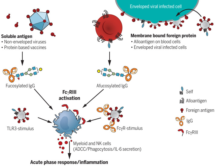
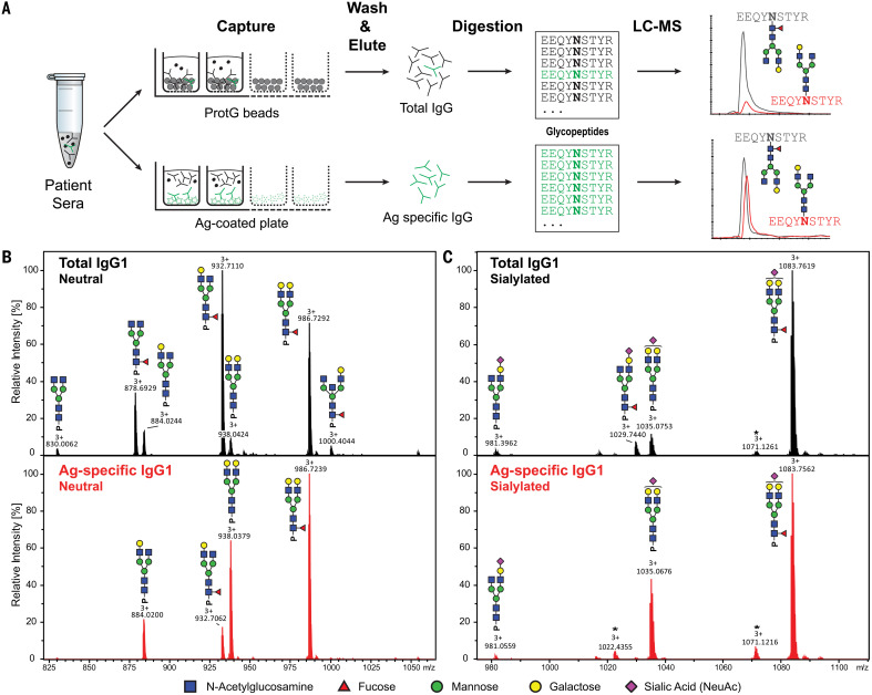
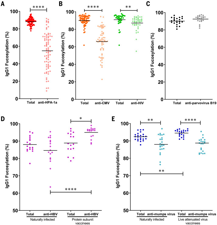
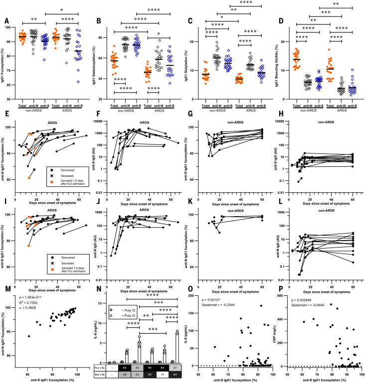
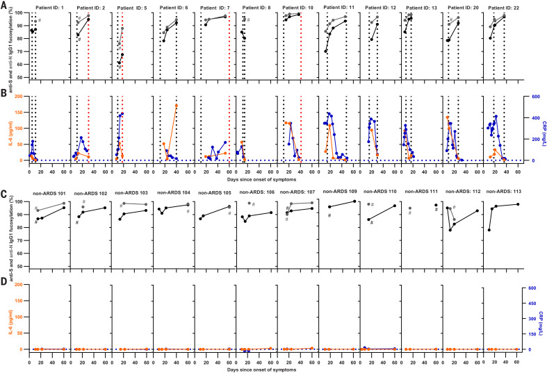
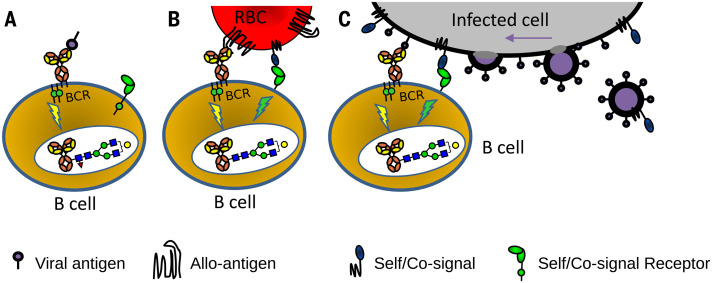
Immunoglobulin G (IgG) antibodies are crucial for protection against invading pathogens. A highly conserved N-linked glycan within the IgG-Fc tail, which is essential for IgG function, shows variable composition in humans. Afucosylated IgG variants are already used in anticancer therapeutic antibodies for their increased activity through Fc receptors (FcγRIIIa). Here, we report that afucosylated IgG (approximately 6% of total IgG in humans) are specifically formed against enveloped viruses but generally not against other antigens. This mediates stronger FcγRIIIa responses but also amplifies brewing cytokine storms and immune-mediated pathologies. Critically ill COVID-19 patients, but not those with mild symptoms, had high concentrations of afucosylated IgG antibodies against severe acute respiratory syndrome coronavirus 2 (SARS-CoV-2), amplifying proinflammatory cytokine release and acute phase responses. Thus, antibody glycosylation plays a critical role in immune responses to enveloped viruses, including COVID-19.


Figures

None
Afucosylated IgG response requires membrane context and results in strong FcγRIII-mediated activity.
Only membrane association on host cells endows foreign antigens to trigger the B cell receptor in the context of other self receptors, leading to an afucosylated IgG response. The elevated FcγRIII binding and activity of afucosylated IgG can in some cases be protective, but for SARS-CoV-2, this triggers excessive inflammation during a natural infection.
Fig. 1
Fig. 1. Flowchart of antibody-specific IgG1 glycosylation analysis by use of mass spectrometry.
(A) Antibodies were captured from sera by using Protein G beads and antigen-coated 96-well plates, resulting in total and antigen-specific IgG fractions, respectively. Thereafter, isolated IgG were digested with trypsin, and the resulting glycopeptides were analyzed by means of nano–LC-MS. (B and C) Representative mass spectra of glycopeptides encompassing the Fc glycosylation site Asn297. (B) Neutral and (C) sialylated IgG1 glycopeptides are shown from a single patient’s total (top, black) and antigen-specific (bottom, red) IgG1 fraction. Asterisks indicate non-Fc glycopeptides.
Fig. 2
Fig. 2. Foreign membrane protein antigens, such as envelope proteins of (attenuated) enveloped viruses or alloantigens, can trigger afucosylated IgG responses.
(A to E) IgG1-Fc fucosylation levels of total (solid circles) and antigen-specific (open circles) antibodies are shown for each differently color-coded group of antigens: (A) alloantigen HPA-1a; (B) viral envelope antigens from CMV and HIV; (C) nonenveloped viral antigens from parvovirus B19; (D) HBsAg, in individuals (left) naturally infected with HBV or (right) vaccinated with recombinant soluble HBsAg; and (E) mumps virus antigens in individuals (left) naturally infected with mumps virus or (right) vaccinated with live attenuated mumps virus. Each circle represents a biological replicate [(A) n = 80 for anti-HPA-1a, (B) n = 65 for CMV and n = 40 for HIV, (C) n = 22 for B19, (D) n = 17 for naturally infected individuals for HBV and n = 17 for HBV vaccinated individuals, and (E) n = 24 naturally infected individuals for mumps virus and n = 21 for mumps vaccinated individuals] of a representative LC-MS run (examples of technical replicates are provided in fig. S1C). Statistical analyses were performed as paired t tests for (A), (B), and (C), and a mixed-model two-way ANOVA with Bonferroni correction of post hoc t tests for comparing Fc fucosylation between groups was performed for (D) and (E). Only statistically significant differences are shown. *P < 0.05, **P < 0.01, and ****P < 0.0001.
Fig. 3
Fig. 3. Fc fucosylation levels of anti-S IgG1 are significantly decreased in critically ill COVID-19 patients.
(A) Fc fucosylation, (B) galactosylation, (C) sialylation, and (D) bisection degree of anti-S, anti-N, and total IgG1 from ARDS patients and non-ARDS donors clearing the infection asymptomatically or with mild symptoms from the initial screen. (E to L) Longitudinal IgG1-Fc fucosylation and IgG quantity for [(E) to (H)] anti-S and [(I) to (L)] anti-N in [(E), (F), (I), and (J)] ARDS patients and [(G), (H), (K), and (L)] non-ARDS cases. (M) Correlation between anti-N and anti-S IgG1-Fc fucosylation. (N) Representative examples of IL-6 release from macrophages triggered by FcγR through stimulation with glycoengineered IgG complexes with or without polyinosinic:polycytidylic acid [poly(I:C)]. (O) Correlation between plasma IL-6 concentrations and degrees of anti-S IgG1-Fc fucosylation. (P) Correlation between plasma CRP concentrations and degrees of anti-S IgG1-Fc fucosylation. Each circle represents a biological replicate: n = 20 for ARDS, n = 23 for non-ARDS [(A) to (D)], n = 17 and n = 14 for longitudinal ARDS and non-ARDS, respectively [(E) to (L)]. Examples of technical replicates for LC-MS data are shown in fig. S1C. [(F), (H), (J), and (L)] IgG data are representative ELISA values calibrated against a standard pool from two technical experiments. For all available paired data used in (M), n = 40. (N) IL-6 production by macrophages was measured with ELISA, with each dot (n = 3) representing a technical replicate. All six biological replicates are shown in fig. S13. CRP and IL-6 numbers were obtained from clinical parameters and IL-6 data by Meso Scale Discovery, using all available paired data [(O) n = 82 and (P) n = 53]. Statistical analyses were performed as a mixed-model two-way ANOVA with Bonferroni correction of post hoc t tests for comparing glycosylation traits and cytokine secretion between groups. Spearman’s correlations were performed in (O) and (P). To test the correlation between Fc fucosylation levels for anti-S and anti-N, a Pearson’s correlation was performed. Only statistically significant differences are shown. *P < 0.05, **P < 0.01, ***P < 0.001, ****P < 0.0001.
Fig. 4
Fig. 4. Longitudinal changes of anti–SARS-CoV-2 IgG1 Fc fucosylation, CRP, and IL-6.
(A and C) Anti-S IgG fucosylation and anti-N IgG fucosylation and (B and D) IL-6 and CRP amounts in [(A) and (B)] an ARDS cohort and [(C) and (D)] a non-ARDS cohort. (A) to (D) represent longitudinal biological replicates of [(A) and (C)] a LC-MS run (examples of technical replicates are available in fig. S1C), CRP obtained from clinical parameters, and [(B) and (D)] IL-6 data by using a validated Meso Scale Discovery assay (n = 12 for ARDS and n = 14 for non-ARDS, with 2 to 16 longitudinal replicates per patient as indicated). Additional non-ARDS samples are provided in fig. S15. Hash signs (#) denote samples before these points were below the limit of detection for IgG1-glycosylation analyses. Vertical dotted lines in (A) and (B) indicate the time of ICU admission and ICU discharge (black) or death (red), whereas dotted horizontal lines in (B) and (D) indicate IL-6 and CRP detection limits.
Fig. 5
Fig. 5. Hypothetical model explaining how different antigen contexts could produce altered immune signaling that gives rise to altered IgG glycosylation.
(A) Immune responses to soluble protein antigen: B cell receptor (BCR; a membrane Ig) is activated, resulting in the production of normal fucosylated antibodies. (B) For immune responses to alloantigens, paternal alloantigens on a red blood cell (RBC) are recognized by the BCR and possibly by other undescribed immune regulatory receptor-ligand pair(s) that provide a signal for recognition of self. (C) For immune responses to enveloped viral infection and attenuated viruses, the recognition of enveloped virus–infected cells by B cells would be similar as for the recognition of cellular alloantigens (B). The initial recognition may potentially occur toward enveloped virus–infected cells and/or after viral assembly (far right). The proposed signaling in (B) and (C) causes altered glyco-programming of the B cells, culminating in a distinct IgG response characterized by a low Fc fucosylation (red triangle, fucose) and enhanced ADCC. This model potentially explains both why immune responses to soluble proteins, nonenveloped viruses, and cellular pathogens such as bacteria are different from responses to enveloped viruses (and attenuated viruses). Furthermore, it may explain why immune responses to alloantigens immunologically resemble those of enveloped viral infections.

References

    1. Biermann M. H. C., Griffante G., Podolska M. J., Boeltz S., Stürmer J., Muñoz L. E., Bilyy R., Herrmann M., Sweet but dangerous—The role of immunoglobulin G glycosylation in autoimmunity and inflammation. Lupus 25, 934–942 (2016). 10.1177/0961203316640368 - DOI - PubMed
    1. Jefferis R., Glycosylation as a strategy to improve antibody-based therapeutics. Nat. Rev. Drug Discov. 8, 226–234 (2009). 10.1038/nrd2804 - DOI - PubMed
    1. Vidarsson G., Dekkers G., Rispens T., IgG subclasses and allotypes: From structure to effector functions. Front. Immunol. 5, 520 (2014). 10.3389/fimmu.2014.00520 - DOI - PMC - PubMed
    1. Dekkers G., Rispens T., Vidarsson G., Novel concepts of altered immunoglobulin G galactosylation in autoimmune diseases. Front. Immunol. 9, 553 (2018). 10.3389/fimmu.2018.00553 - DOI - PMC - PubMed
    1. Shields R. L., Lai J., Keck R., O’Connell L. Y., Hong K., Meng Y. G., Weikert S. H. A., Presta L. G., Lack of fucose on human IgG1 N-linked oligosaccharide improves binding to human Fcgamma RIII and antibody-dependent cellular toxicity. J. Biol. Chem. 277, 26733–26740 (2002). 10.1074/jbc.M202069200 - DOI - PubMed

Publication types

MeSH terms