. 2020 Dec 23;5(6):e01193-20.
doi: 10.1128/mSphere.01193-20.

The Two-Component System CopRS Maintains Subfemtomolar Levels of Free Copper in the Periplasm of Pseudomonas aeruginosa Using a Phosphatase-Based Mechanism

Affiliations

The Two-Component System CopRS Maintains Subfemtomolar Levels of Free Copper in the Periplasm of Pseudomonas aeruginosa Using a Phosphatase-Based Mechanism

Lorena Novoa-Aponte et al. mSphere. .

Abstract

Two-component systems control periplasmic Cu+ homeostasis in Gram-negative bacteria. In characterized systems such as Escherichia coli CusRS, upon Cu+ binding to the periplasmic sensing region of CusS, a cytoplasmic phosphotransfer domain of the sensor phosphorylates the response regulator CusR. This drives the expression of efflux transporters, chaperones, and redox enzymes to ameliorate metal toxic effects. Here, we show that the Pseudomonas aeruginosa two-component sensor histidine kinase CopS exhibits a Cu-dependent phosphatase activity that maintains CopR in a nonphosphorylated state when the periplasmic Cu levels are below the activation threshold of CopS. Upon Cu+ binding to the sensor, the phosphatase activity is blocked and the phosphorylated CopR activates transcription of the CopRS regulon. Supporting the model, mutagenesis experiments revealed that the ΔcopS strain exhibits maximal expression of the CopRS regulon, lower intracellular Cu+ levels, and increased Cu tolerance compared to wild-type cells. The invariant phosphoacceptor residue His235 of CopS was not required for the phosphatase activity itself but was necessary for its Cu dependency. To sense the metal, the periplasmic domain of CopS binds two Cu+ ions at its dimeric interface. Homology modeling of CopS based on CusS structure (four Ag+ binding sites) clearly supports the different binding stoichiometries in the two systems. Interestingly, CopS binds Cu+/2+ with 3 × 10-14 M affinity, pointing to the absence of free (hydrated) Cu+/2+ in the periplasm.IMPORTANCE Copper is a micronutrient required as cofactor in redox enzymes. When free, copper is toxic, mismetallating proteins and generating damaging free radicals. Consequently, copper overload is a strategy that eukaryotic cells use to combat pathogens. Bacteria have developed copper-sensing transcription factors to control copper homeostasis. The cell envelope is the first compartment that has to cope with copper stress. Dedicated two-component systems control the periplasmic response to metal overload. This paper shows that the sensor kinase of the copper-sensing two-component system present in Pseudomonadales exhibits a signal-dependent phosphatase activity controlling the activation of its cognate response regulator, distinct from previously described periplasmic Cu sensors. Importantly, the data show that the system is activated by copper levels compatible with the absence of free copper in the cell periplasm. These observations emphasize the diversity of molecular mechanisms that have evolved in bacteria to manage the copper cellular distribution.

Keywords: Pseudomonas aeruginosa; copper; homeostasis; periplasm; two-component regulatory systems.


Figures

FIG 1
FIG 1
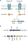
Transcriptional control mediated by TCSs. (A) Activation dynamics of canonical TCSs exemplified with the E. coli Cu-sensing CusRS. (B) Scheme of the TCS P. aeruginosa CopRS regulon. Promoter regions recognized by CopR (yellow rectangles) and transcription direction (red arrowheads) are shown. Overlapping arrows indicate that the start codon of second gene overlaps the stop codon of first gene in both pcoAB and copRS operons.
FIG 2
FIG 2
Cu tolerance of ΔcopR and ΔcopS mutant strains. Growth rate of WT, ΔcopR, ΔcopS (PW5705 and PW5706), ΔcopA1, and CopR and CopS complemented strains in the absence or the presence of increasing (0 to 4 mM) concentrations of CuSO4. Data are the mean ± SEM from at least three independent experiments.
FIG 3
FIG 3
Whole-cell Cu levels in WT, ΔcopR, ΔcopS, ΔcopA1, and CopR and CopS complemented strains under normal growth conditions (i.e., no additional CuSO4 added) (A) and after 10 min exposure to 2 mM CuSO4 (B) or 4 mM CuSO4 (C). Data are the mean ± SEM from three independent experiments. Significant differences from values with the WT strain as determined by unpaired two-tailed Student’s t test are *, P < 0.05; **, P < 0.01; ***, P < 0.001; ****, P < 0.0001.
FIG 4
FIG 4
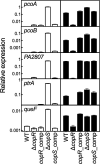
Expression of genes in the CopRS regulon in WT, ΔcopR, ΔcopS, and corresponding complemented strains in the absence (white) and the presence (black) of 0.5 mM CuSO4 (5-min treatment). Transcript levels of pcoA, pcoB, PA2807, ptrA, and queF genes are plotted relative to that of the housekeeping gene PA4268. Data are the mean ± SEM from three independent experiments.
FIG 5
FIG 5
Cu tolerance of ΔcopR and ΔcopS mutant strains complemented with CopR and CopS mutant proteins lacking the phosphorylatable residues. (A) Growth rate of the ΔcopR mutant complemented with copRD51A or copRD51E in the absence or the presence of increasing (0 to 4 mM) concentrations of CuSO4. (B) Growth rate of the ΔcopS mutant complemented with copSH235A in the presence of 0 to 4 mM CuSO4. Data are the mean ± SEM from three independent experiments.
FIG 6
FIG 6
Expression of pcoB in ΔcopR and ΔcopS mutant strains complemented with CopR and CopS lacking the phosphorylatable residues. pcoB expression was determined in the absence (white) and the presence (black) of 2 mM CuSO4 (5-min treatment) in the indicated strains. The ΔcopR mutant was complemented with copR coding for substitutions Asp51Ala and Asp51Glu. The ΔcopS mutant was complemented with the copS gene coding for substitution His235Ala. Transcript levels of pcoB are plotted relative to the housekeeping gene PA4268. Data are the mean ± SEM from three independent experiments.
FIG 7
FIG 7
Structural superposition of the periplasmic Cu+ binding loop of P. aeruginosa CopS (gray) and E. coli CusS (yellow). The structure of CopS was modeled using the CusS structure as the template (PDB ID: 5KU5 [43]). An overall root mean square deviation of 0.791 Å (Cα atoms) was calculated for the superposition of CopS and CusS structures. Conserved Cu binding sites at the dimeric interface (His41, Phe42, and His140) are shown as sticks in the structural model and highlighted in yellow in the sequence alignment. The Cu+ binding sites within the CusS orange loops (framed in rectangle in the alignment) are not conserved in CopS.
FIG 8
FIG 8
Determination of the dissociation constants KD of the periplasmic Cu binding loop of CopS(34–151). (A) Spectrophotometric titration of 100 μM BCA and 18.7 μM Cu+ with 10 to 50 μM His-tagged CopS(34–151). The arrow indicates the decrease in absorbance at 562 nm upon protein addition. The inset shows the fitting of the data set to equation 2 with a KD of (2.77 ± 0.07) × 10−14 M (R2 0.992). Two Cu sites per CopS dimer are assumed. (B) Spectrophotometric titration of 10 μM PAR and 4 μM Cu2+ with 2 to 20 μM Strep-tagged CopS(34–151). The arrows indicate the increase in absorbance at 415 nm and the decrease at 562 nm upon protein addition. The inset shows the fitting of the data set to equation 4 with a KD of (3.3 ± 0.1) × 10−14 M (R2 0.984).
FIG 9
FIG 9
Model of the phosphatase-based mechanism of the P. aeruginosa CopRS. Phosphatase On: when periplasmic free Cu remains under the subfemtomolar level, the CopS phosphatase activity maintains low levels of phosphorylated CopR, shutting off the transcriptional response to high periplasmic Cu. Phosphatase Off: upon Cu binding, CopS autophosphorylates at His235. This turns off the CopS phosphatase activity, allowing the accumulation of phosphorylated CopR and triggering the expression of the CopRS regulon (i.e., pcoA, pcoB, queF, PA2807, and ptrA).

Similar articles

Cited by

References

    1. Argüello JM, Raimunda D, Padilla-Benavides T. 2013. Mechanisms of copper homeostasis in bacteria. Front Cell Infect Microbiol 3:73. doi:10.3389/fcimb.2013.00073. - DOI - PMC - PubMed
    1. Fraústo da Silva JJR, Williams RJP. 2001. The biological chemistry of the elements: the inorganic chemistry of life, 2nd ed Oxford University Press, Oxford, United Kingdom.
    1. Macomber L, Imlay JA. 2009. The iron-sulfur clusters of dehydratases are primary intracellular targets of copper toxicity. Proc Natl Acad Sci U S A 106:8344–8349. doi:10.1073/pnas.0812808106. - DOI - PMC - PubMed
    1. Dupont CL, Grass G, Rensing C. 2011. Copper toxicity and the origin of bacterial resistance-new insights and applications. Metallomics 3:1109–1118. doi:10.1039/c1mt00107h. - DOI - PubMed
    1. Osman D, Cavet JS. 2008. Copper homeostasis in bacteria. Adv Appl Microbiol 65:217–247. doi:10.1016/S0065-2164(08)00608-4. - DOI - PubMed

Publication types

MeSH terms

LinkOut - more resources