Review
. 2020 Dec 23;34(2):e00205-20.
doi: 10.1128/CMR.00205-20. Print 2021 Mar 17.

Staphylococcus lugdunensis: a Skin Commensal with Invasive Pathogenic Potential

Affiliations
Review

Staphylococcus lugdunensis: a Skin Commensal with Invasive Pathogenic Potential

Simon Heilbronner et al. Clin Microbiol Rev. .

Abstract

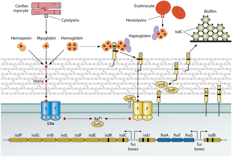
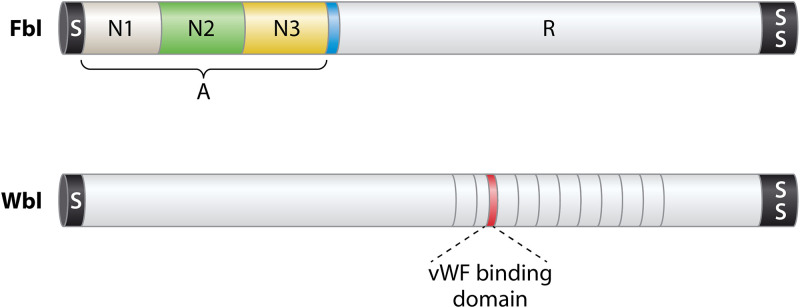
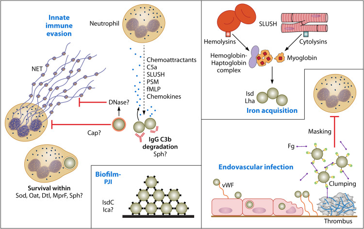
Staphylococcus lugdunensis is a species of coagulase-negative staphylococcus (CoNS) that causes serious infections in humans akin to those of S. aureus It was often misidentified as S. aureus, but this has been rectified by recent routine use of matrix-assisted laser desorption ionization-time of flight mass spectrometry (MALDI-TOF MS) in diagnostic laboratories. It encodes a diverse array of virulence factors for adhesion, cytotoxicity, and innate immune evasion, but these are less diverse than those encoded by S. aureus It expresses an iron-regulated surface determinant (Isd) system combined with a novel energy-coupling factor (ECF) mechanism for extracting heme from hemoproteins. Small cytolytic S. lugdunensis synergistic hemolysins (SLUSH), peptides related to phenol-soluble modulins of S. aureus, act synergistically with β-toxin to lyse erythrocytes. S. lugdunensis expresses a novel peptide antibiotic, lugdunin, that can influence the nasal and skin microbiota. Endovascular infections are initiated by bacterial adherence to fibrinogen promoted by a homologue of Staphylococcus aureus clumping factor A and to von Willebrand factor on damaged endothelium by an uncharacterized mechanism. S. lugdunensis survives within mature phagolysosomes of macrophages without growing and is released only following apoptosis. This differs fundamentally from S. aureus, which actively grows and expresses bicomponent leukotoxins that cause membrane damage and could contribute to survival in the infected host. S. lugdunensis is being investigated as a probiotic to eradicate S. aureus from the nares of carriers. However, this is contraindicated by its innate virulence. Studies to obtain a deeper understanding of S. lugdunensis colonization, virulence, and microbiome interactions are therefore warranted.

Keywords: endocarditis; fibrinogen; heme; invasive infection; macrophage; probiotic; surface proteins; von Willebrand factor.


Figures

FIG 1
FIG 1
Iron acquisition from heme. The lower part of the diagram shows the genetic organization of the isd locus of S. lugdunensis. The locations of fur boxes and the directions of transcription following induction by iron limitation are shown by arrows. The upper part of the diagram shows the cell membrane and cell wall peptidoglycan and the locations of the Isd proteins (right side) and the Lha proteins (left side). Hemoproteins are shown with their bound heme molecules (red rectangles), including four in hemoglobin and one in myoglobin and hemopexin. Myoglobin and hemoglobin are released from myocytes and erythrocytes, respectively, by the action of cytolytic toxins. Black dashes in genes and proteins indicate NEAT domains. IsdB NEAT1 can bind hemoglobin and the haptoglobin-hemoglobin complex. The passage of heme across the cell wall and membrane is shown, along with release of Fe3+ by intracellular hemoxygenase. The predicted second hemoxygenase is not shown. IsdC proteins on different cells also promote biofilm formation.
FIG 2
FIG 2
Cell wall-anchored surface proteins. A schematic diagram showing the two CWA proteins apart from the Isd proteins (Fig. 1) that have been analyzed at the molecular level is presented. The preproteins have a signal sequence (S) that promotes secretion across the membrane via the Sec apparatus at the N terminus and a sorting signal (SS) at the C terminus for anchorage to peptidoglycan. The A domain of Fbl is composed of three separately folded subdomains, with N2 and N3 being the minimum required for fibrinogen binding. Region R comprises SDSDSA repeats, which form a flexible stalk. The minimum putative vWF binding domain of Wbl is shown in red.
FIG 3
FIG 3
Roles of potential virulence factors. A summary of factors that might contribute to pathogenesis of infection based on knowledge of S. aureus innate immune evasion and virulence mechanisms is presented. Upon initiation of infection, many chemoattractants generated by the bacteria and nearby host cells stimulate neutrophils to migrate from the bloodstream toward the site of infection. Interference with opsonophagocytosis is probably mediated by expression of a capsule. Secreted proteases might promote opsonin degradation. DNase could degrade neutrophil extracellular traps (NETs) and prevent entrapment and killing by antimicrobial substances. Once phagocytosis has occurred, several mechanisms likely contribute to resistance to neutrophil killing mechanisms. S. lugdunensis strains form biofilm, which likely contributes to prosthetic joint infections (PJI). IsdC-promoted cell accumulation is shown. Endovascular infections could be initiated by an as-yet-unknown surface component (not vWbl) binding to surface-attached vWF on inflamed endothelial cells. Bacteria can invade endothelial cells in vitro. This might trigger endothelial cell damage. Bacteria attach to vWF bound to collagen in the exposed extracellular matrix. The ability to bind fibrinogen/fibrin via Fbl is likely to contribute to immune evasion in the bloodstream by masking opsonins and by facilitating adherence to thrombi on damaged heart valves. Access to iron is crucial. This occurs by cytolysins releasing hemoglobin from erythrocytes and myoglobin from cardiac myocytes.

References

    1. Fleurette J, Bes M, Brun Y, Freney J, Forey F, Coulet M, Reverdy ME, Etienne J. 1989. Clinical isolates of Staphylococcus lugdunensis and S. schleiferi: bacteriological characteristics and susceptibility to antimicrobial agents. Res Microbiol 140:107–118. doi: 10.1016/0923-2508(89)90044-2. - DOI - PubMed
    1. Bieber L, Kahlmeter G. 2010. Staphylococcus lugdunensis in several niches of the normal skin flora. Clin Microbiol Infect 16:385–388. doi: 10.1111/j.1469-0691.2009.02813.x. - DOI - PubMed
    1. Liu CM, Price LB, Hungate BA, Abraham AG, Larsen LA, Christensen K, Stegger M, Skov R, Andersen PS. 2015. Staphylococcus aureus and the ecology of the nasal microbiome. Sci Adv 1:e1400216. doi: 10.1126/sciadv.1400216. - DOI - PMC - PubMed
    1. Becker K, Heilmann C, Peters G. 2014. Coagulase-negative staphylococci. Clin Microbiol Rev 27:870–926. doi: 10.1128/CMR.00109-13. - DOI - PMC - PubMed
    1. Frank KL, Del Pozo JL, Patel R. 2008. From clinical microbiology to infection pathogenesis: how daring to be different works for Staphylococcus lugdunensis. Clin Microbiol Rev 21:111–133. doi: 10.1128/CMR.00036-07. - DOI - PMC - PubMed

Publication types

LinkOut - more resources