. 2021 Jan-Jun:296:100230.
doi: 10.1074/jbc.RA120.016858. Epub 2021 Jan 7.

A new FRET-based platform to track substrate ubiquitination by fluorescence

Affiliations

A new FRET-based platform to track substrate ubiquitination by fluorescence

Kenneth Wu et al. J Biol Chem. 2021 Jan-Jun.

Abstract

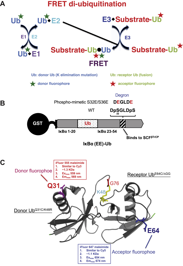
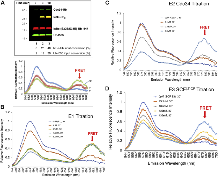
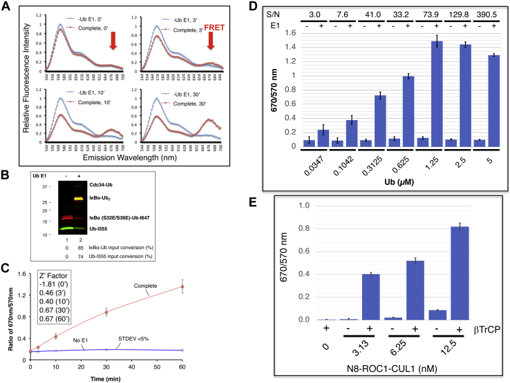
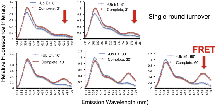
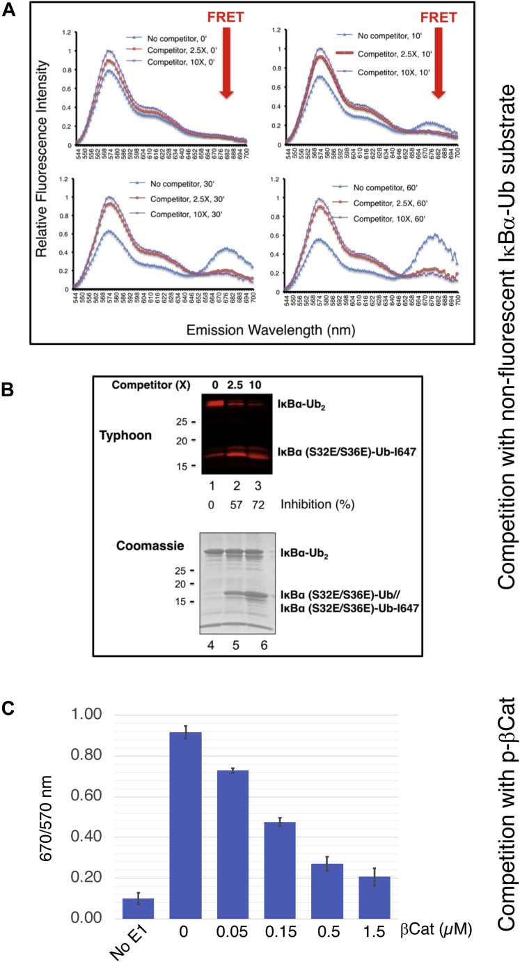
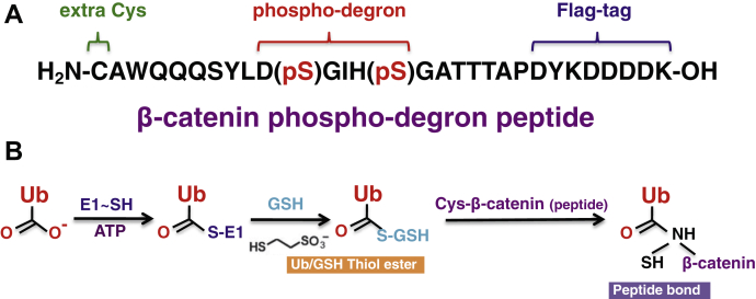
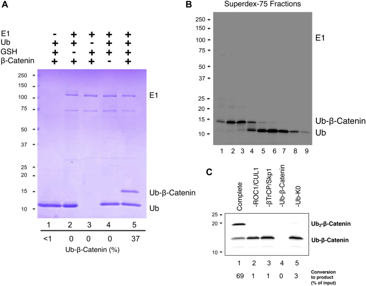
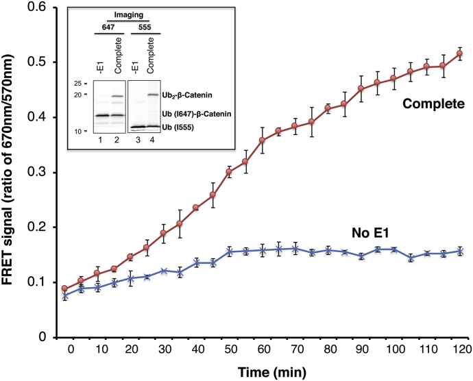
Post-translational modification of protein by ubiquitin (Ub) alters the stability, subcellular location, or function of the target protein, thereby impacting numerous biological processes and directly contributing to myriad cellular defects or disease states, such as cancer. Tracking substrate ubiquitination by fluorescence provides opportunities for advanced reaction dynamics studies and for translational research including drug discovery. However, fluorescence-based techniques in ubiquitination studies remain underexplored at least partly because of challenges associated with Ub chain complexity and requirement for additional substrate modification. Here we describe a general strategy, FRET diubiquitination, to track substrate ubiquitination by fluorescence. This platform produces a uniform di-Ub product depending on specific interactions between a substrate and its cognate E3 Ub ligase. The diubiquitination creates proximity between the Ub-linked donor and acceptor fluorophores, respectively, enabling energy transfer to yield a distinct fluorescent signal. FRET diubiquitination relies on Ub-substrate fusion, which can be implemented using either one of the two validated strategies. Method 1 is the use of recombinant substrate-Ub fusion, applicable to all substrate peptides that can bind to E3. Method 2 is a chemoenzymatic ligation approach that employs synthetic chemistry to fuse Ub with a substrate peptide containing desired modification. Taken together, our new FRET-based diubiquitination system provides a timely technology of potential to advance both basic research and translation sciences.

Keywords: E3 ubiquitin ligase; FRET; chemoenzymatic ligation; kinetics; protein degradation; ubiquitination.


Conflict of interest statement

Conflict of interest The authors declare that they have no conflicts of interest with the contents of this article.

Figures

Figure 1
Figure 1
FRET diubiquitination design.A, general scheme. Diubiquitination is a result of mixing two reaction assemblies. On the right hand, a substrate–receptor Ub fusion, in which Ub is labeled with red-coded acceptor fluorophore, binds to its cognate E3 to form the E3/substrate–Ub complex (assembly 1). On the left hand, a donor Ub, containing a lysine substitution that eliminates Ub chain assembly, is labeled with green-coded donor fluorophore. This donor Ub reacts with E1 and E2 to form E2∼donor Ub thiol ester complex (assembly 2). The combination of assemblies 1 and 2 produces a uniform substrate–di-Ub product, resulting in close proximity between the red and green fluorophores, respectively. When the donor fluorophore is excited, its emission causes the excitation of the acceptor fluorophore, which emits a distinct fluorescent signal. B, Ub fusion with IκBα phosphomimetic degron. IκBα (1–54) is a well-characterized N-terminal fragment of IκBα that contains all the molecular elements required for degradation (19). It possesses a phosphodegron (DpSGLDpS) that binds to E3 SCFβTrCP. To mimic phosphorylation, S32 and S36 are replaced by glutamic acid (E). In addition, IκBα K21 and K22, the authentic receptors for ubiquitination, are replaced by Ub (1–74), which now provides K48 as the attacking lysine residue. This fusion is named IκBα (EE)–Ub. The N-terminal GST tag can be removed by thrombin cleavage. C, fluorophore placement on Ub. Donor Ub Q31 and receptor Ub E64 are substituted by cysteine and labeled with the indicated fluorescent dye. Ub, ubiquitin.
Figure 2
Figure 2
Detection of FRET IκBα diubiquitination.A, FRET IκBα diubiquitination in test tube was carried out as described in Experimental procedures section. After incubation for times as indicated, aliquots of the reaction were subjected to analysis by gel electrophoresis (top) or spectroscopy (bottom). BD, titration of E1 (B), E2 Cdc34 (C), and E3 SCFβTrCP (D).
Figure 3
Figure 3
Tracking FRET IκBα diubiquitination in real time.A, real-time kinetics. The real-time kinetic experiments were carried out as described in Experimental procedures section. The emission spectrum of 544 to 700 nm for the complete reaction and no E1 control are shown. B, gel analysis. The final reaction mixture was subject to SDS-PAGE and Typhoon FLA9500 imaging analysis. C, assessing reliability. Three independent real-time kinetics experiments were carried out, and the results are plotted to reveal standard deviation and calculate Z' factor. The ratio of 670/570 nm represents the FRET efficiency. Note that iFluo 647 (receptor fluorophore) emits at 670 nm, whereas iFluo 555 (donor fluorophore) emits at 570 nm. D, analyte titration. The analyte concentrations are as indicated. The concentrations of enzymes are as follows: E1 (50 nM), E2 Cdc34b (3 μM), and E3 Nedd8–SCFβTrCP (100 nM). S/N = (mean of signal – mean of background)/standard deviation of background. The reaction at 30 °C was monitored on the fluorescence plate reader for 30 min. The 15 min time point (in linear range of the reaction) is used for graph. E, dependency on Skp1–βTrCP and Nedd8–ROC1–CUL1. Skp1–βTrCP (100 nM), donor/receptor Ub (2.5 μM), E2 (50 nM), Cdc34b (1 μM), IκBα (EE)–Ub (E64C)–I647 (2.5 μM), and Ub (K48R/Q31C)–I555 (2.5 μM) were used. The concentrations of Nedd8–ROC1–UL1 are as indicated. The 30 min time point (in linear range of the reaction) is used for graph. Ub, ubiquitin.
Figure 4
Figure 4
Real-time kinetics of FRET IκBα diubiquitination under single-round turnover condition. The reaction was carried out as in Figure 3, AC expect that the preformed substrate–E3 and donor Ub–E2 complexes were treated with apyrase (6 mU/1 μl) for 1 min at 37 °C to deplete ATP. The apyrase-treated reaction mixture was then loaded onto a 384-well plate for incubation on the Synergy-H1 reader set at 30 °C. The reaction was monitored at times as indicated. The emission spectrum of 544 to 700 nm for the complete reaction and no E1 control are shown. Ub, ubiquitin.
Figure 5
Figure 5
Real-time kinetics of FRET IκBα diubiquitination with competitor.AB, the reaction was carried as in Figure 3, AC, except that nonfluorescent IκBα (EE)–Ub (E64C) was added 2.5× or 10× more than fluorescent IκBα (EE)–Ub (E64C)–I647. The reaction was analyzed by spectroscopic analysis (panel A), and gel electrophoresis followed by imaging (top) or Coomassie stain (bottom). The results showed that fluorescent IκBα–Ub2 product was decreased in the presence of the competitor (top, lanes 1–3). Coomassie staining revealed that nonfluorescent IκBα (EE)–Ub (E64C) was able to support ubiquitination, forming nonfluorescent IκBα–Ub2 (bottom, lanes 4–6). C, competition with β-catenin. The concentrations of enzymes and proteins are as follows: donor/receptor Ub (0.5 μM), E1 (50 nM), E2 Cdc34b (1 μM), Skp1–βTrCP (100 nM), Nedd8–ROC1–CUL1 (12 nM), IκBα (EE)–Ub (E64C)–I647 (0.5 μM), and Ub (K48R/Q31C)–I555 (0.5 μM). The reaction at 30 °C was monitored on the fluorescence plate reader for 30 min. The 18 min time point (in linear range of the reaction) is used for graph. Ub, ubiquitin.
Figure 6
Figure 6
Ligation of the β-catenin degron peptide to Ub.A, the β-catenin degron peptide. The β-catenin degron peptide is shown. Phosphodegron motif, N-terminal Cys residue, and Flag-tag are indicated. B, ligation scheme. E1 and GSH were used to promote the ligation of the β-catenin degron peptide to Ub. Ub, ubiquitin.
Figure 7
Figure 7
Preparation of Ub–β-catenin active in ubiquitination.A, ligation reaction. The reaction was carried out as described in Experimental procedures section. Reaction products were imaged by Coomassie stain. The percentage of Ub converted to Ub–β-catenin is indicated. B, purification of β-catenin–Ub. Indicated fractions from chromatography through FPLC superdex-75 were separated by SDS-PAGE followed by imaging iFluor 647 using the Typhoon scanner. Ub–β-catenin and unincorporated Ub were well separated. C, Ub–β-catenin supports ubiquitination. The reaction was carried out as described in Experimental procedures section. Only in complete reaction (lane 1), Ub–β-catenin was conjugated with Ub–K0 to form Ub2–β-catenin. Ub, ubiquitin.
Figure 8
Figure 8
FRET analysis of Ub–β-catenin. The real-time kinetic reaction was carried out as described in Experimental procedures section. The inset shows gel images of the final reaction products detected by scanning with Typhoon FLA9500 for both iFluor 647 and iFluor 555, respectively. Approximately 28% of input Ub (I647)–β-catenin was converted into Ub2–β-catenin. Ub, ubiquitin.

References

    1. Hershko A., Ciechanover A. The ubiquitin system. Annu. Rev. Biochem. 1998;67:425–479. - PubMed
    1. Rajalingam K., Dikic I. SnapShot: expanding the ubiquitin code. Cell. 2016;164:1074–1074.e1. - PubMed
    1. Morreale F.E., Walden H. Types of ubiquitin ligases. Cell. 2016;165:248–248.e1. - PubMed
    1. Baek K., Krist D.T., Prabu J.R., Hill S., Klügel M., Neumaier L.M., von Gronau S., Kleiger G., Schulman B.A. NEDD8 nucleates a multivalent cullin-RING-UBE2D ubiquitin ligation assembly. Nature. 2020;578:461–466. - PMC - PubMed
    1. Skaar J.R., Pagan J.K., Pagano M. SnapShot: F box proteins I. Cell. 2009;137:1160–1160.e1. - PubMed

Publication types

MeSH terms