. 2020 Dec 23;203(6):e00604-20.
doi: 10.1128/JB.00604-20. Online ahead of print.

The conserved translation factor LepA is required for optimal synthesis of a porin family in Mycobacterium smegmatis

Affiliations

The conserved translation factor LepA is required for optimal synthesis of a porin family in Mycobacterium smegmatis

Skye R S Fishbein et al. J Bacteriol. .

Abstract

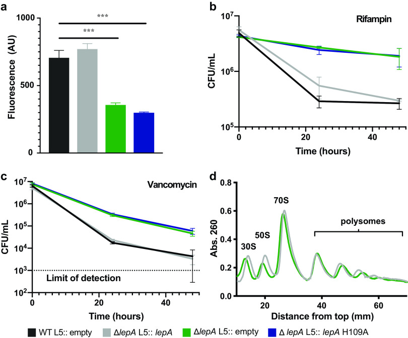
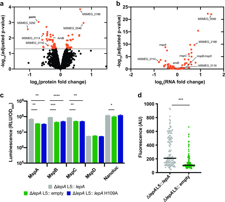
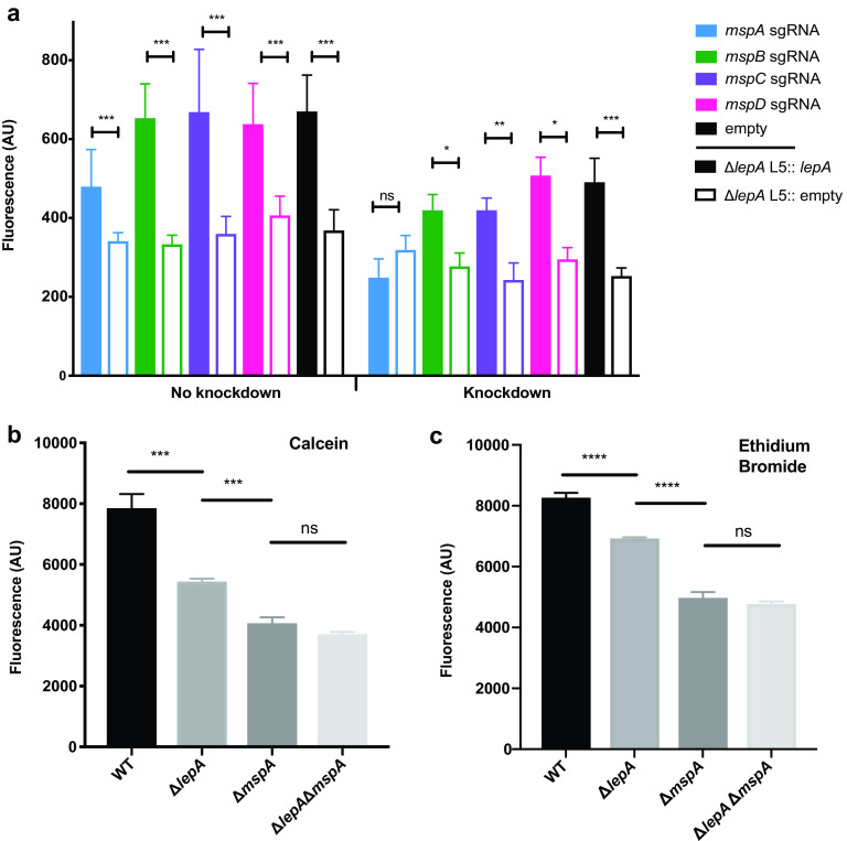
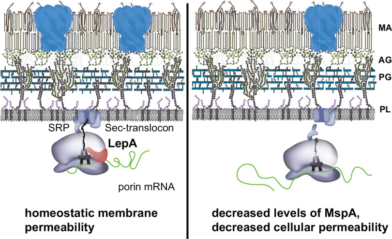
The recalcitrance of mycobacteria to antibiotic therapy is in part due to its ability to build proteins into a multi-layer cell wall. Proper synthesis of both cell wall constituents and associated proteins is crucial to maintaining cell integrity, and intimately tied to antibiotic susceptibility. How mycobacteria properly synthesize the membrane-associated proteome, however, remains poorly understood. Recently, we found that loss of lepA in Mycobacterium smegmatis (Msm) altered tolerance to rifampin, a drug that targets a non-ribosomal cellular process. LepA is a ribosome-associated GTPase found in bacteria, mitochondria, and chloroplasts, yet its physiological contribution to cellular processes is not clear. To uncover the determinants of LepA-mediated drug tolerance, we characterized the whole-cell proteomes and transcriptomes of a lepA deletion mutant relative to strains with lepA We find that LepA is important for the steady-state abundance of a number of membrane-associated proteins, including an outer membrane porin, MspA, which is integral to nutrient uptake and drug susceptibility. Loss of LepA leads to a decreased amount of porin in the membrane which leads to the drug tolerance phenotype of the lepA mutant. In mycobacteria, the translation factor LepA modulates mycobacterial membrane homeostasis, which in turn affects antibiotic tolerance.ImportanceThe mycobacterial cell wall is a promising target for new antibiotics due to the abundance of important membrane-associated proteins. Defining mechanisms of synthesis of the membrane proteome will be critical to uncovering and validating drug targets. We found that LepA, a universally conserved translation factor, controls the synthesis of a number of major membrane proteins in M. smegmatis LepA primarily controls synthesis of the major porin MspA. Loss of LepA results in decreased permeability through the loss of this porin, including permeability to antibiotics like rifampin and vancomycin. In mycobacteria, regulation from the ribosome is critical for the maintenance of membrane homeostasis and, importantly, antibiotic susceptibility.


Figures

FIG 1
FIG 1
Loss of ribosome factor LepA causes altered drug tolerance in mycobacteria. (a) Calcein staining across M. smegmatis strains with different lepA alleles. Values indicate mean calcein fluorescence across three replicates with error bars indicating standard deviation. ***, P < 0.001, calculated using a two-sided Student t test. (b and c) M. smegmatis lepA strains were treated with 10× MICs of rifampin and vancomycin, and cell survival was measured by CFU per milliliter. All values are mean values with error bars indicating standard deviations across three biological replicates. (d) Analysis of ribosome populations by sucrose density centrifugation and fractionation. Distance 0 corresponds to the lightest sucrose fraction. Data in panels a to d are representative of multiple experiments.
FIG 2
FIG 2
Whole-cell profiling finds mycobacterial porins altered by loss of LepA. (a) Proteins altered by loss of LepA. Log fold changes of protein represent mean reporter ion intensities in ΔlepA normalized by mean reporter ion intensities from the strains containing lepAlepA L5::lepA and wild type [WT] L5::empty). Orange dots indicate protein candidates that were significantly altered by loss of LepA. “Porin” indicates the collection of peptides that map to 4 proteins: MspA, MspB, MspC, and MspD. P values for proteomic ratios were calculated using Student’s two-sided t test and adjusted for multiple testing using the Benjamini-Hochberg correction with an α of 0.05. (b) Corresponding transcriptional changes in the subset of proteins significantly altered by LepA. Log fold changes and adjusted P values for RNA levels were generated using DEseq2.1.8 to analyze the same comparison between strains as in panel a. (c) Luminescence of porin reporters in M. smegmatis strains. Mean luminescence is depicted with error bars representing standard deviation of three biological replicates. ***, P < 0.001; **, P < 0.01; *, P < 0.05, calculated using a one-way ANOVA, where each group was compared to the complemented strain, with a Bonferroni correction for multiple testing. (d) Quantification of average fluorescence across a single cell (n = 100) from strains expressing MspA-mRFP. ***, P < 0.001, calculated using a Mann-Whitney test. Data in panels c and d are representative of multiple experiments.
FIG 3
FIG 3
Loss of LepA causes membrane defects primarily through control of MspA. (a) Calcein staining of lepA-porin genotypes. Knockdown of each porin was assessed in lepA strains. Mean fluorescence is depicted with error bars indicating standard deviations across three biological replicates. Sets of colored bars denote porin-specific knockdown strains. “Empty” refers to strains containing the control vector with the aTc-inducible CRISPRi system and no target-specific small guide RNA (sgRNA). ***, P < 0.001; **, P < 0.01; *, P < 0.05; ns, not significant; calculated using a two-sided Student t test. (b) Calcein fluorescence across M. smegmatis strains, with error bars indicating standard deviations across three biological replicates. (c) EtBr fluorescence across M. smegmatis strains, with error bars indicating standard deviations across three biological replicates. ****, P < 0.0001; ***, P < 0.001; ns, not significant, calculated using a one-way ANOVA. Data in panels a to c are representative of multiple experiments.
FIG 4
FIG 4
Model of LepA modulation of porin translation. LepA aids translation of outer membrane porins, most notably MspA, and determines permeability and drug susceptibility in M. smegmatis. MA, mycolic acids; AG, arabinogalactan; PG, peptidoglycan; PL, phospholipids; SRP, signal recognition particle.

Similar articles

Cited by

References

    1. Kerantzas CA, Jacobs WR, Jr. 2017. Origins of combination therapy for tuberculosis: lessons for future antimicrobial development and application. mBio 8:e01586-16. doi:10.1128/mBio.01586-16. - DOI - PMC - PubMed
    1. Harding E. 2020. WHO global progress report on tuberculosis elimination. Lancet Respir Med 8:19. doi:10.1016/S2213-2600(19)30418-7. - DOI - PubMed
    1. Sarathy J, Dartois V, Dick T, Gengenbacher M. 2013. Reduced drug uptake in phenotypically resistant nutrient-starved nonreplicating Mycobacterium tuberculosis. Antimicrob Agents Chemother 57:1648–1653. doi:10.1128/AAC.02202-12. - DOI - PMC - PubMed
    1. Jackson M, Raynaud C, Laneelle MA, Guilhot C, Laurent-Winter C, Ensergueix D, Gicquel B, Daffe M. 1999. Inactivation of the antigen 85C gene profoundly affects the mycolate content and alters the permeability of the Mycobacterium tuberculosis cell envelope. Mol Microbiol 31:1573–1587. doi:10.1046/j.1365-2958.1999.01310.x. - DOI - PubMed
    1. Dulberger CL, Rubin EJ, Boutte CC. 2020. The mycobacterial cell envelope—a moving target. Nat Rev Microbiol 18:47–59. doi:10.1038/s41579-019-0273-7. - DOI - PubMed