Review
. 2021 Feb 16;96(7):327-341.
doi: 10.1212/WNL.0000000000011413. Epub 2020 Dec 22.

7T Epilepsy Task Force Consensus Recommendations on the Use of 7T MRI in Clinical Practice

Affiliations
Review

7T Epilepsy Task Force Consensus Recommendations on the Use of 7T MRI in Clinical Practice

Giske Opheim et al. Neurology. .

Abstract

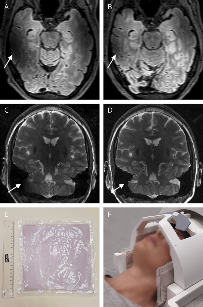
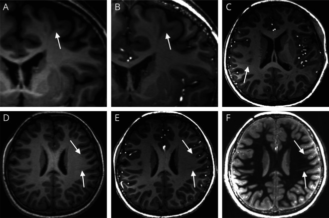
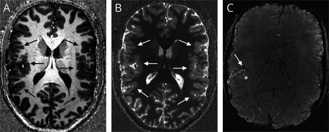
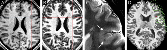
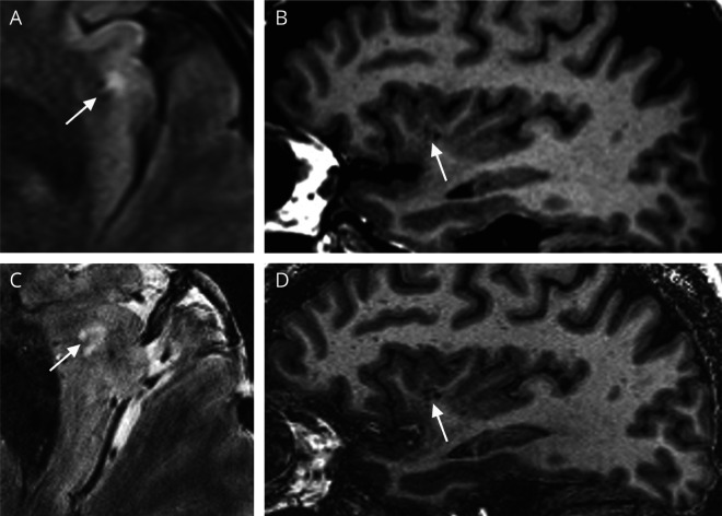
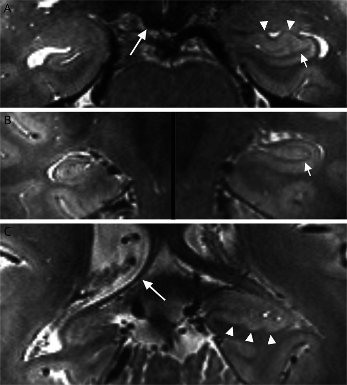
Identifying a structural brain lesion on MRI has important implications in epilepsy and is the most important factor that correlates with seizure freedom after surgery in patients with drug-resistant focal onset epilepsy. However, at conventional magnetic field strengths (1.5 and 3T), only approximately 60%-85% of MRI examinations reveal such lesions. Over the last decade, studies have demonstrated the added value of 7T MRI in patients with and without known epileptogenic lesions from 1.5 and/or 3T. However, translation of 7T MRI to clinical practice is still challenging, particularly in centers new to 7T, and there is a need for practical recommendations on targeted use of 7T MRI in the clinical management of patients with epilepsy. The 7T Epilepsy Task Force-an international group representing 21 7T MRI centers with experience from scanning over 2,000 patients with epilepsy-would hereby like to share its experience with the neurology community regarding the appropriate clinical indications, patient selection and preparation, acquisition protocols and setup, technical challenges, and radiologic guidelines for 7T MRI in patients with epilepsy. This article mainly addresses structural imaging; in addition, it presents multiple nonstructural MRI techniques that benefit from 7T and hold promise as future directions in epilepsy. Answering to the increased availability of 7T MRI as an approved tool for diagnostic purposes, this article aims to provide guidance on clinical 7T MRI epilepsy management by giving recommendations on referral, suitable 7T MRI protocols, and image interpretation.


Figures

Figure 1
Figure 1. Use of Dielectric Pads
Illustration of the effect of dielectric pads on (A and B) 3D fluid-attenuated inversion recovery (FLAIR, 0.7 mm isotropic resolution) and (C and D) T2-weighted sequences (0.3 × 0.3 × 1.5 mm resolution). In (A and C), no pads are used, whereas in (B and D) they are. Arrows indicate corresponding areas before and after signal improvement (A versus B, C versus D). The dielectric pads used in this case are 19 × 19 cm (E); pad placement for obtaining images (B and D) is demonstrated in image (F). Of note, optimal pad placement depends on the head size and shape.
Figure 2
Figure 2. Example of Tuberous Sclerosis Complex at 7T
3T T1-weighted 0.9 mm isotropic MPRAGE (A and D), 7T T1-weighted 0.6 mm isotropic MPRAGE (B–D), and 0.8 mm isotropic white matter-suppressed T1-weighted images (F) in an 11-year-old girl diagnosed with tuberous sclerosis complex (TSC). Cortical tubers were found throughout the brain both at 3T and 7T MRI (arrow in A and B). Radial migration bands, however, were much more difficult to visualize; subtle radial bands could be identified at 7T in the left frontal and parietal lobe (arrows in E and F), which were only retrospectively seen at 3T (D). In addition, more detailed structures surrounding both tubers and radial bands, as well as previously unidentified subtle TSC abnormalities such as a small cyst associated with a radial band in the right parietal lobe (C), were seen only at 7T images. This detailed delineation of TSC abnormalities may improve surgical resection, thereby increasing the likelihood of a seizure-free postoperative outcome. Courtesy of Kaibao Sun, PhD, Center for MR Research, University of Illinois at Chicago, Chicago, IL, USA. Data were acquired during his employment at the State Key Lab. of Brain and Cognitive Science, Beijing MRI Center for Brain Research, Institute of Biophysics, Chinese Academy of Sciences, Beijing, China.
Figure 3
Figure 3. Example of Polymicrogyria at 7T
7T T1-weighted MP2RAGE (A) and MP2RAGE T1-map (B) images illustrate thickening of the perisylvian cortex (arrows in A and B) in an 18-year-old patient who had known polymicrogyria as already visualized at 3T MRI; clinical indication for 7T imaging was better lesion delineation. An additional 7T T2*-weighted (SWI) sequence (C) shows a hyperintense cortex associated with veins perpendicular to the cortex (* in C) and a tree-like distribution of vessels (arrow in C).
Figure 4
Figure 4. Example of Focal Cortical Dysplasia at 7T
Axial 3T T1-weighted MPRAGE (A), axial 7T T1-weighted MP2RAGE (B), and zoomed in axial 7T T2*-weighted GRE (C) images of a patient for whom visual review of 7T MRI yielded previously unappreciated subtle findings. The red crosshairs/arrows pinpoint the location of an area of focal cortical dysplasia (FCD), which was detected by visual analysis of 7T images. The vascular changes associated with the FCD can be well appreciated on the T2*-weighted GRE images in panel C (arrows). Detection of this subtle lesion guided subsequent placement of intracranial-EEG (icEEG) with subdural grids and depth electrodes. The icEEG implantation was devised to confirm the epileptogenicity of the subtle lesion and map out the lesion extent and its proximity to eloquent cortex with language function. The subtle lesion location was concordant with ictal onset on the icEEG as shown in the 3D reconstruction of electrode location and 7T MRI, with a 2D axial cut-plane (D). In panel D, green spheres indicate all implanted electrodes and red spheres indicate ictal onset.
Figure 5
Figure 5. Vascular Changes Mimicking Focal Cortical Dysplasia
Example of a lesion suspected to be FCD at 3T but concluded to be vascular changes after reviewing 7T images. The 3T axial FLAIR (A) and 3T sagittal T1-weighted images (B) suggested subcortical FLAIR hyperintensity (arrow on A) and gray-white matter blurring (arrow on B) of the left insular cortex, suspicious for FCD. The patient had an SEEG evaluation to explore the suspected area and other possible areas for seizure generation. The suspected area in the left insula was not involved in seizure onset. 7T T2*-weighted GRE (C) and 7T sagittal T1-weighted images (D) revealed the lesion to be a vascular abnormality causing adjacent gliosis that mimicked the appearance of FCD, as indicated by arrows. Because of convincing evidence from the 7T images, the patient's surgical plan did not include the left insula.
Figure 6
Figure 6. Hippocampal Sclerosis (HS) at 7T
Coronal T2-weighted images at the level of the hippocampal head (A), body (B), and tail (C) show normal appearance of the left hippocampus including a continuous dark band reflecting the stratum radiatum lacunosum moleculare (arrows) and normal digitations along the head and tail (arrowheads). In contrast, the right hippocampus shows features of HS, including decreased volume, smooth outer counters, and indistinct internal architecture. Note also atrophy of the right mammillary body (long arrow in A) and fornix (long arrow in C).

References

    1. Ryvlin P, Cross JH, Rheims S. Epilepsy surgery in children and adults. Lancet Neurol 2014;13:1114–1126. - PubMed
    1. Téllez-Zenteno JF, Hernández Ronquillo L, Moien-Afshari F, Wiebe S. Surgical outcomes in lesional and non-lesional epilepsy: a systematic review and meta-analysis. Epilepsy Res 2010;89:310–318. - PubMed
    1. van der Zwaag W, Schäfer A, Marques JP, Turner R, Trampel R. Recent applications of UHF-MRI in the study of human brain function and structure: a review. NMR Biomed 2016;29:1274–1288. - PubMed
    1. Trattnig S, Bogner W, Gruber S, et al. . Clinical applications at ultrahigh field (7 T). Where does it make the difference?. NMR Biomed 2016;29:1316–1334. - PubMed
    1. Feldman RE, Delman BN, Pawha PS, et al. . 7T MRI in epilepsy patients with previously normal clinical MRI exams compared against healthy controls. PLoS One 2019;14:e0213642. - PMC - PubMed