Review
. 2020 Dec 23;85(1):e00110-20.
doi: 10.1128/MMBR.00110-20. Print 2021 Feb 17.

Alternative DNA Structures In Vivo: Molecular Evidence and Remaining Questions

Affiliations
Review

Alternative DNA Structures In Vivo: Molecular Evidence and Remaining Questions

Lucie Poggi et al. Microbiol Mol Biol Rev. .

Erratum in

Abstract

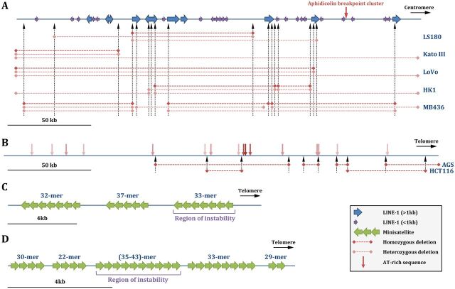
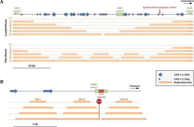
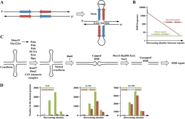
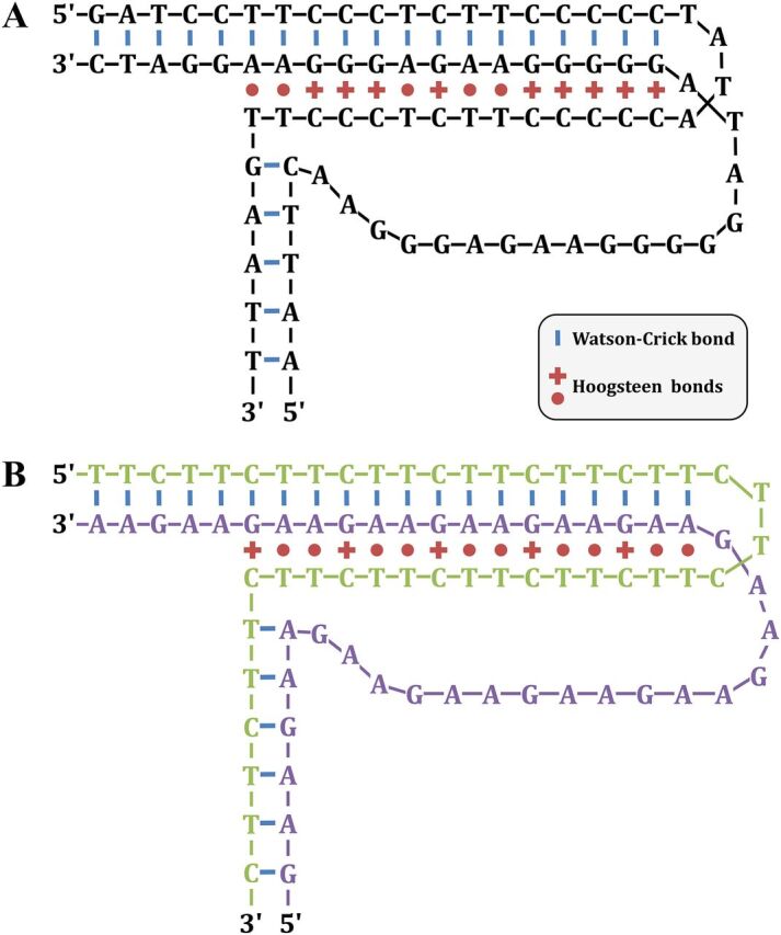
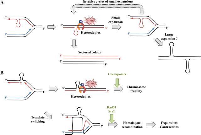
Duplex DNA naturally folds into a right-handed double helix in physiological conditions. Some sequences of unusual base composition may nevertheless form alternative structures, as was shown for many repeated sequences in vitro However, evidence for the formation of noncanonical structures in living cells is difficult to gather. It mainly relies on genetic assays demonstrating their function in vivo or through genetic instability reflecting particular properties of such structures. Efforts were made to reveal their existence directly in a living cell, mainly by generating antibodies specific to secondary structures or using chemical ligands selected for their affinity to these structures. Among secondary structure-forming DNAs are G-quadruplexes, human fragile sites containing minisatellites, AT-rich regions, inverted repeats able to form cruciform structures, hairpin-forming CAG/CTG triplet repeats, and triple helices formed by homopurine-homopyrimidine GAA/TTC trinucleotide repeats. Many of these alternative structures are involved in human pathologies, such as neurological or developmental disorders, as in the case of trinucleotide repeats, or cancers triggered by translocations linked to fragile sites. This review will discuss and highlight evidence supporting the formation of alternative DNA structures in vivo and will emphasize the role of the mismatch repair machinery in binding mispaired DNA duplexes, triggering genetic instability.

Keywords: DNA hairpin; G quadruplex; cruciform; fragile sites; mismatch repair; palindromes; secondary structures; trinucleotide repeats.


Figures

FIG 1
FIG 1
G quadruplexes. (A) Genetic factors stabilizing G quadruplexes. The general consensus is shown (G3 Nx G3 Nx G3 Nx), with the positions of the two lateral loops and the central loop. A stable G4 structure is formed in the presence of potassium ions if the central loop is shorter than 5 nucleotides and if lateral loops are made of one or two pyrimidine residues. If loops are longer or made of adenine residues, the G4 structure is less stable. The helicase Pif1 is known to unwind G4 in S. cerevisiae (29). (B) Stabilization of the Pu24T quadruplex by PhenDC3. Top view of the top guanine tetrad showing non-Watson-Crick bounds as orange dashed lines. Addition of PhenDC3 stabilizes the structure by stacking with the G4. (Based on data from reference .)
FIG 2
FIG 2
Common and rare fragile sites. (A) The common FRA3B fragile site. Complete and incomplete L1 retrotransposons are shown by blue and purple arrows, respectively. Vertical dotted arrows indicate known deletion junctions, whose extents are shown by horizontal dashed lines, as heterozygous or homozygous deletions, in five cancer cell lines in which they were mapped. (Based on data from reference .) (B) The common FRA16D fragile site. AT-rich sequences are indicated by vertical pink arrows, with the darker shade corresponding to a higher flexibility index. Vertical dotted arrows indicate known deletion junctions, whose extents are shown by horizontal dotted lines, in two cancer cell lines in which they were mapped. (Based on data from reference .) (C) The rare FRA16B fragile site. It contains three AT-rich minisatellites, shown by green arrows. The most telomeric repeat is unstable, and its expansion triggers fragile site expression (58). (D) The rare FRA10B fragile site. It contains five AT-rich minisatellites, shown by green arrows. The internal repeat is unstable and prone to expansions. (Based on data from reference .)
FIG 3
FIG 3
Replicating fragile sites. (A) FRA3B. In lymphoblasts, replication forks travel from large distances outside the fragile site inner core, and there is no activation of internal origins. At the end of the S phase, the whole locus is frequently underreplicated and therefore prone to breakage. In fibroblasts, the activation of several internal replication origins allows completion of replication of the locus, decreasing its fragility. (Based on data from reference .) (B) FRAXA. Replication proceeds from three origins within the locus. The fork traveling from ORI II is stalled by the CGG trinucleotide repeat expansion on the lagging strand template. ORI III, traveling in the other orientation, is less affected since the CGG sequence is on the leading strand template (67, 68).
FIG 4
FIG 4
Inverted repeats and cruciforms. (A) The unstructured palindrome is shown as complementary pink and blue arrows. In certain conditions, it may adopt a cruciform structure containing two stems and two loops of variable lengths that form a four-way junction whose structure is similar to a Holliday junction. The stability of a cruciform depends on both the stem and loop lengths. (B) Double-strand break (DSB) frequency according to the identity between direct or inverted tandem repeats in yeast. More identical repeats are more prone to form stable cruciforms, therefore inducing more frequent DSB. (Based on data from reference .) (C) Genetic factors involved in cruciform processing in yeast. The proteins involved in the transition between the nicked form and the capped DSB are not clearly characterized, but ligase IV (DNL4 in yeast) does not play a role in this reaction (96). (D) The number of direct or inverted Alu repeats in the human genome. The distances between repeats were classified into close (0 to 20 nucleotides [nt], left panel), medium (21 to 100 nt, middle panel), and distant (101 to 500 nt, right panel). In each panel, repeats are classified from left to right in order of decreasing identity (>90%, 81 to 90%, 71 to 80%, and 61 to 70% identity). Close inverted repeats are counterselected because they may form stable cruciforms (left panel), whereas this selection is less pronounced at longer distances. (Based on data from https://www.niehs.nih.gov/research/resources/databases/alu/index.cfm.)
FIG 5
FIG 5
H-DNA triplex structures. (A) A polypurine-polypyrimidine H-DNA structure is shown. (Based on data from reference .) (B) A similar structure formed by GAA/TTC trinucleotide repeats. The GAA strand and the TTC strand are colored in green and purple, respectively, to make their visualization easier.
FIG 6
FIG 6
Role of the mismatch repair system in CAG/CTG trinucleotide repeat expansion and fragility. (A) Hairpin formed on the newly synthesized lagging strand. The damaged fork is recognized by the MMR but cannot be fixed, thus leading to a small expansion during the next S phase. Successive cycles of small expansions may occur, ultimately leading to a large expansion. Alternatively, another mechanism may directly lead to large expansions, such as those observed in some human disorders. Unrepaired heteroduplex DNA is observed in the progeny as two cell populations with different repeat tract lengths (see the text for details). (B) Hairpin formed on the lagging-strand template. The damaged fork is recognized by the mismatch repair system (MMR) and may lead to chromosomal fragility at the next S phase if checkpoints are bypassed or if the damage cannot be fixed. Template switching is a possible pathway to repair and restart the fork but may lead to trinucleotide repeat expansions and contractions by homologous recombination under the control of the Rad51 recombinase and the Srs2 helicase in yeast (see the text for details). Note that the hairpin was drawn on the lagging strand (or on its template), but the model can perfectly be reversed if its formation happens on the leading strand (or on its template).

References

    1. Franklin RE, Gosling RG. 1953. Molecular configuration in sodium thymonucleate. Nature 171:740–741. doi: 10.1038/171740a0. - DOI - PubMed
    1. Watson JD, Crick FHC. 1953. Molecular structure of nucleic acids: a structure for deoxyribose nucleic acid. Nature 171:737–738. doi: 10.1038/171737a0. - DOI - PubMed
    1. Chargaff E, Magasanik B, Vischer E, Green C, Doniger R, Elson D. 1950. Nucleotide composition of pentose nucleic acids from yeast and mammalian tissues. J Biol Chem 186:51–67. - PubMed
    1. Hoogsteen K. 1963. The crystal and molecular structure of a hydrogen-bonded complex between 1-methylthymine and 9-methyladenine. Acta Cryst 16:907–916. doi: 10.1107/S0365110X63002437. - DOI
    1. Arnott S, Fuller W, Hodgson A, Prutton I. 1968. Molecular conformations and structure transitions of RNA complementary helices and their possible biological significance. Nature 220:561–564. doi: 10.1038/220561a0. - DOI - PubMed

Publication types

LinkOut - more resources