. 2020 Dec 22;5(6):e00604-20.
doi: 10.1128/mSystems.00604-20.

Acinetobacter baumannii Targets Human Carcinoembryonic Antigen-Related Cell Adhesion Molecules (CEACAMs) for Invasion of Pneumocytes

Affiliations

Acinetobacter baumannii Targets Human Carcinoembryonic Antigen-Related Cell Adhesion Molecules (CEACAMs) for Invasion of Pneumocytes

Cecilia Ambrosi et al. mSystems. .

Abstract

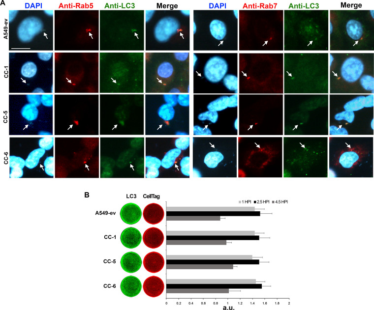
Multidrug-resistant Acinetobacter baumannii is regarded as a life-threatening pathogen mainly associated with nosocomial and community-acquired pneumonia. Here, we show that A. baumannii can bind the human carcinoembryonic antigen-related cell adhesion molecule (CEACAM) receptors CEACAM1, CEACAM5, and CEACAM6. This specific interaction enhances A. baumannii internalization in membrane-bound vacuoles, promptly decorated with Rab5, Rab7, and lipidated microtubule-associated protein light chain 3 (LC3). Dissecting intracellular signaling pathways revealed that infected pneumocytes trigger interleukin-8 (IL-8) secretion via the extracellular signal-regulated kinase (ERK)1/2 and nuclear factor-kappa B (NF-κB) signaling pathways for A. baumannii clearance. However, in CEACAM1-L-expressing cells, IL-8 secretion lasts only 24 h, possibly due to an A. baumannii-dependent effect on the CEACAM1-L intracellular domain. Conversely, the glycosylphosphatidylinositol-anchored CEACAM5 and CEACAM6 activate the c-Jun NH2-terminal kinase (JNK)1/2-Rubicon-NOX2 pathway, suggestive of LC3-associated phagocytosis. Overall, our data show for the first time novel mechanisms of adhesion to and invasion of pneumocytes by A. baumannii via CEACAM-dependent signaling pathways that eventually lead to bacterial killing. These findings suggest that CEACAM upregulation could put patients at increased risk of lower respiratory tract infection by A. baumannii IMPORTANCE This work shows for the first time that Acinetobacter baumannii binds to carcinoembryonic antigen-related cell adhesion molecule 1 (CEACAM1), CEACAM5, and CEACAM6. This binding significantly enhances A. baumannii internalization within alveolar host cell epithelia. Intracellular trafficking involves typical Rab5 and Rab7 vacuolar proteins as well as light chain 3 (LC3) and slowly progresses to bacterial killing by endosome acidification. CEACAM engagement by A. baumannii leads to distinct and specific downstream signaling pathways. The CEACAM1 pathway finely tunes interleukin-8 (IL-8) secretion, whereas CEACAM5 and CEACAM6 mediate LC3-associated phagocytosis. The present study provides new insights into A. baumannii-host interactions and could represent a promising therapeutic strategy to reduce pulmonary infections caused by this pathogen.

Keywords: Acinetobacter baumannii; MAPKs; Rubicon; bacterial adhesion/invasion; carcinoembryonic antigen‐related cell adhesion molecules.


Figures

FIG 1
FIG 1
A. baumannii binds to CEACAM receptors. Strain AB5075 grown to exponential phase was incubated with 10 µg of the indicated recombinant proteins and subjected to pulldown. (A) Pulldown samples (PD) were resolved by Tricine-PAGE and visualized by Western blotting using the anti-CEACAM antibodies (MAb 6G5j and C5-1X) and HRP-conjugated secondary antibodies. Purified proteins (50 ng) were loaded as controls (Ctrl). The recombinant CEACAM1 protein lacking the complete N-domain (CEACAM1ΔN) was also assayed. One representative experiment of four is shown. (B) Pulldown bacteria incubated with MAb 6G5j and TRITC-labeled secondary antibodies were centrifuged on polylysine-treated coverslips and mounted for bright-field (BF) and fluorescence microscopic analyses. One representative experiment of four is shown.
FIG 2
FIG 2
Expression of CEACAM receptors increases A. baumannii adherence to and invasion of lung epithelial cells. Stably transfected A549 cell lines expressing CEACAM1 (CC-1), CEACAM5 (CC-5), CEACAM6 (CC-6), and CEACAM8 (CC-8) as well as the control cell line transfected with the empty vector (A549-ev) were infected with strain AB5075 at an MOI of 1 for the adhesion time (2.5 h). (A) Following extensive washing, the number of adherent bacteria was assessed by counting the CFU/ml. Each adherence assay was performed in duplicate wells, and all data are given as the mean ± standard deviation (SD) from five independent experiments. Asterisks represent P values evaluated by one-way analysis of variance (ANOVA); **, P < 0.01. (B) Representative images of parallel cell monolayers stained in red with phalloidin or immune-labeled with the anti-CEACAM antibody (MAb 6G5j), respectively, followed by DAPI staining (blue). Arrows indicate examples of CEACAMs colocalized with bacteria. Two different magnifications are presented. Images were acquired with the digital FireWire color camera DFX300. Scale bars = 10 µm. (C) A549, CC-1, CC-5, and CC-6 were infected as described above. Following the adhesion time (2.5 h), colistin sulfate (10 µg/ml) was added to kill extracellular bacteria. After a further 24, 48, 72, and 96 h of incubation, the number of intracellular bacteria was assessed by counting the CFU/ml. Each invasion assay was performed in duplicate wells, and all data are given as the mean ± standard deviation (SD) from five independent experiments. Asterisks represent P values evaluated by one-way ANOVA; *, P < 0.05; **, P < 0.01.
FIG 3
FIG 3
A. baumannii colocalizes with endocytic markers and LC3. Stably transfected A549 cell lines expressing CEACAM1 (CC-1), CEACAM5 (CC-5), and CEACAM6 (CC-6) as well as the control cell line transfected with the empty vector (A549-ev) were infected with strain AB5075 at an MOI of 1 for the adhesion time (2.5 h), followed by a further 2 h of incubation in the presence of colistin sulfate (10 µg/ml) to kill extracellular bacteria. (A) Cell monolayers were fixed and processed for immunofluorescence labeling with anti-Rab5, -Rab7, and -LC3 antibodies and were DAPI stained. Representative images were acquired with the digital FireWire color camera DFX300. Scale bar = 10 µm. (B) Parallel cell monolayers infected as described above were fixed after 1 and 2.5 h and a further 2 h of incubation in the presence of colistin sulfate prior to being hybridized with anti-LC3 antibody (green). CellTag 700 staining (red) was used to quantify cell monolayers. Plates were scanned using an Odyssey infrared imager, and signals of the integrated fluorescence intensity were acquired using LI-COR Image Studio 3.1 software. HPI, hours postinfection. Results are expressed as arbitrary units (a.u.) and represent means ± standard deviation (SD) of four independent experiments done in triplicate.
FIG 4
FIG 4
Acidification has detrimental effects on A. baumannii viability. (A) Stably transfected A549 cell lines expressing CEACAM1 (CC-1), CEACAM5 (CC-5), and CEACAM6 (CC-6) as well as the control cell line transfected with the empty vector (A549-ev) were treated with the V-ATPase inhibitor bafilomycin A1 (100 nM) (B+) or left untreated (B–) prior to being infected with strain AB5075 at an MOI of 1. Following the adhesion time (2.5 h) and a further 2 h in the presence of colistin sulfate to kill extracellular bacteria, infected cell monolayers were lysed, and the number of intracellular bacteria was assessed by counting the CFU/ml. Each invasion assay was performed in duplicate wells, and all data are given as the mean ± SD from four independent experiments. Asterisks represent P values evaluated with Student’s t test, *, P < 0.05; **, P < 0.01. (B) Strain AB5075 was grown in LB medium under neutral and acidic pH conditions. At the indicated time points, the number of viable bacteria was assessed by counting the CFU/ml. (C) Stably transfected A549 cell lines, CC-1, CC-5, and CC-6 as well as the control cell line A549-ev were infected with strain AB5075 at an MOI of 1. Following the adhesion time (2.5 h), colistin sulfate (10 µg/ml) was added. After a further 72 h of incubation, Lysotracker red was used to stain acidic vesicles. Cell monolayers were fixed and DAPI stained. Representative images were acquired with the digital FireWire color camera DFX300. Scale bar = 10 µm.
FIG 5
FIG 5
CEACAMs transduce different signaling pathways upon A. baumannii engagement. Stably transfected A549 cell lines expressing CEACAM1 (CC-1), CEACAM5 (CC-5), and CEACAM6 (CC-6) as well as the control cell line transfected with the empty vector (A549-ev) were infected with strain AB5075 at an MOI of 1 for the indicated times. (A to D) At each time point (hours postinfection [HPI]), whole-cell lysates in duplicate were prepared and used in Western blot analysis to detect (A) phospho (P)-ERK, ERK, (B) P-p38, p38, (C) P-JNK, JNK, and (D) P-NF-κB, NF-κB using specific antibodies. Images are representative, whereas bars depict the calculated ratios of phosphorylated/total protein content quantified by densitometry (ImageJ software) and expressed as arbitrary units (a.u.). (E) After 24, 48, and 72 h of infection, IL-8 production was measured in cell supernatants by sandwich enzyme-linked immunosorbent assay (ELISA). Data are given as the mean ± standard deviation (SD) from four independent experiments. Asterisks represent P values evaluated by one-way ANOVA, *, P < 0.05; **, P < 0.01.
FIG 6
FIG 6
The CEACAM5 and CEACAM6 signaling pathway increases NOX2 and Rubicon expression upon A. baumannii engagement. Stably transfected A549 cell lines expressing CEACAM1 (CC-1), CEACAM5 (CC-5), and CEACAM6 (CC-6) as well as the control cell line transfected with the empty vector (A549-ev) were infected with strain AB5075 at an MOI of 1 for the indicated times. (A and B) At each time point (hours postinfection [HPI]), whole-cell lysates in duplicate were prepared and used in Western blot analysis to detect (A) NOX2 and (B) Rubicon using specific antibodies, while glyceraldehyde-3-phosphate dehydrogenase (GAPDH) was used as a loading control. Images are representative, whereas bars depict the calculated ratios of NOX2/GADPH and Rubicon/GAPDH quantified by densitometry (ImageJ software) and expressed as arbitrary units (a.u.). Data are given as the mean ± standard deviation (SD) from four independent experiments. Asterisks represent P values evaluated by one-way ANOVA; *, P < 0.05; **, P < 0.01. (C) CC-5 and CC-6 cell monolayers were infected with strain AB5075 at an MOI of 1 for the adhesion time (2.5 h), followed by a further 2 h of incubation in the presence of colistin sulfate (10 µg/ml) to kill extracellular bacteria. Cell monolayers were fixed and processed for immunofluorescence labeling with the anti-Rubicon antibody and were DAPI stained. Representative images were acquired with the DS-Qi2 monochrome camera. Scale bar = 10 µm.
FIG 7
FIG 7
Working model of the signaling cascades triggered by the interaction of A. baumannii with CEACAM receptors expressed by transfected lung epithelial cells. The adhesion of A. baumannii cells to CEACAM receptors allows bacterial internalization within membrane-bound vacuoles that promptly become decorated by Rab5, Rab7, and LC3. Vacuoles become progressively acidic by recruitment of the V-ATPase, which pumps protons (H+) into the lumen using cytosolic ATP as an energy source. Moreover, phosphatidylinositol 3-kinase (PI3K) generates 3′-phosphoinositides, which accumulate in membrane-bound vacuoles to favor the acidification of vesicles. Activation of p38 enhances the retrieval of Rab5 via the activity of the guanine-nucleotide dissociation inhibitor (GDI), thereby increasing endocytic rates (67). (A) Proposed model for CEACAM1-mediated signaling process. Binding of A. baumannii to CEACAM1 as well as Toll-like receptors 2 (TLR2) and TLR4 triggers Erk1/2 phosphorylation, which in turn, activates NF-κB. Following nuclear translocation, NF-κB promotes transcription of the IL-8 gene, thereby allowing a large amount of IL-8 secretion at 24 h postinfection (52). However, IL-8 secretion drops significantly in the A549 cell line expressing CEACAM1 at later time points, likely due to an inhibitory activity of its immune-receptor tyrosine-based inhibitory motif (ITIM) on the TLR2 signaling cascade. (B) Proposed model for the CEACAM5- and CEACAM6-mediated signaling process. Binding of A. baumannii to CEACAM5 or CEACAM6 as well as TLR2 and TLR4 triggers JNK1/2 phosphorylation, resulting in the recruitment of Rubicon, a component of the PI3K complex. Rubicon stabilizes the NOX2 complex, allowing it to produce reactive oxygen species (ROS) in the microbe-containing vacuole membranes (55). Both signaling pathways involving the intracellular trafficking of A. baumannii vesicles lead to acidification and lysosomal trafficking and, eventually, to microbial degradation. For simplicity, Rubicon is shown as a single protein. Individual components are not to scale.

References

    1. Harding CM, Hennon SW, Feldman MF. 2018. Uncovering the mechanisms of Acinetobacter baumannii virulence. Nat Rev Microbiol 16:91–102. doi:10.1038/nrmicro.2017.148. - DOI - PMC - PubMed
    1. Dexter C, Murray GL, Paulsen IT, Peleg AY. 2015. Community-acquired Acinetobacter baumannii: clinical characteristics, epidemiology and pathogenesis. Expert Rev Anti Infect Ther 13:567–573. doi:10.1586/14787210.2015.1025055. - DOI - PubMed
    1. Xie R, Zhang XD, Zhao Q, Peng B, Zheng J. 2018. Analysis of global prevalence of antibiotic resistance in Acinetobacter baumannii infections disclosed a faster increase in OECD countries. Emerg Microbes Infect 7:31. doi:10.1038/s41426-018-0038-9. - DOI - PMC - PubMed
    1. Dijkshoorn L, Nemec A, Seifert H. 2007. An increasing threat in hospitals: multidrug-resistant Acinetobacter baumannii. Nat Rev Microbiol 5:939–951. doi:10.1038/nrmicro1789. - DOI - PubMed
    1. Ambrosi C, Aleandri M, Giordano A, Scribano D, Marazzato M, Zagaglia C, Conte MP, Palamara AT. 2016. Molecular characterisation of extensively drug-resistant Acinetobacter baumannii: first report of a new sequence type in Italy. J Glob Antimicrob Resist 7:154–156. doi:10.1016/j.jgar.2016.10.002. - DOI - PubMed