. 2021 Jan 12;118(2):e2021785118.
doi: 10.1073/pnas.2021785118.

Structure of SARS-CoV-2 ORF8, a rapidly evolving immune evasion protein

Affiliations

Structure of SARS-CoV-2 ORF8, a rapidly evolving immune evasion protein

Thomas G Flower et al. Proc Natl Acad Sci U S A. .

Abstract

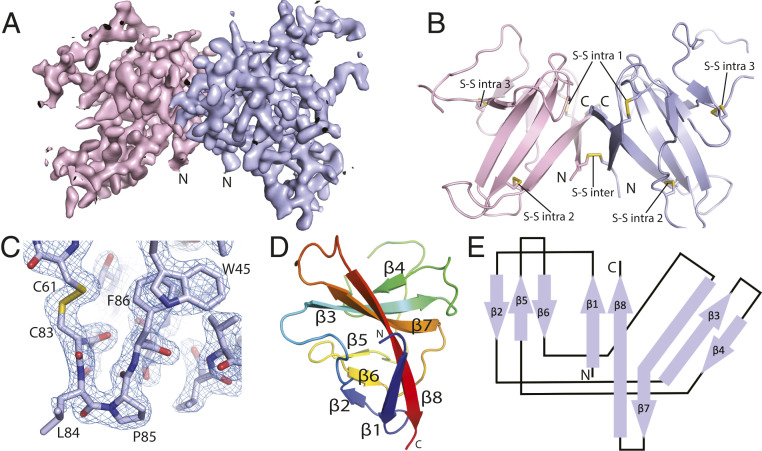
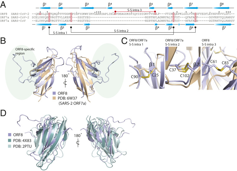
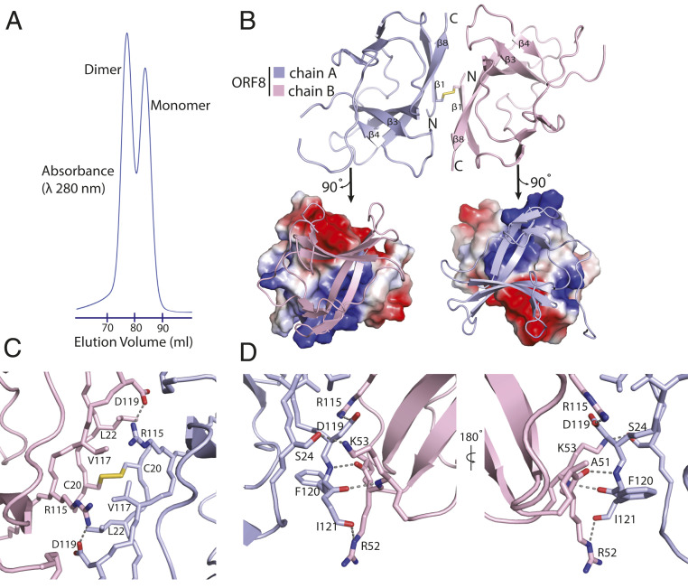
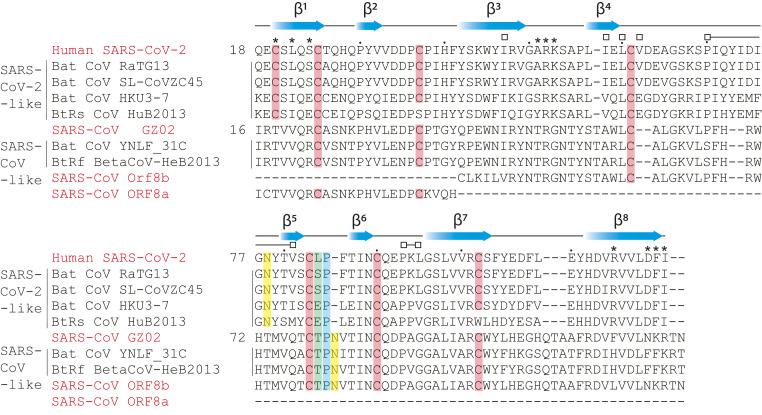
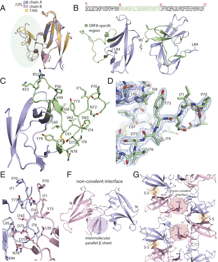
The molecular basis for the severity and rapid spread of the COVID-19 disease caused by severe acute respiratory syndrome coronavirus 2 (SARS-CoV-2) is largely unknown. ORF8 is a rapidly evolving accessory protein that has been proposed to interfere with immune responses. The crystal structure of SARS-CoV-2 ORF8 was determined at 2.04-Å resolution by X-ray crystallography. The structure reveals a ∼60-residue core similar to SARS-CoV-2 ORF7a, with the addition of two dimerization interfaces unique to SARS-CoV-2 ORF8. A covalent disulfide-linked dimer is formed through an N-terminal sequence specific to SARS-CoV-2, while a separate noncovalent interface is formed by another SARS-CoV-2-specific sequence, 73YIDI76 Together, the presence of these interfaces shows how SARS-CoV-2 ORF8 can form unique large-scale assemblies not possible for SARS-CoV, potentially mediating unique immune suppression and evasion activities.

Keywords: COVID-19; SARS-CoV-2; X-ray crystallography.


Conflict of interest statement

The authors declare no competing interest.

Figures

Fig. 1.
Fig. 1.
Crystal structure of SARS-CoV-2 ORF8. (A) A 2Fo-Fc electron density map of SARS-CoV-2 ORF8 crystallographic dimer determined to 2.04 Å (chain A, light blue; chain B, light pink). (B) Cartoon representation of the SARS-CoV-2 ORF8 crystallographic dimer. Disulfide bonds are modeled showing both intermolecular and intramolecular bond pairs. (C) Representative 2Fo-Fc density of the Cys83−Leu84−Pro85 turn motif. The map is contoured at 2σ and represented as a blue mesh. (D) Cartoon representation of the SARS-CoV-2 ORF8 monomer. β-strands are labeled β1 to β8, and chain is colored by rainbow gradient for clarity. (E) Topographic representation of the ORF8 monomer showing antiparallel β-sheets formed by β1 to β8.
Fig. 2.
Fig. 2.
SARS-CoV-2 ORF8 adopts an Ig-like fold. (A) Structure-guided sequence alignment of CoV-2 ORF8 with SARS-CoV and SARS-CoV-2 ORF7a. Secondary structure assignments (blue cartoon arrows) correspond to the structures of SARS-CoV-2 ORF8 (top) and SARS-CoV-2 ORF7a (bottom). Cysteine residues involved in disulfide formation are highlighted (salmon). The conserved cysteine−cysteine linkages between ORF7a and ORF8 are shown (bottom: black), as well as the unique ORF8 intramolecular cysteine−cysteine linkage (top: red). (B) Alignment of SARS-CoV-2 ORF8 and SARS-CoV-2 ORF7a (PDB ID code 6W37). Alignment produced a DALI server Z score = 4.3 and an rmsd = 2.5 Å. Unique region of ORF8 structure is highlighted (light green). (C) SARS-CoV-2 ORF8 and ORF7a intramolecular disulfide bonds. The disulfides structurally conserved between the two proteins are shown, as well as the ORF8-specific intramolecular disulfide bond. (D) Alignment of CoV-2 ORF8 and other representative Ig-like fold proteins (PDB ID codes Dscam1, 4X83; CD244, and 2PTU). Alignments produced a DALI server Z score = 6.2 and 6.1 and an rmsd = 2.4 and 3.5, respectively.
Fig. 3.
Fig. 3.
SARS-CoV-2 ORF8 forms a disulfide-linked homodimer. (A) SEC elution profile from a HiLoad 16/600 Superdex 75 column showing peaks corresponding to dimeric and monomeric ORF8 species. Absorbance is measured as a product of elution volume. (B) The top half of the panel communicates a cartoon representation of the asymmetric unit containing a single copy of the ORF8 dimer. The intermolecular disulfide bridge is formed between two cysteines, both corresponding to position 20 in the primary sequence. N and C termini are labeled accordingly. The bottom half shows an electrostatic potential surface representation of each monomer generated at neutral pH with positive, negative, and neutral charges colored blue, red, and gray, respectively. (C) Detailed view of the dimeric interface, centered on the intermolecular disulfide bridge. (D) The edge of the dimeric interface is stabilized by multiple hydrogen bonds. The opposite side of the interface displays a near-identical arrangement. Key residues are labeled, and hydrogen bonds and salt bridges are shown as dashed lines.
Fig. 4.
Fig. 4.
Conserved and unique features of SARS-CoV and SARS-CoV-2 ORF8. Primary sequence alignment of SARS-CoV-2 ORF8, SARS-CoV ORF8, and closely related ORF8 homologs found in bat betacoronavirus strains (3). Secondary structure assignments (blue cartoon arrows) correspond to the SARS-CoV-2 ORF8 structure. Putative and conserved biochemical features are highlighted: cysteines−disulfides (salmon), cis-proline (blue), residue 84 (green; SARS-CoV-2 ORF8 numbering), N-glycosylation site (yellow; predicted site for SARS-CoV-2) (12). Asterisks designate residues contributing to the “covalent” dimer interface. Open squares designate residues contributing to the alternate dimeric interface. Dots designate intervals of 10 aa according to SARS-CoV-2 ORF8 numbering.
Fig. 5.
Fig. 5.
SARS-CoV-2 ORF8 contains a large, unstructured insertion. (A) Structural alignment of ORF8 chains A and B of the disulfide-linked dimer (rmsd = 0.29) and comparison with PDB ID code 7JX6 chain A (rmsd = 0.42). The region corresponding to the ORF8-specific region is highlighted in green. (B) (Top) Primary sequence of the SARS-CoV-2 ORF8 construct used in this study is shown. The ORF8-specific region is highlighted in green. (Bottom) A cartoon representation of the monomer with the ORF8-specific region colored green. (C) A close-up of the ORF8-specific region is annotated. Notable residues are shown as sticks and labeled accordingly. (D) Stick representation of the insertion with 2Fo-Fc electron density map. The map is contoured at 2σ and represented as a blue mesh. (E) The crystallographic contact between ORF8 chains A and B form a noncovalent interface highlighted by an extensive array of hydrophobic residues. The residues are annotated and shown in stick form. (F) The noncovalent interface between ORF8 chains A and B forms a short, parallel β-sheet. (G) Cartoon representation of alternating covalent disulfide and noncovalent interfaces in the ORF8 crystal lattice.

Update of

References

    1. Lau S. K., et al. , Severe acute respiratory syndrome (SARS) coronavirus ORF8 protein is acquired from SARS-related coronavirus from greater horseshoe bats through recombination. J. Virol. 89, 10532–10547 (2015). - PMC - PubMed
    1. Cui J., Li F., Shi Z. L., Origin and evolution of pathogenic coronaviruses. Nat. Rev. Microbiol. 17, 181–192 (2019). - PMC - PubMed
    1. Lu R., et al. , Genomic characterisation and epidemiology of 2019 novel coronavirus: Implications for virus origins and receptor binding. Lancet 395, 565–574 (2020). - PMC - PubMed
    1. Ceraolo C., Giorgi F. M., Genomic variance of the 2019-nCoV coronavirus. J. Med. Virol. 92, 522–528 (2020). - PMC - PubMed
    1. Mohammad S., et al. , SARS-CoV-2 ORF8 and SARS-CoV ORF8ab: Genomic divergence and functional convergence. Pathogens 9, 677 (2020). - PMC - PubMed

Publication types