. 2021 Feb 24;95(6):e01962-20.
doi: 10.1128/JVI.01962-20. Print 2021 Feb 24.

In-Tree Behavior of Diverse Viruses Harbored in the Chestnut Blight Fungus, Cryphonectria parasitica

Affiliations

In-Tree Behavior of Diverse Viruses Harbored in the Chestnut Blight Fungus, Cryphonectria parasitica

Nobuhiro Suzuki et al. J Virol. .

Abstract

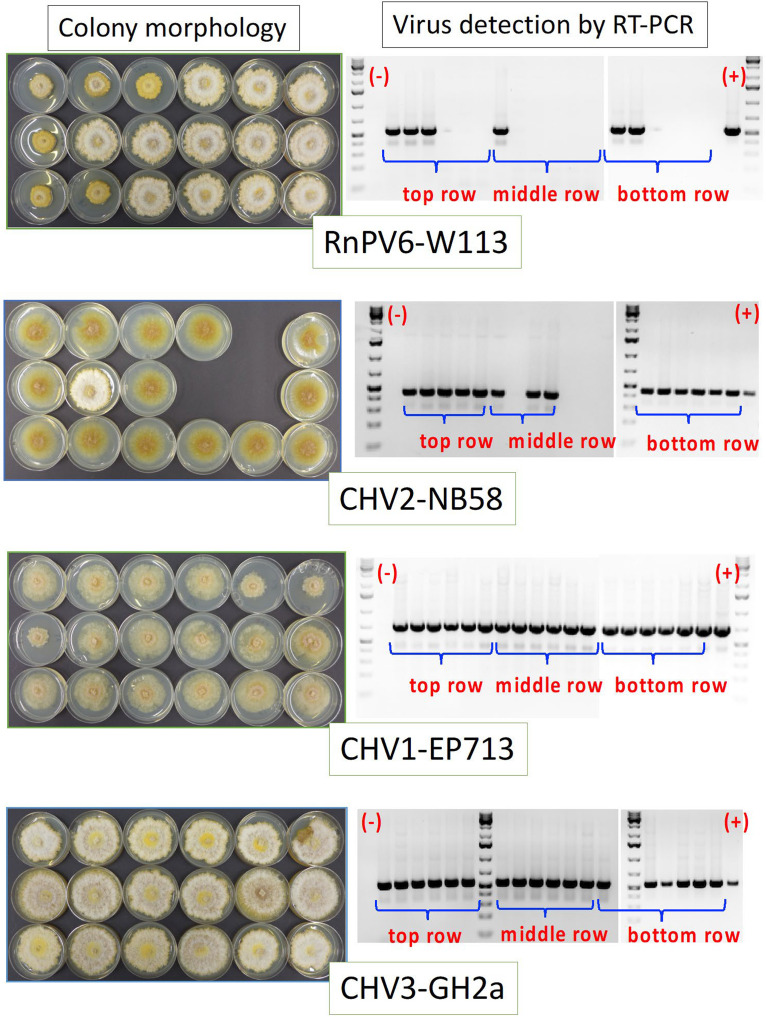
The ascomycete Cryphonectria parasitica causes destructive chestnut blight. Biological control of the fungus by virus infection (hypovirulence) has been shown to be an effective control strategy against chestnut blight in Europe. To provide biocontrol effects, viruses must be able to induce hypovirulence and spread efficiently in chestnut trees. Field studies using living trees to date have focused on a selected family of viruses called hypoviruses, especially prototypic hypovirus CHV1, but there are now known to be many other viruses that infect C. parasitica Here, we tested seven different viruses for their hypovirulence induction, biocontrol potential, and transmission properties between two vegetatively compatible but molecularly distinguishable fungal strains in trees. The test included cytosolically and mitochondrially replicating viruses with positive-sense single-stranded RNA or double-stranded RNA genomes. The seven viruses showed different in planta behaviors and were classified into four groups. Group I, including CHV1, had great biocontrol potential and could protect trees by efficiently spreading and converting virulent to hypovirulent cankers in the trees. Group II could induce high levels of hypovirulence but showed much smaller biocontrol potential, likely because of inefficient virus transmission. Group III showed poor performance in hypovirulence induction and biocontrol, while efficiently being transmitted in the infected trees. Group IV could induce hypovirulence and spread efficiently but showed poor biocontrol potential. Nuclear and mitochondrial genotyping of fungal isolates obtained from the treated cankers confirmed virus transmission between the two fungal strains in most isolates. These results are discussed in view of dynamic interactions in the tripartite pathosystem.IMPORTANCE The ascomycete Cryphonectria parasitica causes destructive chestnut blight, which is controllable by hypovirulence-conferring viruses infecting the fungus. The tripartite chestnut/C. parasitica/virus pathosystem involves the dynamic interactions of their genetic elements, i.e., virus transmission and lateral transfer of nuclear and mitochondrial genomes between fungal strains via anastomosis occurring in trees. Here, we tested diverse RNA viruses for their hypovirulence induction, biocontrol potential, and transmission properties between two vegetatively compatible but molecularly distinguishable fungal strains in live chestnut trees. The tested viruses, which are different in genome type (single-stranded or double-stranded RNA) and organization, replication site (cytosol or mitochondria), virus form (encapsidated or capsidless) and/or symptomatology, have been unexplored in the aforementioned aspects under controlled conditions. This study showed intriguing different in-tree behaviors of the seven viruses and suggested that to exert significant biocontrol effects, viruses must be able to induce hypovirulence and spread efficiently in the fungus infecting the chestnut trees.

Keywords: Cryphonectria parasitica; chestnut blight fungus; hypovirus; mitovirus; mycovirus; reovirus; virus spread.


Figures

FIG 1
FIG 1
Colony morphology of PC7 and EP155 infected with different viruses. EP155 (top row) and PC7 (bottom row) infected by the seven virus strains were prepared as described in Materials and Methods. Introduced viruses, listed in Table 1, are shown at the top and bottom. Colonies were grown on PDA for 1 week on the benchtop at approximately 23°C and photographed. Virus-free EP155 and PC7 were grown in parallel.
FIG 2
FIG 2
Schematic representation of the experimental procedures to assess biocontrol potential and in-tree spread of viruses. For both assays, the European virulent strain PC7 was inoculated into European chestnut trees (shown by blue circles) with the aid of a cork borer (A). Two weeks postinoculation, the trees were challenged (shown by yellow circles) with EP155 colonies infected with different viruses (Table 1) at one (for assay II, virus spread) or eight sites (for assay III, biocontrol) on the periphery of virulent cankers (shown by brownish ovals) induced by PC7 (B) with the aid of a cork borer. For fungal isolation, bark plugs were obtained from a total of 18 sites (for virus spread assay) or 4 sites inside the original canker area plus 4 sites in expanded areas (for the biocontrol assay) for challenge inoculation with CHV3-GH2a-, RnPV1-W113-, CpMV1-NB631-, and MyRV1-9B21-infected EP155 (C). For challenge inoculation with the remaining virus-infected colonies, only four inside plugs were utilized, because the original virulent cankers became inactive and failed to expand. Bark sampling was performed 3 and 6 weeks (for the virus spread assay) and 2 months (for the biocontrol assay) after challenge inoculation with the aid of a bone marrow needle. Isolated fungi were examined for virus infection and nuclear and mitochondrial genotypes.
FIG 3
FIG 3
Virulence of EP155 infected by different viruses. Three trees per virus-infected strain were inoculated. Virulence levels were expressed by areas of cankers induced by the respective fungal strains that were measured 2 months postinoculation. Virus-free EP155 was also inoculated into three chestnut trees in parallel. (A) Mean canker areas and standard deviations calculated from the values in panel B. (B) Canker areas measured for three trees per virus-infected strain 2 months postinoculation. Measurements made at different time points are shown in Table 3.
FIG 4
FIG 4
Virus transmission in chestnut trees. As shown in Fig. 2, 3-year-old chestnut trees were first inoculated with a virus-free virulent fungal strain, PC7. EP155 strains each infected by the respective viruses were inoculated at the lengthwise growing edge of the canker 2 weeks after the first inoculation. Eighteen bark plugs were taken and placed on water agar containing streptomycin to isolate fungal strains. After a few days, mycelia were transferred to PDA plates. Fungal isolates obtained 3 weeks postchallenge inoculation from cankers treated with RnPV6-W113-, CHV2-NB58-, CHV1-EP713-, and CHV3-GH2a-infected EP155 were cultured on PDA for 1 week on the benchtop and photographed (left). RT-PCR analysis of the fungal isolates recovered from bark samples was carried out (right). Direct colony RT-PCR with toothpicks was employed to examine recovered fungal isolates for virus infection. Amplified cDNA fragments were electrophoresed in 1.2% agarose gel in the 1× TBE buffer system (89 mM Tris-borate, 89 mM boric acid, 2.5 mM EDTA [pH 8.3]). GeneRuler 1 kb Plus DNA ladders (Thermo Fisher Scientific) were used as size standards. Virus transmission rates are summarized in Table 4.
FIG 5
FIG 5
Schematic representation of bark sampling sites for virus-free isolates and isolates with a mixture of the EP155 and PC7 genomes. Incomplete spread of MyRV1-9B21 (A) and RnPV1-W113 (B) and detection of the EP155 nuclear genome in the CHV3-GH2a spread assay (C). As shown in Fig. 1, the blue and yellow filled circles indicate the sites of the primary and challenge inoculations. Gray circles indicate the sites where the respective virus was detected, and white circles indicate where it was not detected. (A) Results of experiment I 6 weeks after challenge inoculation. (B) Results of experiment II 6 weeks after challenge inoculation (Table 4). (C) Genotyping revealed a mixture of the EP155 and PC7 nuclear genomes in a few isolates obtained from the CHV3-GH2a spread experiments I and II (Table 4). Orange symbols indicate the four sampling sites from which isolates with the mixed genotype were recovered. Gray symbols denote the sites where only the PC7 genotype was present.
FIG 6
FIG 6
Biological control effects of different viruses. Three-year-old European chestnut trees were first inoculated with a virus-free virulent fungal strain, PC7, at one site per tree (Fig. 2). After 2 weeks, the trees (five per fungal strain) were challenged by EP155 infected with different viruses (Table 1) inoculated at 8 sites per tree at the periphery of growing cankers. Resulting cankers induced by the first and second challenge inoculations were photographed 8 weeks after the first inoculation. (A) Representative cankers, with the viruses used for the challenge inoculation shown at the bottom. Cankers induced by the challenge inoculation with agar as a negative control in parallel are also shown. The original canker area induced by PC7 at the time point of the challenge inoculation is shown by a white bracket, while the expanded canker area 6 weeks after challenge inoculation is indicated by a yellow bracket. Canker areas were measured 6 weeks after challenge inoculation. (B) Mean canker expansions and standard deviations, calculated by five biological replicates. Biocontrol effects of viruses on the fungal pathogen are greater as canker expansions are smaller.
FIG 7
FIG 7
Chestnut trees surviving and dying of inoculation by PC7. Two representative trees, inoculated with the virulent strain PC7, are shown. The trees were challenged with the CHV1-Δp69-infected fungus (right) or mock-inoculated (agar) (left) and photographed 15 weeks postchallenge inoculation.

Similar articles

Cited by

References

    1. Anagnostakis SL. 1982. Biological control of chestnut blight. Science 215:466–471. doi:10.1126/science.215.4532.466. - DOI - PubMed
    1. Heiniger U, Rigling D. 1994. Biological control of chestnut blight in Europe. Annu Rev Phytopathol 32:581–599. doi:10.1146/annurev.py.32.090194.003053. - DOI
    1. Rigling D, Prospero S. 2018. Cryphonectria parasitica, the causal agent of chestnut blight: invasion history, population biology and disease control. Mol Plant Pathol 19:7–20. doi:10.1111/mpp.12542. - DOI - PMC - PubMed
    1. Nuss DL. 1992. Biological control of chestnut blight: an example of virus-mediated attenuation of fungal pathogenesis. Microbiol Rev 56:561–576. doi:10.1128/MR.56.4.561-576.1992. - DOI - PMC - PubMed
    1. Anagnostakis SL, Chen BS, Geletka LM, Nuss DL. 1998. Hypovirus transmission to ascospore progeny by field-released transgenic hypovirulent strains of Cryphonectria parasitica. Phytopathology 88:598–604. doi:10.1094/PHYTO.1998.88.7.598. - DOI - PubMed

Publication types

Supplementary concepts

LinkOut - more resources